. 2021 Apr;73(4):1290-1306.
doi: 10.1002/hep.31609. Epub 2021 Mar 19.

Identification of a Metabolic, Transcriptomic, and Molecular Signature of Patatin-Like Phospholipase Domain Containing 3-Mediated Acceleration of Steatohepatitis

Affiliations

Identification of a Metabolic, Transcriptomic, and Molecular Signature of Patatin-Like Phospholipase Domain Containing 3-Mediated Acceleration of Steatohepatitis

Bubu A Banini et al. Hepatology. 2021 Apr.

Abstract

Background and aims: The mechanisms by which the I148M mutant variant of the patatin-like phospholipase domain-containing 3 (PNPLA3I148M ) drives development of nonalcoholic steatohepatitis (NASH) are not known. The aim of this study was to obtain insights on mechanisms underlying PNPLA3I148M -induced acceleration of NASH.

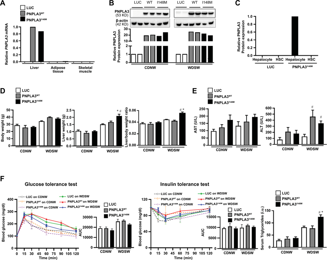
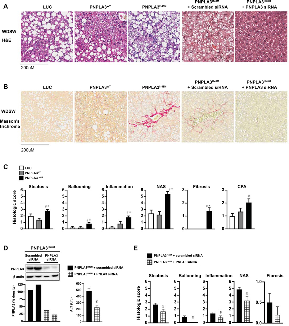
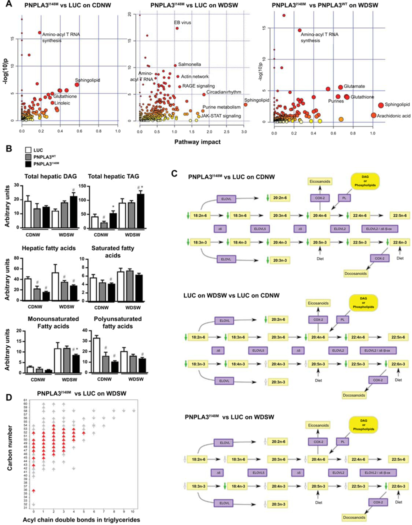
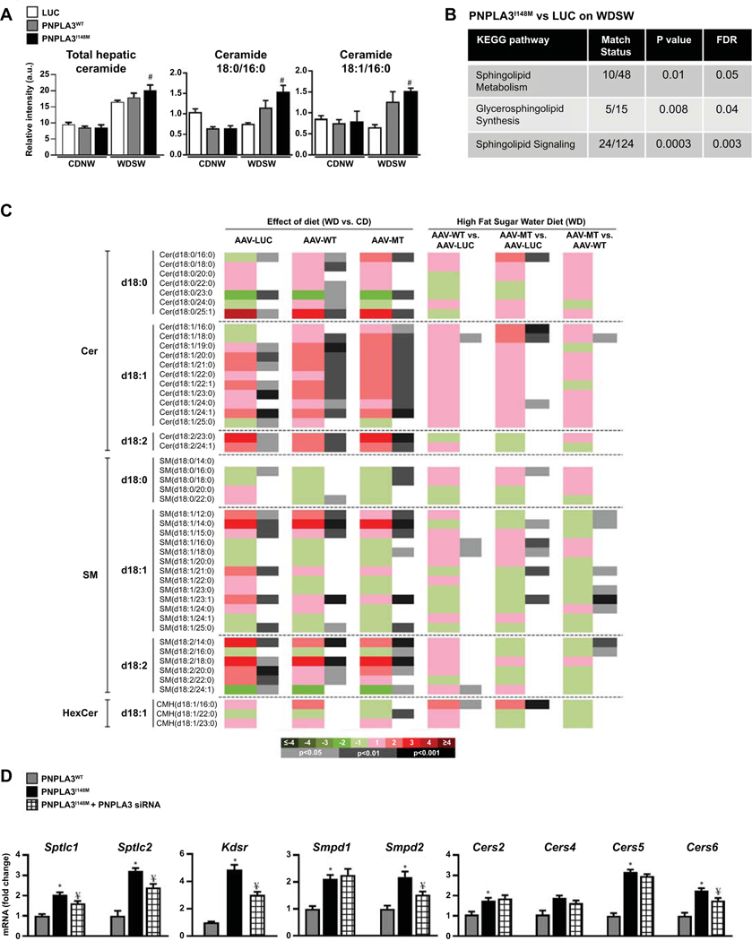
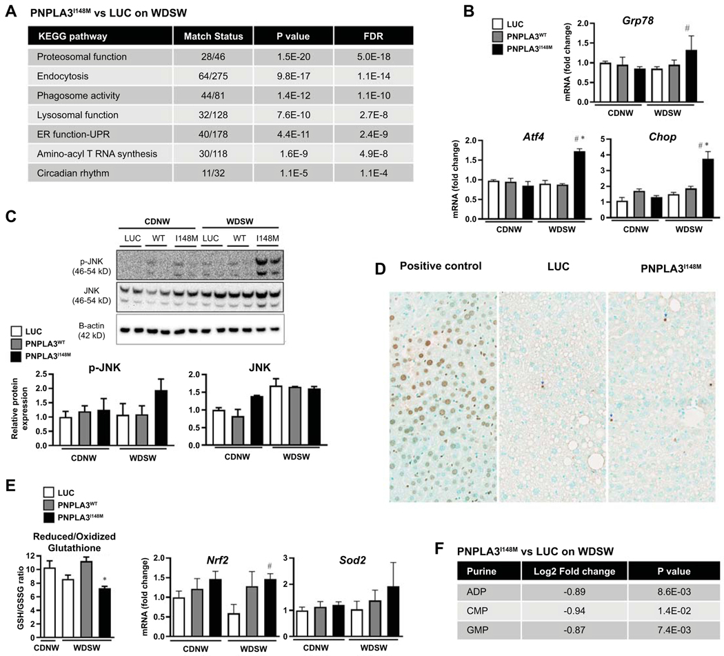
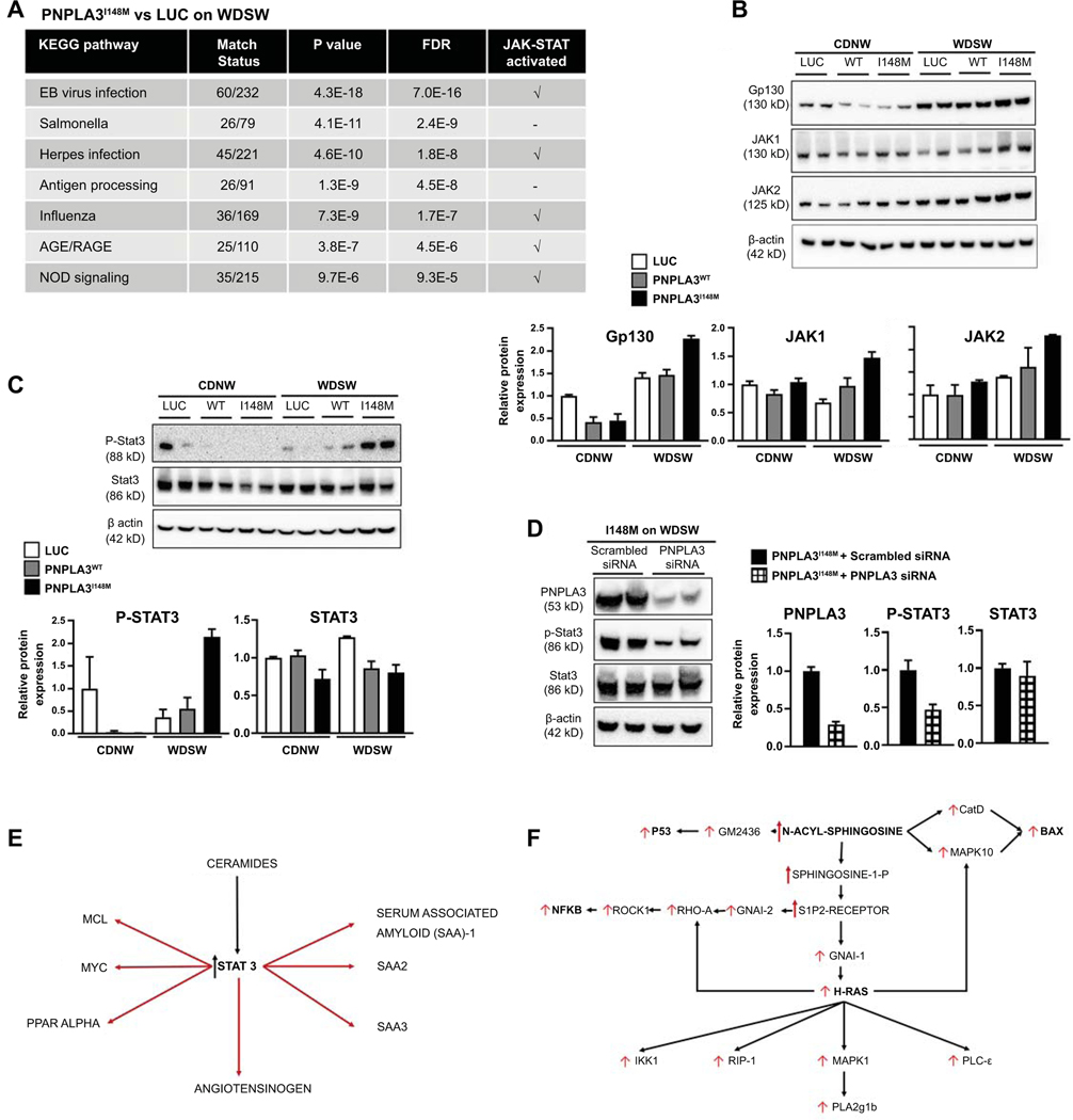
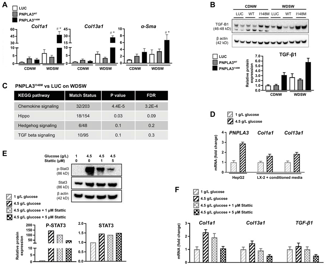
Approach and results: Hepatocyte-specific overexpression of empty vector (luciferase), human wild-type PNPLA3, or PNPLA3I148M was achieved using adeno-associated virus 8 in a diet-induced mouse model of nonalcoholic fatty liver disease followed by chow diet or high-fat Western diet with ad libitum administration of sugar in drinking water (WDSW) for 8 weeks. Under WDSW, PNPLA3I148M overexpression accelerated steatohepatitis with increased steatosis, inflammation ballooning, and fibrosis (P < 0.001 versus other groups for all). Silencing PNPLA3I148M after its initial overexpression abrogated these findings. PNPLA3I148M caused 22:6n3 docosahexanoic acid depletion and increased ceramides under WDSW in addition to increasing triglycerides and diglycerides, especially enriched with unsaturated fatty acids. It also increased oxidative stress and endoplasmic reticulum stress. Increased total ceramides was associated with signature of transducer and activator of transcription 3 (STAT3) activation with downstream activation of multiple immune-inflammatory pathways at a transcriptomic level by network analyses. Silencing PNPLA3I148M reversed STAT3 activation. Conditioned media from HepG2 cells overexpressing PNPLA3I148M increased procollagen mRNA expression in LX2 cells; this was abrogated by hepatocyte STAT3 inhibition.

Conclusions: Under WDSW, PNPLA3I148M overexpression promotes steatosis and NASH by metabolic reprogramming characterized by increased triglycerides and diglycerides, n3 polyunsaturated fatty acid depletion, and increased ceramides with resultant STAT3 phosphorylation and downstream inflammatory pathway activation driving increased stellate cell fibrogenic activity.


Figures

Figure 1:
Figure 1:. AAV-mediated PNPLA3 expression is liver-specific and its effects are dependent on a high fat high sugar diet.
(A) Liver-specific overexpression of human PNPLA3 gene in syngeneic C57Bl6J/129S1svlmJ mice (n=3 mice per group). Adeno associated virus (AAV) containing empty luciferase (luc), PNPLA3WT, or PNPLA3I148M was introduced by retro-orbital injection. Real-time PCR demonstrated high liver expression of both PNPLA3 forms with virtually no expression in other organs. (B) Mice were randomized to CDNW or WDSW ad libitum for 8 weeks after retro-orbital injection of the three AAV constructs. Western blot analysis was performed in liver lysates to determine PNPLA3 protein expression, which was normalized to AAV-luc on CDNW or WDSW. (C) Hepatocytes and stellate cells were isolated from the liver 2 weeks after injection of the AAV constructs. PNPLA3 was expressed in hepatocytes but not stellate cells confirming hepatocyte specific expression. (D) Mice were randomized to CDNW or WDSW after injection of the three AAV constructs. All mice on WDSW gained more weight than mice on WDSW. The liver weight and liver/body weight ratio was significantly increased in PNPLA3I148M mice compared to other groups on WDSW diet. (E) Plasma collected at the time of sacrifice was used to determine aminotransferase levels, with ALT reaching significance in PNPLA3WT and PNPLA3I148M mice on WDSW compared to luc. (F) There were no differences across the AAV-groups with respect to glucose homeostasis and insulin resistance measured by glucose tolerance test and insulin tolerance tests, however PNPLA3I148M mice had significant hypertriglyceridemia on WDSW compared to other groups. The mice data shown represent 8–10 mice per group unless previously indicated. # represents p<0.05 compared to luc on WDSW; * represents p<0.05 compared to PNPLA3WT on WDSW.
Figure 2:
Figure 2:. PNPLA3I148M expression aggravates steatohepatitis and fibrosis which is reversed by PNPLA3I148M silencing.
(A) Liver tissue collected at the time of sacrifice were sectioned for histological analysis; representative Hematoxylin and eosin (H&E) stained sections of liver tissue for mice on WDSW are shown. Whereas Luc and PNPLA3WT mice had a fatty liver, PNPLA3I148M mice developed steatohepatitis with fibrosis. The specificity of these findings were confirmed through siRNA-mediated silencing of PNPLA3I148M which resulted in only fatty liver despite feeding on WDSW, in contrast to PNPLA3I148M mice who received scrambled siRNA in which steatohepatitis and fibrosis were re-demonstrated. (B) Sirius Red staining showed that fibrosis developed only in PNPLA3I148M mice with or without scrambled siRNA. (C) Histological scoring of H&E sections showed that the severity of steatosis, ballooning, lobular inflammation, and NAFLD activity score were all significantly higher in PNPLA3I148M mice compared to Luc and PNPLA3WT. Similarly, scoring of Sirius Red stained sections showed that fibrosis stage and collagen proportional area (CPA) were increased only in PNPLA3I148M mice. (D) A Western blot demonstrating effective silencing of PNPLA3 I148M after its initial overexpression in liver tissue is shown. Silencing PNPLA3I148M led to significant reduction in liver inflammation as demonstrated by reduced ALT. (E) Silencing PNPLA3I148M also reduced the severity of steatosis, ballooning, lobular inflammation, NAFLD activity score and fibrosis stage. Histological sections are representative sections and the graphical data represent a total of 8–10 in each group for Luc, PNPLA3WT and PNPLA3I148M and 6 mice each for the experiment with scrambled sequence or siRNA administration. # represents p<0.05 compared to luc on WDSW; * represents p<0.05 compared to PNPLA3WT on WDSW; ¥ represents p<0.05 compared to scrambled siRNA-PNPLA3I148M
Figure 3:
Figure 3:. Metabolic reprogramming induced by overexpression of PNPLA3I148M.
(A) Integrated analysis of metabolites and gene-expression data obtained from metabolomic and RNA seq analysis of liver tissue was performed. Under chow diet, the pathways most significantly altered (largest number of significantly altered metabolites and genes) by PNPLA3I148M (vs Luc) when plotted as a function of pathway impact (number of significantly altered pathways intersecting with differentially expressed genes and metabolites in a specific pathway on network analysis) were PUFA and sphingolipid metabolism (left panel). Under WDSW diet, there was a larger set of pathways, mainly metabolic, that were both significantly altered and had a high impact score (middle panel). The specificity of these changes for PNPLA3I148M was tested by comparison to PNPLA3WT under WDSW diet (right panel). These confirmed that the main impact attributable to mutation of the PNPLA3 gene was a metabolic reprogramming with main effects on PUFA, glutathione and sphingolipid pathways. (B) Total DAG, TAG, FFA, and SFA, MUFA and PUFA are shown and demonstrate an increase in DAG and TAG, and a decrease in all fatty acid species in PNPLA3I148M mice on WDSW compared to the other groups on the same diet. (C) Significantly altered PUFA (shown by green arrows) in PNPLA3I148M vs Luc under chow diet are shown (top panel). Luc mice on WDSW diet vs CDNW demonstrated a major depletion of PUFA by a WDSW diet (middle panel). Additional changes in PUFA in PNPLA3I148M (vs Luc mice) on WDSW diet over and on top of changes induced by the diet itself were seen (bottom panel). Several PUFA were further decreased (shown in non-colored arrows) reaching significance for docosahexanoic acid (green arrow). (D) The depletion of PUFAs was accompanied by an increasing number of triglycerides double bonds. Data for 4–5 mice in each group are shown. # represents p<0.05 compared to luc on the same diet; * represents p<0.05 compared to PNPLA3WT on the same diet.
Figure 4:
Figure 4:. PNPLA3I148M upregulates ceramides signaling.
(A) Total ceramides were increased in all groups on WDSW diet, with PNPLA3I148M on WDSW demonstrating significantly increased in total ceramides including unsaturated and saturated ceramides compared to other groups. (B) KEGG pathway analysis between PNPLA3I148M and luc on WDWS demonstrate upregulation of sphingolipid signaling pathways. (C) A heat-map of altered sphingolipids is shown with effect of diet (WDSW vs CDNW) in Luc, PNPLA3WTand PNPLA3I148M mice (left three columns). Pairwise comparisons of groups on WDSW are also shown (right three columns). Both diet and PNPLA3 expression remodel the sphingolipidome, with the greatest increase in ceramides seen in PNPLA3I148M mice on WDSW diet. (D) Real time PCR analysis of hepatic expression of genes involved in ceramide synthesis shows upregulation of several key genes involved in de novo synthesis, salvage pathway and sphingomyelinase in PNPLA3I148M mice compared to PNPLA3WT on WDSW. Silencing PNPLA3 I148M resulted in downregulation of these genes, indicating that the observed increments were linked to PNPLA3I148M overexpression. Data from 4–5 mice in each group are shown. # represents p<0.05 compared to luc; * represents p<0.05 compared to PNPLA3WT; ¥ represents p<0.05 compared PNPLA3I148M
Figure 5:
Figure 5:. PNPLA3I148M promotes ER stress, oxidative stress and cell death.
(A) KEGG pathway analysis showed significant alteration of key cellular processes involved in ER stress and cell turnover in PNPLA3I148M mice versus Luc on WDSW. (B) Real time PCR confirmed significant upregulation of hepatic genes involved in ER stress, including GRP78, ATF4, and CHOP in mice overexpressing PNPLA3I148M compared to Luc and PNPLA3WT. (C) Despite similar levels of PNPLA3 protein expression, upregulation of ER stress as demonstrated by increase in phosphorylated JNK was specific to PNPLA3I148M, indicating what this was not simply a function of excess PNPLA3 protein synthesis. (D) This was accompanied by increased cell death shown by TUNEL assay in PNPLA3I148M mice. (E) PNPLA3I148M mice on WDSW showed increase in oxidative stress as evidenced by decrease in the reduced/oxidized gluthathione ratio and increased expression of superoxide dismutase and nrf-2 genes. (F) There was also a depletion of multiple purines including ADP, CMP, and GMP in PNPLA3I148M mice compared to luc on WDSW. Data from 4–6 mice in each group are shown.
Figure 6:
Figure 6:. PNPLA3I148M on high fat diet differentially upregulates innate immune system and inflammatory pathways.
(A) KEGG pathway analysis of combined metabolite-gene expression pathways showed significant upregulation of immune-inflammatory pathways, with several pathways related to the innate immune system and adaptive immune system significantly activated in PNPLA3I148M (vs Luc) on WDSW diet. Most of these pathways included JAK-STAT activation as a component. (B and C): Activation of Gp130, JAK and STAT3 signaling was confirmed by Western blot analysis which showed increased protein levels in PNPLA3I148M mice on WDSW compared to the other groups. (D) The specificity of these findings for PNPLA3I148M was further established by STAT3 inactivation following silencing of PNPLA3I148M. (E) Network analysis was performed to identify key metabolites linked to activation status of inflammatory pathways specifically activated by PNPLA3I148M overexpression under WDSW conditions (vs Luc and PNPLA3WT). A strong signal for STAT3 targets coordinately activated with STAT3 (at a transcriptomic level) and specifically linked to accelerated steatohepatitis was noted. (F) Similarly, multiple injury-inflammatory targets were activated coordinately with ceramides (specifically those immediately upstream of n-acyl sphingosine) that were linked to the accelerated histological phenotype of NASH in PNPLA3I148M. Data from 4–6 mice in each group are shown.
Figure 7:
Figure 7:. PNPLA3I148M upregulates fibrogenic pathways which is reversed by silencing.
(A) Realtime PCR of liver tissue showed upregulation of procollagen I, III and α-smooth muscle actin mRNA in PNPLA3I148M mice compared to luc and PNPLA3WT under WDSW conditions. (B) Western blot analysis showed upregulation of TGF-β protein levels in PNPLA3I148M mice on WDSW. (C) KEGG pathway analysis showed differential upregulation of signaling pathways related to fibrosis, with chemokine and hippo pathway signaling reaching statistical significance. (D) Cross-talk between hepatocytes overexpressing PNPLA3I148 and hepatic stellate cells was explored in vitro. HepG2 cells, known to express PNPLA3I148, were exposed to high glucose which resulted in upregulation of PNPLA3I148 (left panel). Conditioned media from HepG2 cells treated with high or low glucose resulted in dose-dependent upregulation of procollagen I and 3 mRNA by LX2 cells (middle and right panel). (E) Treatment of HepG2 cells with a selective Stat3 inhibitor, Stattic, resulted in a dose-dependent decrease in Stat3 activation despite exposure to high glucose. (F) Selective Stat3 inhibition resulted in a corresponding downregulation of procollagen 1 and 3 and TGF-β1 mRNA in HepG2 cells.

Similar articles

Cited by

References

    1. Estes C, Razavi H, Loomba R, Younossi Z, Sanyal AJ. Modeling the epidemic of nonalcoholic fatty liver disease demonstrates an exponential increase in burden of disease. Hepatology 2018;67:123–133. - PMC - PubMed
    1. Friedman SL, Neuschwander-Tetri BA, Rinella M, Sanyal AJ. Mechanisms of NAFLD development and therapeutic strategies. Nat Med 2018;24:908–922. - PMC - PubMed
    1. Kleiner DE, Brunt EM, Wilson LA, Behling C, Guy C, Contos M, Cummings O, et al. Association of Histologic Disease Activity With Progression of Nonalcoholic Fatty Liver Disease. JAMA Netw Open 2019;2:e1912565. - PMC - PubMed
    1. Romeo S, Kozlitina J, Xing C, Pertsemlidis A, Cox D, Pennacchio LA, Boerwinkle E, et al. Genetic variation in PNPLA3 confers susceptibility to nonalcoholic fatty liver disease. Nat Genet 2008;40:1461–1465. - PMC - PubMed
    1. Valenti L, Al-Serri A, Daly AK, Galmozzi E, Rametta R, Dongiovanni P, Nobili V, et al. Homozygosity for the patatin-like phospholipase-3/adiponutrin I148M polymorphism influences liver fibrosis in patients with nonalcoholic fatty liver disease. Hepatology 2010;51:1209–1217. - PubMed

Publication types

MeSH terms