

Versican: A Dynamic Regulator of the Extracellular Matrix
- PMID: 33131383
- PMCID: PMC7649968
- DOI: 10.1369/0022155420953922
Versican: A Dynamic Regulator of the Extracellular Matrix
Abstract
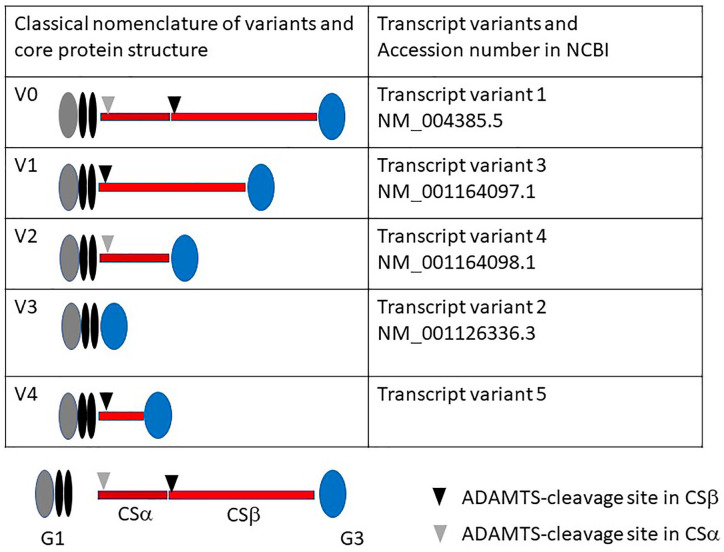
Versican is a large chondroitin sulfate/dermatan sulfate proteoglycan belonging to the aggrecan/lectican family. In adults, this proteoglycan serves as a structural macromolecule of the extracellular matrix in the brain and large blood vessels. In contrast, versican is transiently expressed at high levels during development and under pathological conditions when the extracellular matrix dramatically changes, including in the inflammation and repair process. There are many reports showing the upregulation of versican in cancer, which correlates with cancer aggressiveness. Versican has four classical splice variants, and all the variants contain G1 and G3 domains at N- and C-termini, respectively. There are two glycosaminoglycan attachment domains CSα and CSβ. The largest V0 variant contains both CSα and CSβ, V1 contains CSβ, V2 contains CSα, and the shortest G3 variant has neither of them. Versican degradation is initiated by cleavage at a site in the CSβ domain by ADAMTS (a disintegrin and metalloproteinase with thrombospondin motifs) proteinases. The N-terminal fragment containing the G1 domain has been reported to exert various biological functions, although its mechanisms of action have not yet been elucidated. In this review, we describe the role of versican in inflammation and cancer and also address the biological function of versikine.
Keywords: extracellular matrix; glycosaminoglycan; hyaluronan; matrikine; microenvironment; proteoglycan.
Conflict of interest statement
Figures
Similar articles
-
Isolation and Purification of Versican and Analysis of Versican Proteolysis.Methods Mol Biol. 2022;2303:559-578. doi: 10.1007/978-1-0716-1398-6_43. Methods Mol Biol. 2022. PMID: 34626407
-
Accumulation of versican facilitates wound healing: Implication of its initial ADAMTS-cleavage site.Matrix Biol. 2020 May;87:77-93. doi: 10.1016/j.matbio.2019.10.006. Epub 2019 Oct 26. Matrix Biol. 2020. PMID: 31669737
-
The multiple, complex roles of versican and its proteolytic turnover by ADAMTS proteases during embryogenesis.Matrix Biol. 2014 Apr;35:34-41. doi: 10.1016/j.matbio.2014.01.005. Epub 2014 Jan 18. Matrix Biol. 2014. PMID: 24444773 Free PMC article. Review.
-
Determinants of versican-V1 proteoglycan processing by the metalloproteinase ADAMTS5.J Biol Chem. 2014 Oct 3;289(40):27859-73. doi: 10.1074/jbc.M114.573287. Epub 2014 Aug 13. J Biol Chem. 2014. PMID: 25122765 Free PMC article.
-
Aggrecan and versican: two brothers close or apart.Am J Physiol Cell Physiol. 2022 May 1;322(5):C967-C976. doi: 10.1152/ajpcell.00081.2022. Epub 2022 Apr 6. Am J Physiol Cell Physiol. 2022. PMID: 35385326 Review.
Cited by
-
Multimodal Evaluation and Management of Wagner Syndrome-Three Patients from an Affected Family.Genes (Basel). 2024 Sep 8;15(9):1178. doi: 10.3390/genes15091178. Genes (Basel). 2024. PMID: 39336769 Free PMC article.
-
First Characterization of ADAMTS-4 in Kidney Tissue and Plasma of Patients with Chronic Kidney Disease-A Potential Novel Diagnostic Indicator.Diagnostics (Basel). 2022 Mar 7;12(3):648. doi: 10.3390/diagnostics12030648. Diagnostics (Basel). 2022. PMID: 35328201 Free PMC article.
-
Single-Cell Gene Network Analysis and Transcriptional Landscape of MYCN-Amplified Neuroblastoma Cell Lines.Biomolecules. 2021 Jan 28;11(2):177. doi: 10.3390/biom11020177. Biomolecules. 2021. PMID: 33525507 Free PMC article.
-
Targeting Versican as a Potential Immunotherapeutic Strategy in the Treatment of Cancer.Front Oncol. 2021 Aug 30;11:712807. doi: 10.3389/fonc.2021.712807. eCollection 2021. Front Oncol. 2021. PMID: 34527586 Free PMC article. Review.
-
VCAN in the extracellular matrix drives glioma recurrence by enhancing cell proliferation and migration.Front Neurosci. 2024 Nov 1;18:1501906. doi: 10.3389/fnins.2024.1501906. eCollection 2024. Front Neurosci. 2024. PMID: 39554845 Free PMC article.
References
-
- Karamanos NK, Piperigkou Z, Theocharis AD, Watanabe H, Franchi M, Baud S, Brezillon S, Gotte M, Passi A, Vigetti D, Ricard-Blum S, Sanderson RD, Neill T, Iozzo RV. Proteoglycan chemical diversity drives multifunctional cell regulation and therapeutics. Chem Rev. 2018;118(18):9152–232. doi:10.1021/acs.chemrev.8b00354. - DOI - PubMed
Publication types
MeSH terms
Substances
LinkOut - more resources
Full Text Sources
Medical
