

Review
doi: 10.1016/j.tins.2020.10.010.
Epub 2020 Oct 22.
PET Imaging as a Tool for Assessing COVID-19 Brain Changes
Affiliations
- PMID: 33131922
- PMCID: PMC7580682
- DOI: 10.1016/j.tins.2020.10.010
Item in Clipboard
Review
PET Imaging as a Tool for Assessing COVID-19 Brain Changes
Trends Neurosci.
2020 Dec.
Abstract
A substantial fraction of coronavirus disease 2019 (COVID-19) patients experience neurological manifestations. Nevertheless, brain changes caused by severe acute respiratory syndrome coronavirus 2 (SARS-CoV-2) remain largely unknown. Here, we provide a brief overview of positron emission tomography (PET) applications that could advance current understanding of CNS pathophysiological alterations associated with SARS-CoV-2 infection.
Keywords: PET radiotracers; SARS-CoV-2; neurological manifestations.
Copyright © 2020 Elsevier Ltd. All rights reserved.
Figures
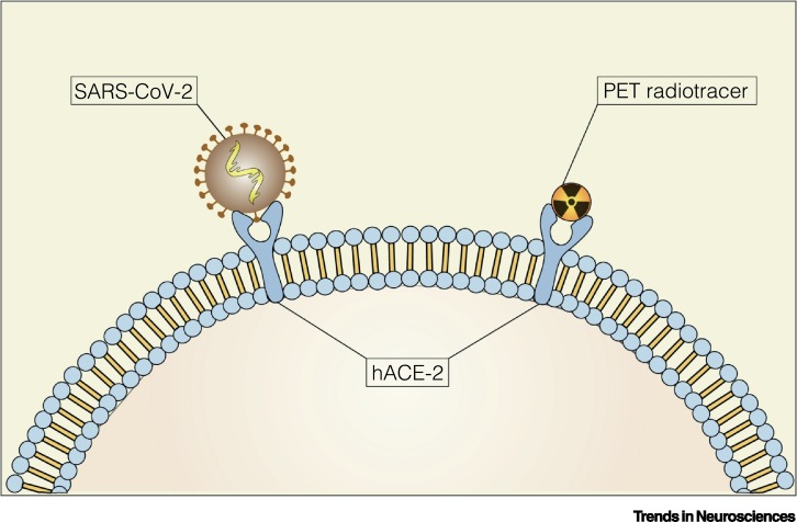
A Potential Positron Emission Tomography (PET) Radiotracer Targeting Human Angiotensin-Converting Enzyme 2 (hACE2). Schematic representation of a potential PET radiotracer and severe acute respiratory syndrome coronavirus 2 (SARS-CoV-2) binding to the hACE2.
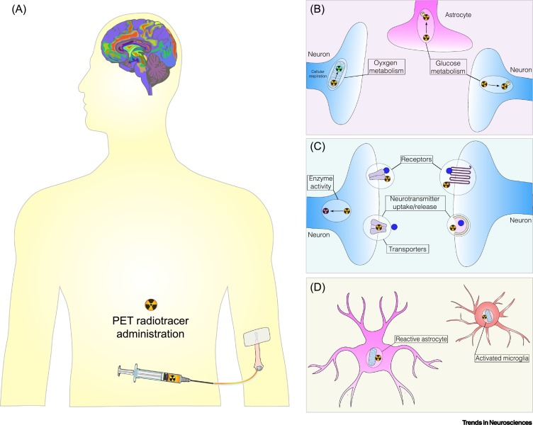
Assessing Coronavirus Disease 2019 (COVID-19) Brain Changes Using Positron Emission Tomography (PET) Imaging. (A) PET radiotracers are injected intravenously at tracer concentrations and a PET scanner records the distribution of radioactivity in the brain, which is reconstructed, analyzed, and read by imaging experts. PET radiotracers, commonly used in brain imaging research, can be used to uncover brain changes associated with severe acute respiratory syndrome coronavirus 2 (SARS-CoV-2) in the domains of (B) metabolic parameters, such as glucose and oxygen metabolism; (C) neurotransmission systems, by estimating neurotransmitter uptake/release/synthesis, enzyme activity, and receptor/transporter availability; and (D) brain cell types affected: neurons, astrocytes, or microglia.
References
Publication types
MeSH terms
LinkOut - more resources
Full Text Sources
Medical
Miscellaneous
