. 2020 Nov;8(2):e001540.
doi: 10.1136/bmjdrc-2020-001540.

Pancreatic GLP-1r binding potential is reduced in insulin-resistant pigs

Affiliations

Pancreatic GLP-1r binding potential is reduced in insulin-resistant pigs

Charles-Henri Malbert et al. BMJ Open Diabetes Res Care. 2020 Nov.

Abstract

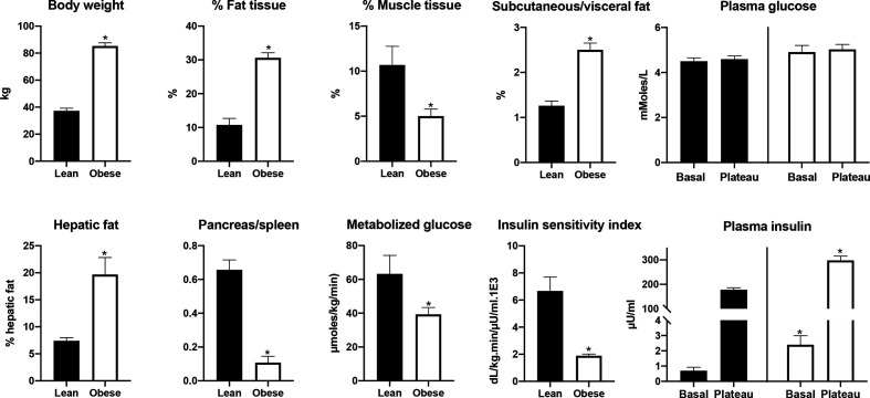
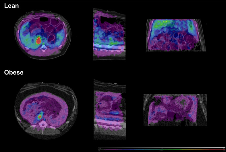
Introduction: The insulinotropic capacity of exogenous glucagon like peptide-1 (GLP-1) is reduced in type 2 diabetes and the insulin-resistant obese. We have tested the hypothesis that this response is the consequence of a reduced pancreatic GLP-1 receptor (GLP-1r) density in insulin-resistant obese animals.

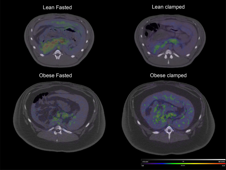
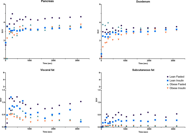
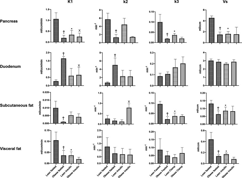
Research design and methods: GLP-1r density was measured in lean and insulin-resistant adult miniature pigs after the administration of a 68Ga-labeled GLP-1r agonist. The effect of hyperinsulinemia on GLP-1r was assessed using sequential positron emission tomography (PET), both in the fasted state and during a clamp. The impact of tissue perfusion, which could account for changes in GLP-1r agonist uptake, was also investigated using 68Ga-DOTA imaging.

Results: GLP-1r binding potential in the obese pancreas was reduced by 75% compared with lean animals. Similar reductions were evident for fat tissue, but not for the duodenum. In the lean group, induced hyperinsulinemia reduced pancreatic GLP-1r density to a level comparable with that of the obese group. The reduction in blood to tissue transfer of the GLP-1r ligand paralleled that of tissue perfusion estimated using 68Ga-DOTA.

Conclusions: These observations establish that a reduction in abdominal tissue perfusion and a lower GLP-1r density account for the diminished insulinotropic effect of GLP-1 agonists in type 2 diabetes.

Keywords: animal experimentation; glucagon-like peptide 1; insulin resistance; positron-emission tomography.


Conflict of interest statement

Competing interests: None declared.

Figures

Figure 1
Figure 1
Differences in phenotypic and metabolic features between the lean and obese groups. Note the absence of hyperglycemia in the obese while the fasting insulin is already elevated. Fat and muscle tissue percentage and the ratio between subcutaneous and visceral fat were quantified using a CT image of the abdomen. Hepatic fat was calculated from the ratio of X-ray attenuation between the liver and the spleen. Fat infiltration in the pancreas was estimated using the same method, but in the absence of a published correlation is presented only as ratio between the two attenuations. Metabolized glucose and insulin sensitivity were calculated from the clamp data. *Indicates a significant difference at p<0.05 between groups.
Figure 2
Figure 2
Hybrid image representing Vs (binding potential) coded PET image over CT anatomical image. Note the clear uptake of the GLP-1r radiolabel in the lean fasted animal and the reduction in Vs during clamp. This Vs value was similar to that observed in the obese animal both in the fasted condition and during the clamp. The scale was identical between the four conditions. Vs images were calculated using the Logan graphical method. GLP-1r, glucagon like peptide-1 receptor; Vs, specific volume of distribution.
Figure 3
Figure 3
Time activity curves describing [68Ga]Ga-DO3A-exendin-4 uptake in the pancreas, duodenum, visceral, and subcutaneous fat for fasting and clamp conditions in lean and obese animals. For simplicity, SE and several time intervals at the beginning of the curves are not presented. The uptake by the stomach is also not presented since it does not follow a 2-tissue compartment model. Note the absence of an effect of obesity and/or of hyperinsulinemia produced by the clamp on duodenal uptake, while obesity and hyperglycemia both reduce it. SUV, standard uptake value.
Figure 4
Figure 4
Transfer parameters of the 2-tissue compartment model describing the tissue uptake of [68Ga]Ga-DO3A-exendin-4. The value of the binding potential is also presented as Vs value. Note that all tissues, with the exception of the duodenum, exhibit a reduction in K1 as a consequence of obesity, a feature reproduced in lean animals by the administration of exogenous insulin. The same evolution is also evident for k3 parameter representing the rate of association of the ligand with the specific binding sites. †Significant difference from lean fasted, *significant difference from lean fasted, X significant difference from obese fasted. Vs, specific volume of distribution.
Figure 5
Figure 5
Hybrid image representing K1 coded PET image of 68Ga-DOTA distribution over CT anatomical image. The PET image was coded pixel-wise for K1 then filtered with a Gaussian filter for presentation. Note that K1 was markedly reduced in the obese demonstrating an overall reduction in tissue perfusion for both the duodenum and the pancreas.

Similar articles

Cited by

References

    1. Nauck MA, Meier JJ. The incretin effect in healthy individuals and those with type 2 diabetes: physiology, pathophysiology, and response to therapeutic interventions. Lancet Diabetes Endocrinol 2016;4:525–36. 10.1016/S2213-8587(15)00482-9 - DOI - PubMed
    1. Drucker DJ. Mechanisms of action and therapeutic application of glucagon-like peptide-1. Cell Metab 2018;27:740–56. 10.1016/j.cmet.2018.03.001 - DOI - PubMed
    1. Monami M, Dicembrini I, Nreu B, et al. . Predictors of response to glucagon-like peptide-1 receptor agonists: a meta-analysis and systematic review of randomized controlled trials. Acta Diabetol 2017;54:1101–14. 10.1007/s00592-017-1054-2 - DOI - PubMed
    1. Marathe CS, Rayner CK, Jones KL, et al. . Relationships between gastric emptying, postprandial glycemia, and incretin hormones. Diabetes Care 2013;36:1396–405. 10.2337/dc12-1609 - DOI - PMC - PubMed
    1. Gasbjerg LS, Bergmann NC, Stensen S, et al. . Evaluation of the incretin effect in humans using GIP and GLP-1 receptor antagonists. Peptides 2020;125:170183. 10.1016/j.peptides.2019.170183 - DOI - PubMed

Publication types