

Human placental hydrolysate promotes the long-term culture of hepatocyte-like cells derived from canine bone marrow
- PMID: 33132358
- PMCID: PMC7804030
- DOI: 10.1292/jvms.20-0320
Human placental hydrolysate promotes the long-term culture of hepatocyte-like cells derived from canine bone marrow
Abstract
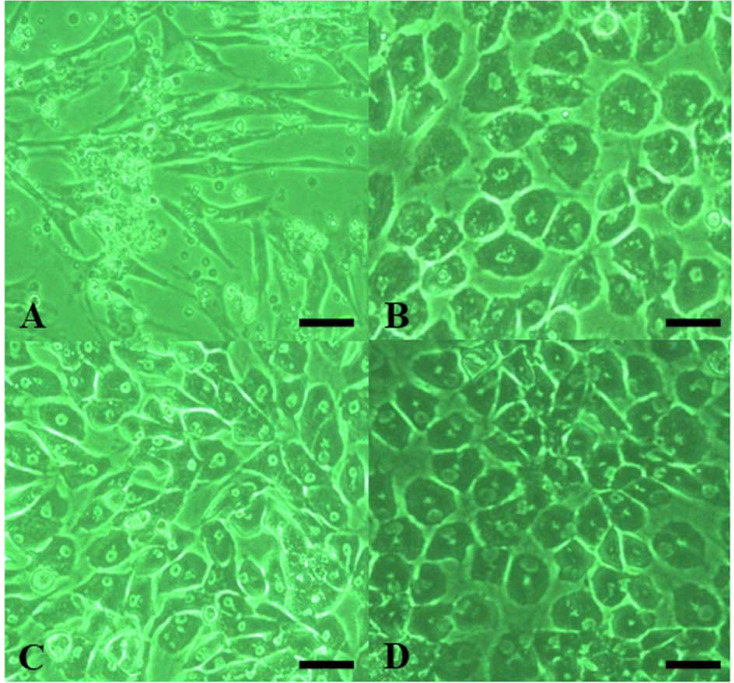
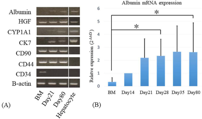
Long-term culture of canine artificial hepatocytes has not been established. We hypothesized that human placental hydrolysate (hPH) may support the long-term culture of differentiated hepatocyte-like cells. Canine bone marrow cells were cultured using modified hepatocyte growth medium supplemented with hPH. Quantitative reverse transcription polymerase chain reaction (RT-PCR) and immunocytochemical analysis for albumin, qualitative RT-PCR for cytochrome P450 1A1 (CYP1A1), hepatocyte growth factor (HGF), Cytokeratin 7 (CK7), CD90, CD44, and CD34, and functional analyses of CYP450 activity and low-density lipoprotein (LDL) uptake were performed. Cultured hepatocyte-like cells were able to maintain hepatocyte characteristics, including morphology, albumin synthesis, CYP450 activity, and LDL uptake for 80 days. Thus, hPH may be a potential facilitator for the long-term culture of hepatocyte-like cells. Clinicopathologically, this culture protocol of artificial hepatocytes will contribute to liver function evaluation.
Keywords: bone marrow; dog; hepatocyte; long-term culture; placenta.
Conflict of interest statement
The authors have nothing to disclose.
Figures


Similar articles
-
Canine bone marrow cells differentiate into hepatocyte-like cells and placental hydrolysate is a potential inducer.Res Vet Sci. 2009 Aug;87(1):1-6. doi: 10.1016/j.rvsc.2008.11.008. Epub 2009 Jan 1. Res Vet Sci. 2009. PMID: 19121529
-
Conversion of mesenchymal stem cells into a canine hepatocyte-like cells by Foxa1 and Hnf4a.Regen Ther. 2020 Feb 20;14:165-176. doi: 10.1016/j.reth.2020.01.003. eCollection 2020 Jun. Regen Ther. 2020. PMID: 32123700 Free PMC article.
-
Selection, proliferation and differentiation of bone marrow-derived liver stem cells with a culture system containing cholestatic serum in vitro.World J Gastroenterol. 2004 Nov 15;10(22):3308-12. doi: 10.3748/wjg.v10.i22.3308. World J Gastroenterol. 2004. PMID: 15484306 Free PMC article.
-
Establishment of large canine hepatocyte spheroids by mixing vascular endothelial cells and canine adipose-derived mesenchymal stem cells.Regen Ther. 2021 Dec 29;19:1-8. doi: 10.1016/j.reth.2021.11.007. eCollection 2022 Mar. Regen Ther. 2021. PMID: 35024388 Free PMC article.
-
A fat option for the pig: hepatocytic differentiated mesenchymal stem cells for translational research.Exp Cell Res. 2014 Feb 15;321(2):267-75. doi: 10.1016/j.yexcr.2013.10.018. Epub 2013 Nov 4. Exp Cell Res. 2014. PMID: 24200501
Cited by
-
Human Placenta Hydrolysate Protects Against Acetaminophen-Induced Liver Injury in Mice.Biomedicines. 2025 May 18;13(5):1219. doi: 10.3390/biomedicines13051219. Biomedicines. 2025. PMID: 40427046 Free PMC article.
References
MeSH terms
LinkOut - more resources
Full Text Sources
Research Materials
Miscellaneous