A case of internal trapping to a thrombosed giant rapidly growing aneurysm at the posterior cerebral artery
- PMID: 33132439
- PMCID: PMC7548258
- DOI: 10.18999/nagjms.82.3.557
A case of internal trapping to a thrombosed giant rapidly growing aneurysm at the posterior cerebral artery
Abstract
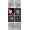
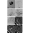
We describe a case of internal trapping including the vasa vasorum for a thrombosed giant rapidly growing posterior cerebral artery aneurysm and performing a detailed analysis. A 48-year-old woman was followed up in our hospital for a thrombosed large posterior cerebral artery aneurysm located in the P2 segment. She initially presented after experiencing a sudden headache on two occasions. Head computed tomography and magnetic resonance imaging indicated a larger aneurysm than before. Digital subtraction angiography with balloon occlusion test was assessed, and internal trapping was sequentially conducted. We detected that the vasa vasorum originated from the posterior temporal artery. Therefore, we embolized the posterior temporal artery including the vasa vasorum using N-butyl-2-cyanoacrylate and Lipiodol. Next, the anterior temporal artery was embolized with N-butyl-2-cyanoacrylate and Lipiodol, posterior temporal artery P3 segment and the aneurysm and finally the proximal P2 segment were embolized with coils. Final vertebral and internal carotid angiography showed complete obliteration of the aneurysm. On the day after the procedure her paresis worsened and she developed left upper quadrantanopia, however was finally discharged with no hemiparesis. We reported a case of a rapidly growing thrombosed giant posterior cerebral artery aneurysm treated by parent artery occlusion including the vasa vasorum with detailed image analysis.
Keywords: parent artery occlusion; posterior cerebral aneurysm; thrombosed giant aneurysm; vasa vasorum.
Conflict of interest statement
Declaration of interest: none. This summary statement ultimately will be published if the article is accepted.
Figures


Similar articles
-
Continued growth of and increased symptoms from a thrombosed giant aneurysm of the vertebral artery after complete endovascular occlusion and trapping: the role of vasa vasorum. Case report.J Neurosurg. 2003 Feb;98(2):407-13. doi: 10.3171/jns.2003.98.2.0407. J Neurosurg. 2003. PMID: 12593631
-
Symptomatic enlargement of an occluded giant carotido-ophthalmic aneurysm after endovascular treatment: the vasa vasorum theory.Acta Neurochir (Wien). 2009 Sep;151(9):1153-8. doi: 10.1007/s00701-009-0270-0. Epub 2009 Apr 3. Acta Neurochir (Wien). 2009. PMID: 19343269
-
Antegrade recanalization of completely embolized internal carotid artery after treatment of a giant intracavernous aneurysm: a case report.Surg Neurol. 1999 Dec;52(6):611-6. doi: 10.1016/s0090-3019(99)00131-7. Surg Neurol. 1999. PMID: 10660029
-
Multistage "Hybrid" (Open and Endovascular) Surgical Treatment of Vertebral Artery-Thrombosed Giant Aneurysm by Trapping and Thrombectomy.World Neurosurg. 2018 Jun;114:144-150. doi: 10.1016/j.wneu.2018.03.055. Epub 2018 Mar 16. World Neurosurg. 2018. PMID: 29551721 Review.
-
[Giant partially thrombosed aneurysm of the vertebral artery: a case report and literature review].Zh Vopr Neirokhir Im N N Burdenko. 2016;80(5):106-115. doi: 10.17116/neiro2016805106-115. Zh Vopr Neirokhir Im N N Burdenko. 2016. PMID: 28635695 Review. Russian.
References
-
- Drake CG, Amacher AL. Aneurysms of the posterior cerebral artery. J Neurosurg. 1969;30:468–474. - PubMed
-
- Ferrante L, Acqui M, Trillò G, Lunardi P, Fortuna A. Aneurysms of the posterior cerebral artery: do they present specific characteristics? Acta Neurochir (Wien). 1996;138:840–852. - PubMed
-
- Hamada J, Morioka M, Yano S, Todaka T, Kai Y, Kuratsu J. Clinical features of aneurysms of the posterior cerebral artery: a 15-year experience with 21 cases. Neurosurgery. 2005;56:662–670. - PubMed
-
- Kawashima M, Rhoton AL Jr, Tanriover N, Ulm AJ, Yasuda A, Fujii K. Microsurgical anatomy of cerebral revascularization. Part II: posterior circulation. J Neurosurg. 2005;102:132–147. - PubMed
Publication types
MeSH terms
LinkOut - more resources
Full Text Sources
Medical
