

Case Reports
doi: 10.18999/nagjms.82.3.579.
A Japanese female with chronic mild anemia and primary iron overloading disease
Affiliations
- PMID: 33132441
- PMCID: PMC7548253
- DOI: 10.18999/nagjms.82.3.579
Item in Clipboard
Case Reports
A Japanese female with chronic mild anemia and primary iron overloading disease
Nagoya J Med Sci.
2020 Aug.
Abstract
A 65-year-old woman died of congestive heart failure and diabetes mellitus. She had a history of mild anemia since adolescence, but received neither iron supplementation nor transfusion. The cirrhotic liver obtained at autopsy contained a large amount of iron. The heart and pancreas also had excess iron. Her iron overload may be due to excess iron absorption in the gut because of the absence of an iatrogenic background such as transfusion or iron supplementation.
Keywords: anemia; cirrhosis; congestive heart failure; diabetes mellitus; iron overload.
Conflict of interest statement
All authors declare that they have no conflict of interest.
Figures
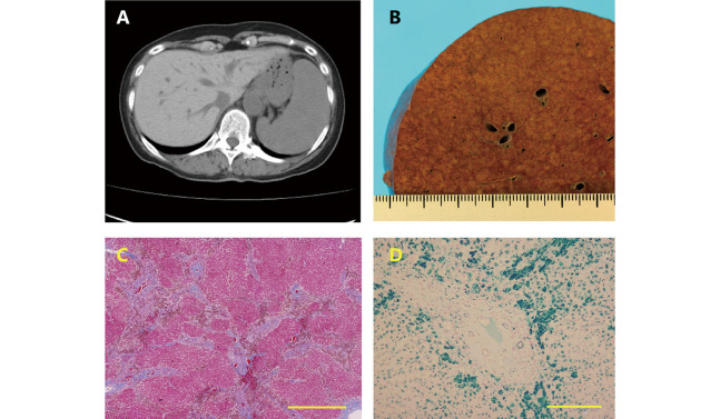
Liver images of patient Fig. 1A: Abdominal CT. Liver parenchyma with a high density indicates iron deposition, and marked splenomegaly has an unknown etiology. Fig. 1B: The autopsied liver: Macroscopy of a cross-section of the liver. The cross section of the liver is representative of cirrhosis, showing nodular parenchyma among wide fibrous tissues. Fig. 1C: The autopsied liver: Masson trichrome stain. The liver consists of parenchyma surrounded by wide fibrous septa, indicating F4 of Laennec cirrhosis. Bar indicates 1.0 mm. Fig. 1D: The autopsy liver: Berlin blue stain. A large amount of iron is seen in the peri-fibrous parenchymal cells. Bar indicates 300 μm.
References
-
- Takatoku M, Uchiyama T, Okamoto S, et al. Retrospective nationwide survey of Japanese patients with transfusion-dependent MDS and aplastic anemia highlights the negative impact of iron overload on morbidity/mortality. Eur J Haematol. 2007;78(6):487–494. doi: 10.1111/j.1600-0609.2007.00842.x. - DOI - PubMed
Publication types
MeSH terms
LinkOut - more resources
Full Text Sources
Medical