

Leukocyte populations and their cell adhesion molecules expression in newborn dromedary camel calves
- PMID: 33132598
- PMCID: PMC7566236
- DOI: 10.14202/vetworld.2020.1863-1869
Leukocyte populations and their cell adhesion molecules expression in newborn dromedary camel calves
Abstract
Background and aim: Different properties of the newborn immune system have been characterized in many species. For the newborn camel calf, however, the phenotype and composition of blood leukocytes have so far not been evaluated. The current study aimed to analyze the distribution of leukocyte subpopulations and their expression pattern of cell adhesion molecules in newborn and adult dromedary camels.
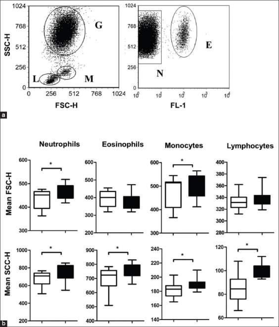
Materials and methods: Blood samples were collected from 17 newborn camel calves and 32 adult camels. For each sample, total leukocytes were separated and analyzed for their composition and cell adhesion molecules expression by flow cytometry.
Results: In comparison to adult camels, newborn camel calves had higher leukocyte numbers and higher numbers of neutrophils, monocytes, and lymphocytes but lower numbers of eosinophils in their blood. Among the lymphocyte populations in calves, the fractions of B cells and γδ T cells were elevated when compared to adults, whereas CD4-positive T cells were reduced. The comparison between camel calves and adult camels revealed significantly lower expression of the cell adhesion molecules CD11a, CD11b, and CD18 on granulocytes, monocytes, and lymphocytes in calves.
Conclusion: Newborn camel calves show a distinct composition and phenotype pattern of blood leukocytes when compared to adult camels. The observed rise in many leukocyte populations in calf blood may be due to reduced migratory activity in calf leukocyte populations.
Keywords: adhesion molecules; flow cytometry; immunophenotype; leukocytes; newborn camel calf.
Copyright: © Gaashan, et al.
Figures


Similar articles
-
Recent Advances in Camel Immunology.Front Immunol. 2021 Jan 25;11:614150. doi: 10.3389/fimmu.2020.614150. eCollection 2020. Front Immunol. 2021. PMID: 33569060 Free PMC article.
-
The Impact of the Animal Housing System on Immune Cell Composition and Function in the Blood of Dromedary Camels.Animals (Basel). 2022 Jan 28;12(3):317. doi: 10.3390/ani12030317. Animals (Basel). 2022. PMID: 35158641 Free PMC article.
-
Dromedary camel CD14high MHCIIhigh monocytes display inflammatory properties and are reduced in newborn camel calves.BMC Vet Res. 2020 Feb 18;16(1):62. doi: 10.1186/s12917-020-02285-8. BMC Vet Res. 2020. PMID: 32070351 Free PMC article.
-
Immunomodulatory Effects of the Cyclooxygenase Inhibitor Lornoxicam on Phenotype and Function of Camel Blood Leukocytes.Animals (Basel). 2021 Jul 6;11(7):2023. doi: 10.3390/ani11072023. Animals (Basel). 2021. PMID: 34359151 Free PMC article.
-
Leukocyte-cell adhesion: a molecular process fundamental in leukocyte physiology.Immunol Rev. 1990 Apr;114:67-108. doi: 10.1111/j.1600-065x.1990.tb00562.x. Immunol Rev. 1990. PMID: 1973408 Review.
Cited by
-
Haematological and biochemical blood reference values for Canary Island camels (Camelus dromedarius), an endangered dromedary species.Saudi J Biol Sci. 2023 Jun;30(6):103677. doi: 10.1016/j.sjbs.2023.103677. Epub 2023 May 4. Saudi J Biol Sci. 2023. PMID: 37213697 Free PMC article.
-
Recent Advances in Camel Immunology.Front Immunol. 2021 Jan 25;11:614150. doi: 10.3389/fimmu.2020.614150. eCollection 2020. Front Immunol. 2021. PMID: 33569060 Free PMC article.
-
Flow Cytometric Analysis of Leukocyte Populations in the Lung Tissue of Dromedary Camels.Vet Sci. 2022 Jun 10;9(6):287. doi: 10.3390/vetsci9060287. Vet Sci. 2022. PMID: 35737339 Free PMC article.
-
The Impact of the Animal Housing System on Immune Cell Composition and Function in the Blood of Dromedary Camels.Animals (Basel). 2022 Jan 28;12(3):317. doi: 10.3390/ani12030317. Animals (Basel). 2022. PMID: 35158641 Free PMC article.
-
Blood Gas, Acid-Base and Electrolyte Analysis in Healthy Dromedary Camel Calves up to 21 Days of Life.Animals (Basel). 2023 Mar 22;13(6):1117. doi: 10.3390/ani13061117. Animals (Basel). 2023. PMID: 36978657 Free PMC article.
References
-
- Salhi I, Bessalah S, Mbarek S.B, Chniter M, Seddik M.M, Khorchani T, Hammadi M. Passive transfer of maternal immunity in the dromedary (Camelus dromedarius), involvement of heavy chain antibodies. Trop. Anim. Health Prod. 2015;47(3):613–618. - PubMed
-
- Bornstein S, Gluecks I.V, Younan M, Thebo P, Mattsson J.G. Isospora orlovi infection in suckling dromedary camel calves (Camelus dromedarius) in Kenya. Vet. Parasitol. 2008;152(3-4):194–201. - PubMed
-
- Wernery U, Ali M, Wernery R, Seifert H.S. Severe heart muscle degeneration caused by Clostridium perfringens Type A in camel calves (Camelus dromedarius) Rev. Elev. Med. Vet. Pays. Trop. 1992;45(3-4):255–259. - PubMed
-
- Kamber R, Farah Z, Rusch P, Hassig M. The supply of newborn camel foals (Camelus dromedarius) with immunoglobulin G. Schweiz. Arch. Tierheilkd. 2000;142(10):581–588. - PubMed
LinkOut - more resources
Full Text Sources
Research Materials