Review
. 2020 Oct 14;26(38):5784-5796.
doi: 10.3748/wjg.v26.i38.5784.

Role of artificial intelligence in the diagnosis of oesophageal neoplasia: 2020 an endoscopic odyssey

Affiliations
Review

Role of artificial intelligence in the diagnosis of oesophageal neoplasia: 2020 an endoscopic odyssey

Mohamed Hussein et al. World J Gastroenterol. .

Abstract

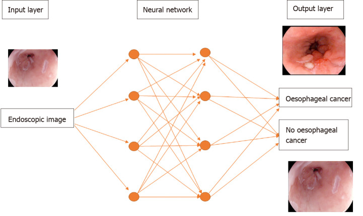
The past decade has seen significant advances in endoscopic imaging and optical enhancements to aid early diagnosis. There is still a treatment gap due to the underdiagnosis of lesions of the oesophagus. Computer aided diagnosis may play an important role in the coming years in providing an adjunct to endoscopists in the early detection and diagnosis of early oesophageal cancers, therefore curative endoscopic therapy can be offered. Research in this area of artificial intelligence is expanding and the future looks promising. In this review article we will review current advances in artificial intelligence in the oesophagus and future directions for development.

Keywords: Artificial intelligence; Barrett's oesophagus; Computer aided diagnosis; Deep learning; Oesophageal neoplasia; Squamous dysplasia.


Conflict of interest statement

Conflict-of-interest statement: Mohamed Hussein: No conflict of interest. Juana Gonzalez-Bueno Puyal: Employee at odin vision. Peter Mountney: Odin Vision employee. Laurence B lovat: Consultancy and minor share holder Odin Vision. Rehan Haidry: Educational grants to support research infrastructure from Medtronic ltd. Cook endoscopy (fellowship support), Pentax Europe, C2 therapeutics, Beamline diagnostic, Fractyl Ltd.

Figures

Figure 1
Figure 1
A deep learning model. Features of an endoscopic image processed through multiple neural layers to produce a predicted diagnosis of oesophageal cancer or no oesophageal cancer present on the image.
Figure 2
Figure 2
Three independent data sets are required to create a machine learning model that can predict an oesophageal cancer diagnosis.
Figure 3
Figure 3
Intrapapillary capillary loops patterns during magnification endoscopy to assess for early squamous cell neoplasia and depth of invasion. M1, M2, M3 = invasion of epithelium, lamina propria and muscularis propria respectively. SM1= superficial submucosal invasion. Citation: Inoue H, Kaga M, Ikeda H, Sato C, Sato H, Minami H, Santi EG, Hayee B, Eleftheriadis N. Magnification endoscopy in esophageal squamous cell carcinoma: a review of the intrapapillary capillary loop classification. Ann Gastroenterol 2015; 28: 41-48. Copyright© The Authors 2015. Published by Hellenic Society of Gastroenterology.
Figure 4
Figure 4
The computer-aided detection system providing real time feedback regarding absence of dysplasia (top row) or presence of dysplasia (bottom row). Citation: de Groof AJ, Struyvenberg MR, Fockens KN, van der Putten J, van der Sommen F, Boers TG, Zinger S, Bisschops R, de With PH, Pouw RE, Curvers WL, Schoon EJ, Bergman JJGHM. Deep learning algorithm detection of Barrett's neoplasia with high accuracy during live endoscopic procedures: a pilot study (with video). Gastrointest Endosc 2020; 91: 1242-1250. Copyright© The Authors 2020. Published by Elsevier.
Figure 5
Figure 5
Volumetric laser endomicroscopy image showing area of overlap (yellow arrow) between the 3 features of dysplasia identified with the colour schemes. A: View looking down into the oesophagus; B: Close up of dysplastic area; C: Forward view of the dysplastic area. A-C: Citation: Trindade AJ, McKinley MJ, Fan C, Leggett CL, Kahn A, Pleskow DK. Endoscopic Surveillance of Barrett's Esophagus Using Volumetric Laser Endomicroscopy With Artificial Intelligence Image Enhancement. Gastroenterology 2019; 157: 303-305. Copyright© The Authors 2019. Published by Elsevier.
Figure 6
Figure 6
Input images on the left and corresponding heat maps on the right illustrating the features recognised by the convolutional neural network when classifying images by recognising the abnormal intrapapillary capillary loops patterns in early squamous cell neoplasia. Citation: Everson M, Herrera L, Li W, Luengo IM, Ahmad O, Banks M, Magee C, Alzoubaidi D, Hsu HM, Graham D, Vercauteren T, Lovat L, Ourselin S, Kashin S, Wang HP, Wang WL, Haidry RJ. Artificial intelligence for the real-time classification of intrapapillary capillary loop patterns in the endoscopic diagnosis of early oesophageal squamous cell carcinoma: A proof-of-concept study. United European Gastroenterol J 2019; 7: 297-306. Copyright© The Authors 2019. Published by SAGE Journals.
Figure 7
Figure 7
Esophageal squamous cell cancer diagnosed by the artificial intelligence system as superficial cancer with SM2 invasion. A and B: Citation: Nakagawa K, Ishihara R, Aoyama K, Ohmori M, Nakahira H, Matsuura N, Shichijo S, Nishida T, Yamada T, Yamaguchi S, Ogiyama H, Egawa S, Kishida O, Tada T. Classification for invasion depth of esophageal squamous cell carcinoma using a deep neural network compared with experienced endoscopists. Gastrointest Endosc 2019; 90: 407-414. Copyright© The Authors 2019. Published by Elsevier.

Similar articles

Cited by

References

    1. Ferlay J, Soerjomataram I, Dikshit R, Eser S, Mathers C, Rebelo M, Parkin DM, Forman D, Bray F. Cancer incidence and mortality worldwide: sources, methods and major patterns in GLOBOCAN 2012. Int J Cancer. 2015;136:E359–E386. - PubMed
    1. Bray F, Ferlay J, Soerjomataram I, Siegel RL, Torre LA, Jemal A. Global cancer statistics 2018: GLOBOCAN estimates of incidence and mortality worldwide for 36 cancers in 185 countries. CA Cancer J Clin. 2018;68:394–424. - PubMed
    1. Guo L, Xiao X, Wu C, Zeng X, Zhang Y, Du J, Bai S, Xie J, Zhang Z, Li Y, Wang X, Cheung O, Sharma M, Liu J, Hu B. Real-time automated diagnosis of precancerous lesions and early esophageal squamous cell carcinoma using a deep learning model (with videos) Gastrointest Endosc. 2020;91:41–51. - PubMed
    1. Menon S, Trudgill N. How commonly is upper gastrointestinal cancer missed at endoscopy? A meta-analysis. Endosc Int Open. 2014;2:E46–E50. - PMC - PubMed
    1. Visrodia K, Singh S, Krishnamoorthi R, Ahlquist DA, Wang KK, Iyer PG, Katzka DA. Magnitude of Missed Esophageal Adenocarcinoma After Barrett's Esophagus Diagnosis: A Systematic Review and Meta-analysis. Gastroenterology. 2016;150:599–607.e7; quiz e14-5. - PMC - PubMed