Review
. 2020 Sep 10:14:283.
doi: 10.3389/fncel.2020.00283. eCollection 2020.

Shifting Developmental Trajectories During Critical Periods of Brain Formation

Affiliations
Review

Shifting Developmental Trajectories During Critical Periods of Brain Formation

Nathalie Dehorter et al. Front Cell Neurosci. .

Abstract

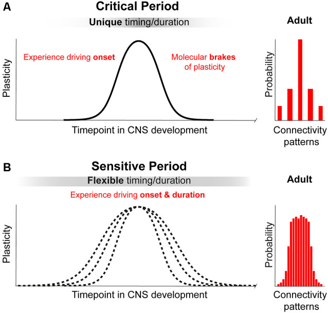
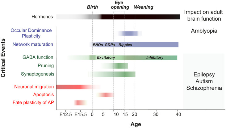
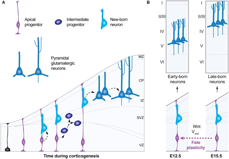
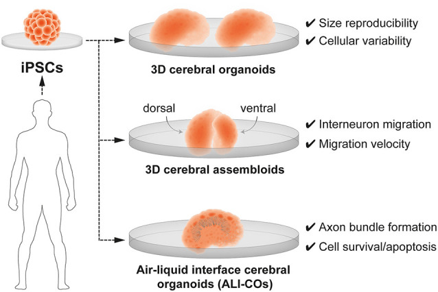
Critical periods of brain development are epochs of heightened plasticity driven by environmental influence necessary for normal brain function. Recent studies are beginning to shed light on the possibility that timely interventions during critical periods hold potential to reorient abnormal developmental trajectories in animal models of neurological and neuropsychiatric disorders. In this review, we re-examine the criteria defining critical periods, highlighting the recently discovered mechanisms of developmental plasticity in health and disease. In addition, we touch upon technological improvements for modeling critical periods in human-derived neural networks in vitro. These scientific advances associated with the use of developmental manipulations in the immature brain of animal models are the basic preclinical systems that will allow the future translatability of timely interventions into clinical applications for neurodevelopmental disorders such as intellectual disability, autism spectrum disorders (ASD) and schizophrenia.

Keywords: brain organoids; critical period; development; neurodevelopmental disorders; plasticity.


Figures

Figure 1
Figure 1
Principles of critical periods compared to sensitive periods of plasticity. (A) Critical periods in brain development represent narrow time windows of heightened plasticity driven by environmental input. Closure of critical periods is achieved through molecular brakes that constrain plasticity and allow for permanent structural consolidation of one of a few possible connectivity patterns. (B) Sensitive periods in brain development represent broad time windows of gradual change in plasticity driven by environmental input. Sensitive periods are not closed by molecular constrains and can be further reopen by changing environmental cues. The consolidation of one of a broad range of possible patterns of connectivity is reversible and remains functionally dynamic.
Figure 2
Figure 2
Critical events during embryonic and postnatal periods of brain development that impact adult brain function. Scheme shows a timeline of the key stages in the development of neuronal networks in the mouse brain (embryonic critical period: E12 to E18; Perinatal critical period: P0-P10; late postnatal critical period: P10-P40). It summarizes the events that occur during brain maturation and leads to long-term alterations. OD, ocular dominance; ENOs, early network oscillations; GDP, giant depolarizing potentials; GABA, switch from excitatory to inhibitory.
Figure 3
Figure 3
Fate plasticity during corticogenesis. (A) Schematic of corticogenesis (left panel) showing three populations of progenitor cells: neuroepithelial cells (in black), apical progenitors (light blue) and intermediate progenitors (dark blue). Apical progenitors (radial glia) within the ventricular zone (VZ) initially expand their population and generate neurons and intermediate progenitors. As corticogenesis proceeds, intermediate progenitors (within the SVZ) as well as apical progenitors generate neurons. (B) Temporal progression of neurogenic stage (right panel) shows apical progenitors around embryonic stage (E) 12.5 that give rise to early born deep-layer pyramidal neurons. In the standard temporal progression of corticogenesis apical progenitors at E15.5 give rise to late-born upper-layer pyramidal neurons. Apical progenitors at E15.5 can change their neurogenic state, a process named “fate plasticity” and give rise to late-born deep-layer pyramidal neurons under specific conditions such as cell hyperpolarization or reactivation of Wnt-signaling. VZ, ventricular zone; SVZ, subventricular zone; IZ, intermediate zone; CP, cortical plate; MZ, marginal zone; Vrest, resting membrane potential.
Figure 4
Figure 4
3D brain organoids for modeling human brain development and neurodevelopmental disorders in vitro. Cerebral organoids are 3D brain models derived from human induced-pluripotent stem cells (iPSCs). 3-D brain organoids generated by Velasco et (; top panel) are consistent in size and reproducibly present a spectrum of cell types. 3-D cerebral assembloids (middle panel) are formed of a pallium-like organoid (dorsal) and a subpallium-like organoid (ventral) and can recapitulate GABAergic interneuron migration. Air-liquid interface cerebral organoids (ALI-Cos; bottom panel) show an improved neural survival and can therefore be suitable to assay critical periods of programmed cell death. Neurons within ALI-COs form axon bundles over long-distances mimicking long-range projections.

Similar articles

Cited by

References

    1. Adhya D., Swarup V., Nagy R., Dutan L., Shum C., Valencia-Alarcón E. P., et al. . (2020). Atypical neurogenesis in induced pluripotent stem cell (iPSC) from autistic individuals. Biol. Psychiatry 3223, 31702–31779. 10.1016/j.biopsych.2020.06.014 - DOI - PMC - PubMed
    1. Aledo-Serrano Á., Mingorance A., Jiménez-Huete A., Toledano R., García-Morales I., Anciones C., et al. . (2020). Genetic epilepsies and COVID-19 pandemic: lessons from the caregiver perspective. Epilepsia 61, 1312–1314. 10.1111/epi.16537 - DOI - PMC - PubMed
    1. Allene C., Cattani A., Ackman J. B., Bonifazi P., Aniksztejn L., Ben-Ari Y., et al. . (2008). Sequential generation of two distinct synapse-driven network patterns in developing neocortex. J. Neurosci. 28, 12851–12863. 10.1523/JNEUROSCI.3733-08.2008 - DOI - PMC - PubMed
    1. Allene C., Cossart R. (2010). Early NMDA receptor-driven waves of activity in the developing neocortex: physiological or pathological network oscillations? J. Physiol. 588, 83–91. 10.1113/jphysiol.2009.178798 - DOI - PMC - PubMed
    1. Antón-Bolaños N., Sempere-Ferràndez A., Guillamón-Vivancos T., Martini F. J., Pérez-Saiz L., Gezelius H., et al. . (2019). Prenatal activity from thalamic neurons governs the emergence of functional cortical maps in mice. Science 364, 987–990. 10.1126/science.aav7617 - DOI - PMC - PubMed

LinkOut - more resources