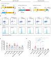
Comparison of CRISPR/Cas Endonucleases for in vivo Retinal Gene Editing
- PMID: 33132845
- PMCID: PMC7511709
- DOI: 10.3389/fncel.2020.570917
Comparison of CRISPR/Cas Endonucleases for in vivo Retinal Gene Editing
Abstract
CRISPR/Cas has opened the prospect of direct gene correction therapy for some inherited retinal diseases. Previous work has demonstrated the utility of adeno-associated virus (AAV) mediated delivery to retinal cells in vivo; however, with the expanding repertoire of CRISPR/Cas endonucleases, it is not clear which of these are most efficacious for retinal editing in vivo. We sought to compare CRISPR/Cas endonuclease activity using both single and dual AAV delivery strategies for gene editing in retinal cells. Plasmids of a dual vector system with SpCas9, SaCas9, Cas12a, CjCas9 and a sgRNA targeting YFP, as well as a single vector system with SaCas9/YFP sgRNA were generated and validated in YFP-expressing HEK293A cell by flow cytometry and the T7E1 assay. Paired CRISPR/Cas endonuclease and its best performing sgRNA was then packaged into an AAV2 capsid derivative, AAV7m8, and injected intravitreally into CMV-Cre:Rosa26-YFP mice. SpCas9 and Cas12a achieved better knockout efficiency than SaCas9 and CjCas9. Moreover, no significant difference in YFP gene editing was found between single and dual CRISPR/SaCas9 vector systems. With a marked reduction of YFP-positive retinal cells, AAV7m8 delivered SpCas9 was found to have the highest knockout efficacy among all investigated endonucleases. We demonstrate that the AAV7m8-mediated delivery of CRISPR/SpCas9 construct achieves the most efficient gene modification in neurosensory retinal cells in vivo.
Keywords: AAV (adeno-associated virus); CRISPR (clustered regularly interspaced short palindromic repeats); gene editing; retina; retinal dystrophy.
Copyright © 2020 Li, Wing, Wang, Luu, Bender, Chen, Wang, Lu, Nguyen Tran, Young, Wong, Pébay, Cook, Hung, Liu and Hewitt.
Figures


Similar articles
-
AAV-Mediated CRISPR/Cas Gene Editing of Retinal Cells In Vivo.Invest Ophthalmol Vis Sci. 2016 Jun 1;57(7):3470-6. doi: 10.1167/iovs.16-19316. Invest Ophthalmol Vis Sci. 2016. PMID: 27367513
-
Utility of Self-Destructing CRISPR/Cas Constructs for Targeted Gene Editing in the Retina.Hum Gene Ther. 2019 Nov;30(11):1349-1360. doi: 10.1089/hum.2019.021. Epub 2019 Oct 25. Hum Gene Ther. 2019. PMID: 31373227
-
Adeno-Associated Vector-Delivered CRISPR/SaCas9 System Reduces Feline Leukemia Virus Production In Vitro.Viruses. 2021 Aug 18;13(8):1636. doi: 10.3390/v13081636. Viruses. 2021. PMID: 34452500 Free PMC article.
-
CRISPR Systems Suitable for Single AAV Vector Delivery.Curr Gene Ther. 2022;22(1):1-14. doi: 10.2174/1566523221666211006120355. Curr Gene Ther. 2022. PMID: 34620062 Review.
-
Bacterial CRISPR/Cas DNA endonucleases: A revolutionary technology that could dramatically impact viral research and treatment.Virology. 2015 May;479-480:213-20. doi: 10.1016/j.virol.2015.02.024. Epub 2015 Mar 7. Virology. 2015. PMID: 25759096 Free PMC article. Review.
Cited by
-
CRISPR Technology for Ocular Angiogenesis.Front Genome Ed. 2020 Dec 22;2:594984. doi: 10.3389/fgeed.2020.594984. eCollection 2020. Front Genome Ed. 2020. PMID: 34713223 Free PMC article. Review.
-
Characterization of RNA editing and gene therapy with a compact CRISPR-Cas13 in the retina.Proc Natl Acad Sci U S A. 2024 Nov 5;121(45):e2408345121. doi: 10.1073/pnas.2408345121. Epub 2024 Oct 30. Proc Natl Acad Sci U S A. 2024. PMID: 39475642 Free PMC article.
-
New Editing Tools for Gene Therapy in Inherited Retinal Dystrophies.CRISPR J. 2022 Jun;5(3):377-388. doi: 10.1089/crispr.2021.0141. Epub 2022 May 3. CRISPR J. 2022. PMID: 35506982 Free PMC article. Review.
-
Genome Editing Inhibits Retinal Angiogenesis in a Mouse Model of Oxygen-Induced Retinopathy.Methods Mol Biol. 2023;2678:207-217. doi: 10.1007/978-1-0716-3255-0_17. Methods Mol Biol. 2023. PMID: 37326717
-
Current and Future Landscape in Genetic Therapies for Leber Hereditary Optic Neuropathy.Cells. 2023 Aug 7;12(15):2013. doi: 10.3390/cells12152013. Cells. 2023. PMID: 37566092 Free PMC article. Review.
References
LinkOut - more resources
Full Text Sources
Other Literature Sources
Research Materials

