. 2020 Oct 8:14:571155.
doi: 10.3389/fncel.2020.571155. eCollection 2020.

Insulin-Like Growth Factor 1 on the Maintenance of Ribbon Synapses in Mouse Cochlear Explant Cultures

Affiliations

Insulin-Like Growth Factor 1 on the Maintenance of Ribbon Synapses in Mouse Cochlear Explant Cultures

Li Gao et al. Front Cell Neurosci. .

Abstract

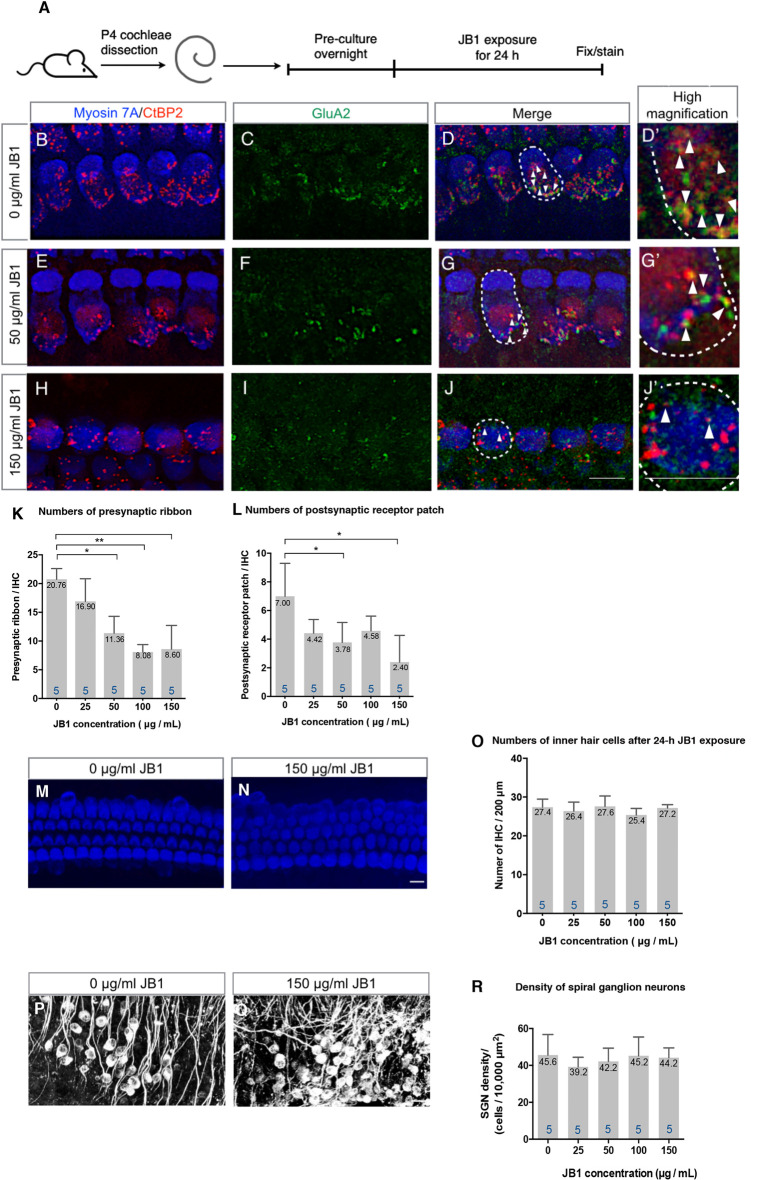
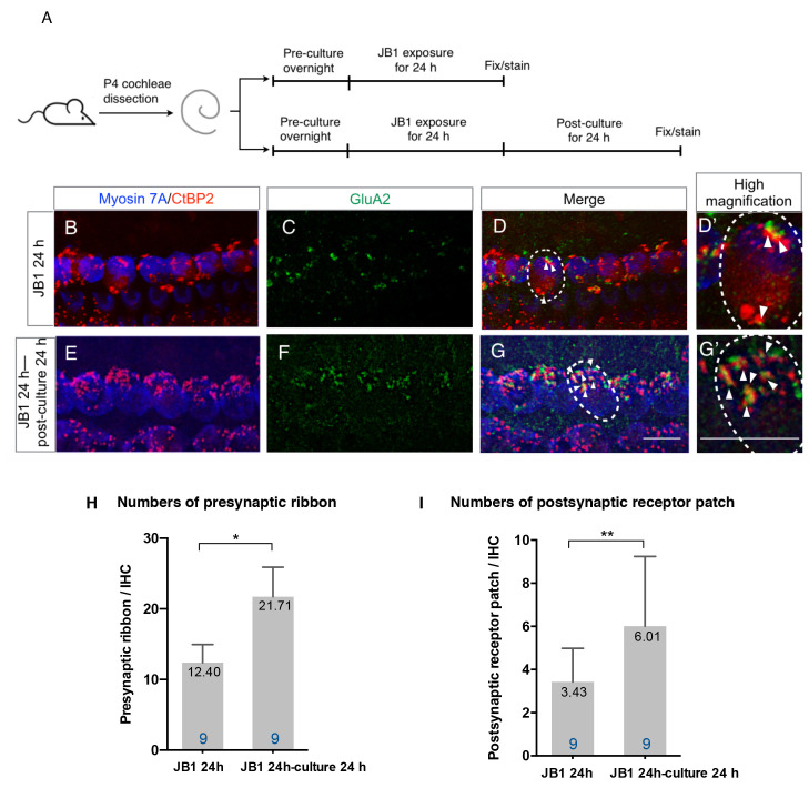
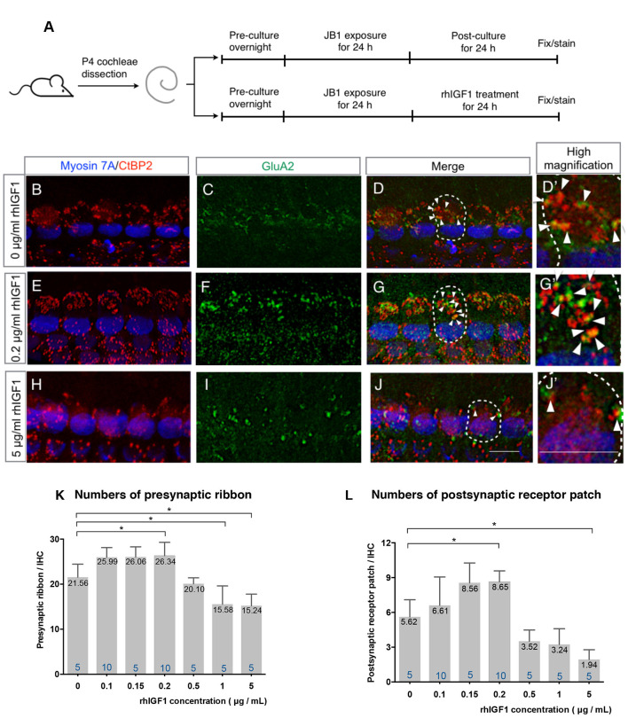
Hearing loss has become one of the most common disabilities worldwide. The synaptic connections between inner hair cells (IHCs) and spiral ganglion neurons have specialized synaptic constructions, termed ribbon synapses, which are important for auditory function. The ribbon synapses in the cochlea are quite vulnerable to various insults. As such, the maintenance of ribbon synapses is important for ensuring hearing function. Insulin-like growth factor 1 (IGF1) plays a critical role in the development and maintenance of the cochlea and has the potential to protect cochlear hair cells from various insults. In this study, we examined the role of IGF1 in the maintenance of ribbon synapses in cochlear explants of postnatal day four mice. We cultured cochlear explants with an IGF1 receptor antagonist, JB1, which is an IGF1 peptide analog. Results showed that exposure to JB1 for 24 h resulted in the loss of ribbon synapses. After an additional 24-h culture without JB1, the number of ribbon synapses spontaneously recovered. The application of exogenous IGF1 showed two different aspects of ribbon synapses. Low doses of exogenous IGF1 promoted the recovery of ribbon synapses, while it compromised the spontaneous recovery of ribbon synapses at high doses. Altogether, these results indicate that the paracrine or autocrine release of IGF1 in the cochlea plays a crucial role in the maintenance of cochlear ribbon synapses.

Keywords: cochlea; inner hair cell; insulin-like growth factor 1; maintenance; ribbon synapse.


Figures

Figure 1
Figure 1
An insulin-like growth factor 1 receptor (IGF1R) antagonist JB1 induced the loss of ribbon synapses in cochlear explant cultures. Cochlear explants of P4 mice were exposed to JB1 at a concentration of 25, 50, 100, or 150 μg/ml for 24 h (A). (B–D) Maximal-intensity projection images with z-stack of ribbon synapses in control specimens that were cultured without JB1. Panels (E–J) are maximal-intensity projection images of the degeneration of ribbon synapses in specimens cultured with JB1 at concentrations of 50 and 150 μg/ml, respectively. Arrows show the postsynaptic receptor patches and dotted lines indicate the location of an IHC. (K) JB1 showed significant effects on both presynaptic ribbons (p < 0.001 by one-way ANOVA) and postsynaptic receptor patches (L; p = 0.004 by one-way ANOVA). Tukey’s post hoc test revealed significant losses at concentrations of 50 (p = 0.001), 100 (p < 0.001), and 150 μg/ml (p < 0.001) in the number of presynaptic ribbons, and at concentrations of 50 (p = 0.032) and 150 μg/ml (p = 0.002) in the number of postsynaptic receptor patches, in comparison with controls which were cultured without JB1. (M–R) No inner hair cell (IHC) or spiral ganglion neuron (SGN) loss was found at any JB1 concentrations (p = 0.428 by one-way ANOVA for IHC, p = 0.730 by one-way ANOVA for SGN). Scale bars: 10 μm. Data are expressed as mean (digits at the top of each bar) ± SD. The digits at the bottom of each bar represent the sample number. *p < 0.05; **p < 0.01 with Tukey’s post hoc test. P4, postnatal day 4; IHC, inner hair cell; SGN, spiral ganglion neuron; SD, standard deviation; ANOVA, analysis of variance.
Figure 2
Figure 2
Spontaneous recovery of ribbon synapses. Cochlear explants from P4 mice were exposed to 50 μg/ml JB1 for 24 h, with or without an additional 24 h of culture (A). (B–D) Maximal-intensity projection images with z-stack of the immunostaining results of specimens immediately after 24-h JB1 exposure. (E–G) Maximal-intensity projection images of the specimens following an additional 24-h culture. Arrows show the postsynaptic receptor patches and dotted lines indicate the location of an IHC. (H) The number of presynaptic ribbons (p < 0.001 by unpaired t-test) and postsynaptic receptor patches (I; p = 0.046 by unpaired t-test) in the specimens were significantly increased after an additional 24-h culture. Scale bars: 10 μm. Data are expressed as mean (digits at the top of each bar) ± SD. The digits at the bottom of each bar represent the sample number. *p < 0.01; **p < 0.05 by an unpaired t-test. P4, postnatal day 4; IHC, inner hair cell; SD, standard deviation.
Figure 3
Figure 3
Effect of exogenous IGF1 on the recovery of ribbon synapses. Cochlear explants from P4 mice were exposed to 50 μg/ml JB1 for 24 h, after which they were incubated with culture media supplemented with rhIGF1 at a concentration of 0.1, 0.15, 0.2, 0.5, or 1, 5 μg/ml for 24 h (A). (B–D) Maximal-intensity projection images with z-stack of the immunostaining images of specimens cultured without rhIGF1. Panels (E–J) are maximal-intensity projection images of the recovery of ribbon synapses in specimens cultured with 50 μg/ml JB1 for 24 h, followed by rhIGF1 treatment for 24 h at concentrations of 0.2 and 5 μg/ml, respectively. Arrows show the postsynaptic receptor patches and dotted lines indicate the location of an IHC. Exogenous rhIGF1 showed significant effects on the number of both presynaptic ribbons (K; p < 0.001 by one-way ANOVA) and postsynaptic receptor patches (L; p < 0.001 by one-way ANOVA). Tukey’s post hoc test revealed significant difference at concentrations of 0.2 (p = 0.034), 1 (p = 0.017), and 5 μg/ml (p = 0.010) in the number of presynaptic ribbons, and at concentrations of 0.2 (p = 0.020) and 5 μg/ml (p = 0.012) in the number of postsynaptic receptor patches, in comparison with controls which were cultured in 0 μg/ml rhIGF1. Scale bars: 10 μm. Data are expressed as mean (digits at the top of each bar) ± SD. The digits at the bottom of each bar represent the sample number. *p < 0.05 by one-way ANOVA, followed by Tukey’s post hoc test. P4, postnatal day 4; IHC, inner hair cell; SD, standard deviation; rhIGF1, recombinant human insulin-like growth factor-1; ANOVA, analysis of variance.

Similar articles

Cited by

References

    1. Allahdadi K. J., de Santana T. A., Santos G. C., Azevedo C. M., Mota R. A., Nonaka C. K., et al. . (2019). IGF-1 overexpression improves mesenchymal stem cell survival and promotes neurological recovery after spinal cord injury. Stem Cell Res. Ther. 10:146. 10.1186/s13287-019-1223-z - DOI - PMC - PubMed
    1. Becker L., Schnee M. E., Niwa M., Sun W., Maxeiner S., Talaei S., et al. . (2018). The presynaptic ribbon maintains vesicle populations at the hair cell afferent fiber synapse. eLife 12:e30241. 10.7554/eLife.30241 - DOI - PMC - PubMed
    1. Bharadwaj H. M., Verhulst S., Shaheen L., Liberman M. C., Shinn-Cunningham B. G. (2014). Cochlear neuropathy and the coding of supra-threshold sound. Front. Syst. Neurosci. 8:26. 10.3389/fnsys.2014.00026 - DOI - PMC - PubMed
    1. Camarero G., Avendano C., Fernandez-Moreno C., Villar A., Contreras J., de Pablo F., et al. . (2001). Delayed inner ear maturation and neuronal loss in postnatal IGF-1-deficient mice. J. Neurosci. 21, 7630–7641. 10.1523/JNEUROSCI.21-19-07630.2001 - DOI - PMC - PubMed
    1. Camarero G., Villar M. A., Contreras J., Fernández-Moreno C., Pichel J. G., Avendaño C., et al. . (2002). Cochlear abnormalities in insulin-like growth factor-1 mouse mutants. Hear. Res. 170, 2–11. 10.1016/S0378-5955(02)00447-1 - DOI - PubMed