

Functional Alterations in the Posterior Insula and Cerebellum in Migraine Without Aura: A Resting-State MRI Study
- PMID: 33132860
- PMCID: PMC7573354
- DOI: 10.3389/fnbeh.2020.567588
Functional Alterations in the Posterior Insula and Cerebellum in Migraine Without Aura: A Resting-State MRI Study
Abstract
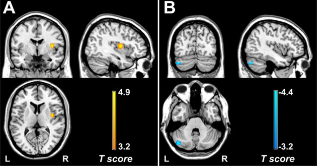
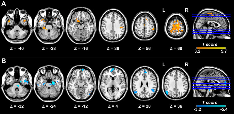
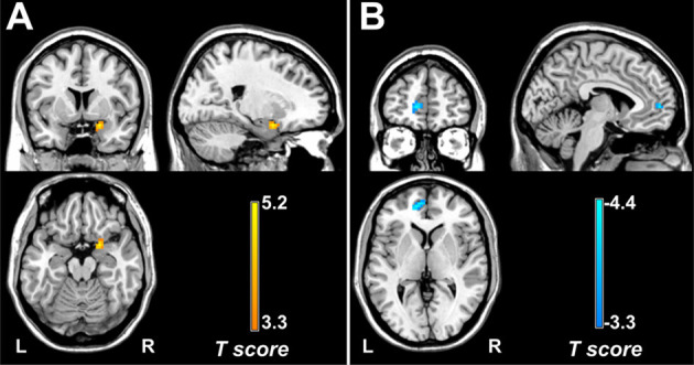
Background: Hypothesis-driven functional connectivity (FC) analyses have revealed abnormal functional interaction of regions or networks involved in pain processing in episodic migraine patients. We aimed to investigate the resting-state FC patterns in episodic migraine by combining data-driven voxel-wise degree centrality (DC) calculation and seed-based FC analysis. Methods: Thirty-nine patients suffering from episodic migraine without aura and 35 healthy controls underwent clinical assessment and functional MRI. DC was analyzed voxel-wise and compared between groups, and FC of regions with DC differences were further examined using a seed-based approach. Results: Compared with the control group, the migraine group showed increased and decreased DC in the right posterior insula and left crus I, respectively. Seed-based FC analyses revealed that migraine patients demonstrated increased right posterior insula connections with the postcentral gyrus, supplementary motor area/paracentral lobule, fusiform gyrus and temporal pole. The left crus I showed decreased FC with regions of the default mode network (DMN), including the medial prefrontal cortex (mPFC), angular gyrus, medial and lateral temporal cortex in patients with migraine. Furthermore, pain intensity positively correlated with DC in the right amygdala/parahippocampal gyrus, and migraine frequency negatively correlated with FC between the left crus I and mPFC. Conclusion: Patients with episodic migraine without aura have increased FC with the right posterior insula and decreased FC within the DMN, which may underlie disturbed sensory integration and cognitive processing of pain. The left crus I-mPFC connectivity may be a useful biomarker for assessing migraine frequency.
Keywords: degree centrality; functional connectivity; functional magnetic resonance imaging; migraine; resting-state.
Copyright © 2020 Ke, Yu, Zhang, Su, Wang, Hu, Dai, Hu, Zhao and Dai.
Figures
Similar articles
-
Altered functional brain network patterns in patients with migraine without aura after transcutaneous auricular vagus nerve stimulation.Sci Rep. 2023 Jun 13;13(1):9604. doi: 10.1038/s41598-023-36437-1. Sci Rep. 2023. PMID: 37311825 Free PMC article.
-
Resting state connectivity between default mode network and insula encodes acute migraine headache.Cephalalgia. 2018 Apr;38(5):846-854. doi: 10.1177/0333102417715230. Epub 2017 Jun 12. Cephalalgia. 2018. PMID: 28605972
-
Abnormal degree centrality in end-stage renal disease (ESRD) patients with cognitive impairment: a resting-state functional MRI study.Brain Imaging Behav. 2021 Jun;15(3):1170-1180. doi: 10.1007/s11682-020-00317-3. Brain Imaging Behav. 2021. PMID: 32902798
-
Resting-state abnormalities in functional connectivity of the default mode network in migraine: A meta-analysis.Front Neurosci. 2023 Mar 1;17:1136790. doi: 10.3389/fnins.2023.1136790. eCollection 2023. Front Neurosci. 2023. PMID: 36937687 Free PMC article. Review.
-
Alterations in degree centrality and functional connectivity in tension-type headache: a resting-state fMRI study.Brain Imaging Behav. 2024 Aug;18(4):819-829. doi: 10.1007/s11682-024-00875-w. Epub 2024 Mar 21. Brain Imaging Behav. 2024. PMID: 38512647 Review.
Cited by
-
Involvement of the cerebellum in structural connectivity enhancement in episodic migraine.J Headache Pain. 2024 Sep 18;25(1):154. doi: 10.1186/s10194-024-01854-8. J Headache Pain. 2024. PMID: 39294590 Free PMC article.
-
Dynamics and concordance alterations of regional brain function indices in vestibular migraine: a resting-state fMRI study.J Headache Pain. 2024 Jan 5;25(1):1. doi: 10.1186/s10194-023-01705-y. J Headache Pain. 2024. PMID: 38178029 Free PMC article.
-
Differential Modulating Effect of Acupuncture in Patients With Migraine Without Aura: A Resting Functional Magnetic Resonance Study.Front Neurol. 2021 May 28;12:680896. doi: 10.3389/fneur.2021.680896. eCollection 2021. Front Neurol. 2021. PMID: 34122321 Free PMC article.
-
Alterations in Effective Connectivity of the Hippocampus in Migraine without Aura.J Pain Res. 2021 Oct 21;14:3333-3343. doi: 10.2147/JPR.S327945. eCollection 2021. J Pain Res. 2021. PMID: 34707401 Free PMC article.
-
A robust multimodal brain MRI-based diagnostic model for migraine: validation across different migraine phases and longitudinal follow-up data.J Headache Pain. 2025 Jan 9;26(1):5. doi: 10.1186/s10194-024-01946-5. J Headache Pain. 2025. PMID: 39789428 Free PMC article.
References
-
- Buckner R. L., Sepulcre J., Talukdar T., Krienen F. M., Liu H., Hedden T., et al. . (2009). Cortical hubs revealed by intrinsic functional connectivity: mapping, assessment of stability and relation to Alzheimer’s disease. J. Neurosci. 29, 1860–1873. 10.1523/jneurosci.5062-08.2009 - DOI - PMC - PubMed
LinkOut - more resources
Full Text Sources
Miscellaneous