

Children With Autism Produce a Unique Pattern of EEG Microstates During an Eyes Closed Resting-State Condition
- PMID: 33132865
- PMCID: PMC7579608
- DOI: 10.3389/fnhum.2020.00288
Children With Autism Produce a Unique Pattern of EEG Microstates During an Eyes Closed Resting-State Condition
Abstract
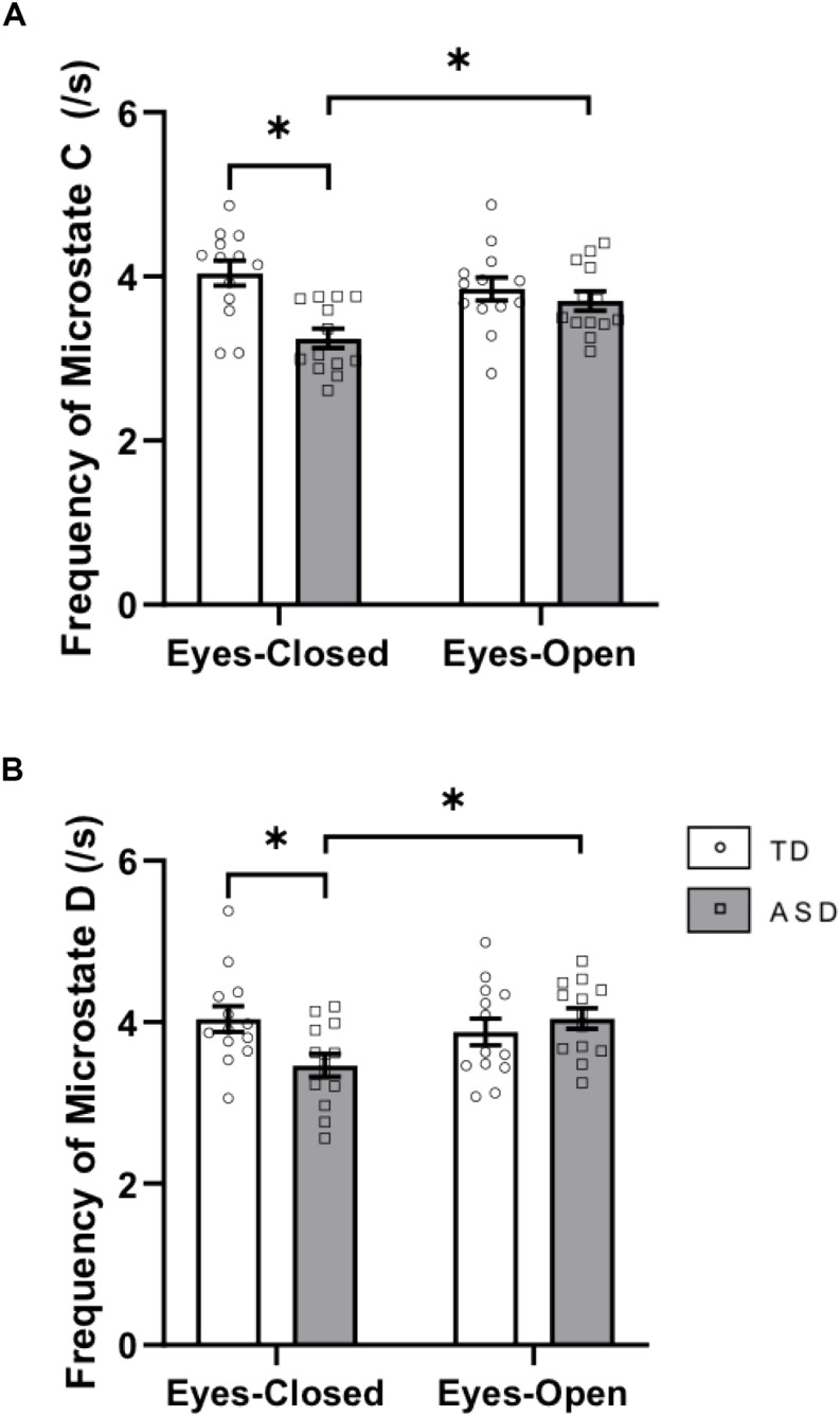
Although fMRI studies have produced considerable evidence for differences in the spatial connectivity of resting-state brain networks in persons with autism spectrum disorder (ASD) relative to typically developing (TD) peers, little is known about the temporal dynamics of these brain networks in ASD. The aim of this study was to examine the EEG microstate architecture in children with ASD as compared to TD at rest in two separate conditions - eyes-closed (EC) and eyes-open (EO). EEG microstate analysis was performed on resting-state data of 13 ASD and 13 TD children matched on age, gender, and IQ. We found that children with ASD and TD peers produced topographically similar canonical microstates at rest. Group differences in the duration and frequency of these microstates were found primarily in the EC resting-state condition. In line with previous fMRI findings that have reported differences in spatial connectivity within the salience network (previously correlated with the activity of microstate C) in ASD, we found that the duration of activation of microstate C was increased, and the frequency of microstate C was decreased in ASD as compared to TD in EC resting-state. Functionally, these results may be reflective of alterations in interoceptive processes in ASD. These results suggest a unique pattern of EEG microstate architecture in ASD relative to TD during resting-states and also that EEG microstate parameters in ASD are susceptible to differences in resting-state conditions.
Keywords: EEG; autism spectrum disorders; microstates; resting-state; salience network.
Copyright © 2020 Nagabhushan Kalburgi, Whitten, Key and Bodfish.
Figures


References
-
- Atluri S., Wong W., Blumberger D. M., Daskalakis Z. J., Farzan F. (2017). 533. Insights from EEG Microstate Analysis on the Pathophysiology of Depression and Mechanisms of Seizure Therapy. Biol. Psych. 81:S216 10.1016/j.biopsych.2017.02.1141 - DOI
Grants and funding
LinkOut - more resources
Full Text Sources