

β-Amyloid Plaque Reduction in the Hippocampus After Focused Ultrasound-Induced Blood-Brain Barrier Opening in Alzheimer's Disease
- PMID: 33132889
- PMCID: PMC7575813
- DOI: 10.3389/fnhum.2020.593672
β-Amyloid Plaque Reduction in the Hippocampus After Focused Ultrasound-Induced Blood-Brain Barrier Opening in Alzheimer's Disease
Abstract
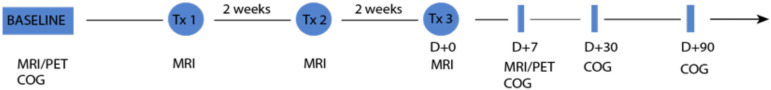
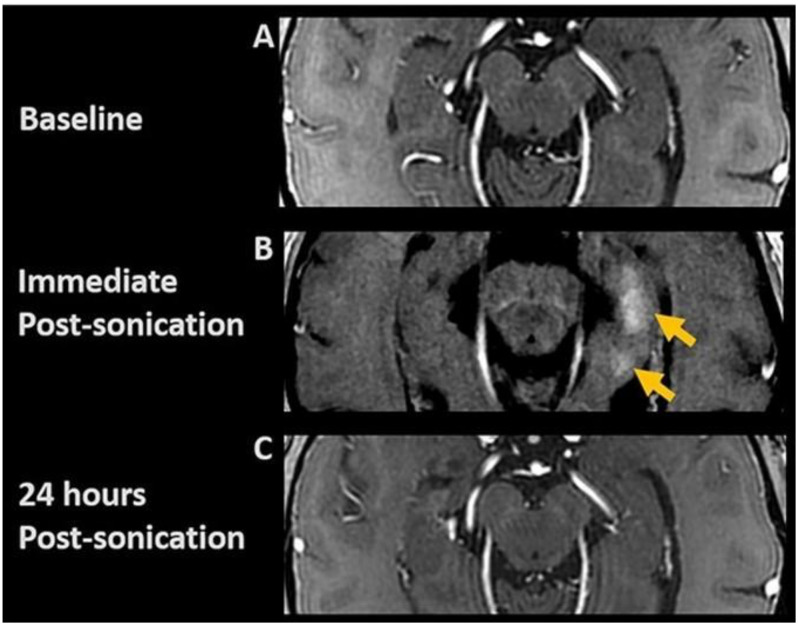
The blood-brain barrier (BBB) limits therapeutic delivery in Alzheimer's disease (AD) and other neurological disorders. Animal models have demonstrated safe BBB opening and reduction in β-amyloid plaque with focused ultrasound (FUS). We recently demonstrated the feasibility, safety, and reversibility of FUS-induced BBB opening in the hippocampus and entorhinal cortex in six participants with early AD. We now report the effect of BBB opening with FUS treatment on β-amyloid plaque. Six participants underwent 18F-Florbetaben PET scan at baseline and 1 week after the completion of the third FUS treatment (60 days interval). PET analysis comparing the hippocampus and entorhinal cortex in the treated and untreated hemispheres revealed a decrease in the ratio of 18F-Florbetaben ligand binding. The standard uptake value ratios (SUVr) reduction ranged from 2.7% to 10% with an average of 5.05% (±2.76) suggesting a decrease in β-amyloid plaque.
Keywords: Alzheimer’s disease; FUS; blood-brain barrier; focused ultrasound; hippocampus; β-amyloid reduction.
Copyright © 2020 D’Haese, Ranjan, Song, Haut, Carpenter, Dieb, Najib, Wang, Mehta, Chazen, Hodder, Claassen, Kaplitt and Rezai.
Figures
References
-
- Burgess A., Dubey S., Yeung S., Hough O., Eterman N., Aubert I., et al. (2014). Alzheimer disease in a mouse model: MR Imaging–guided focused ultrasound targeted to the hippocampus opens the blood-brain barrier and improves pathologic abnormalities and behavior. Radiology 273 736–745. 10.1148/radiol.14140245 - DOI - PMC - PubMed
-
- Chakravorti S., Jermakowicz W. J., Wu C., Li R., Gonzalez R. W., Dawant B. M., et al. (2019). “Evaluation of nonrigid registration around the hippocampus for the construction of statistical maps in a multicenter dataset of epilepsy laser ablation patients,” in Proceedings of the Medical Imaging 2019: Image-Guided Procedures, Robotic Interventions, and Modeling, (San Diego, CA: SPIE Medical Imaging; ), 10.1117/12.2512587 - DOI
-
- Klein G., Sampat M., Staewen D., Scott D., Suhy J. (2015). P1-035: comparative assessment of SUVR methods and reference regions in amyloid PET studies. Alzheimers. Dement. 11 350–350. 10.1016/j.jalz.2015.06.231 - DOI
Grants and funding
LinkOut - more resources
Full Text Sources