. 2020 Oct 8:11:540156.
doi: 10.3389/fneur.2020.540156. eCollection 2020.

AQP4-IgG and MOG-IgG Related Optic Neuritis-Prevalence, Optical Coherence Tomography Findings, and Visual Outcomes: A Systematic Review and Meta-Analysis

Affiliations

AQP4-IgG and MOG-IgG Related Optic Neuritis-Prevalence, Optical Coherence Tomography Findings, and Visual Outcomes: A Systematic Review and Meta-Analysis

Angeliki G Filippatou et al. Front Neurol. .

Abstract

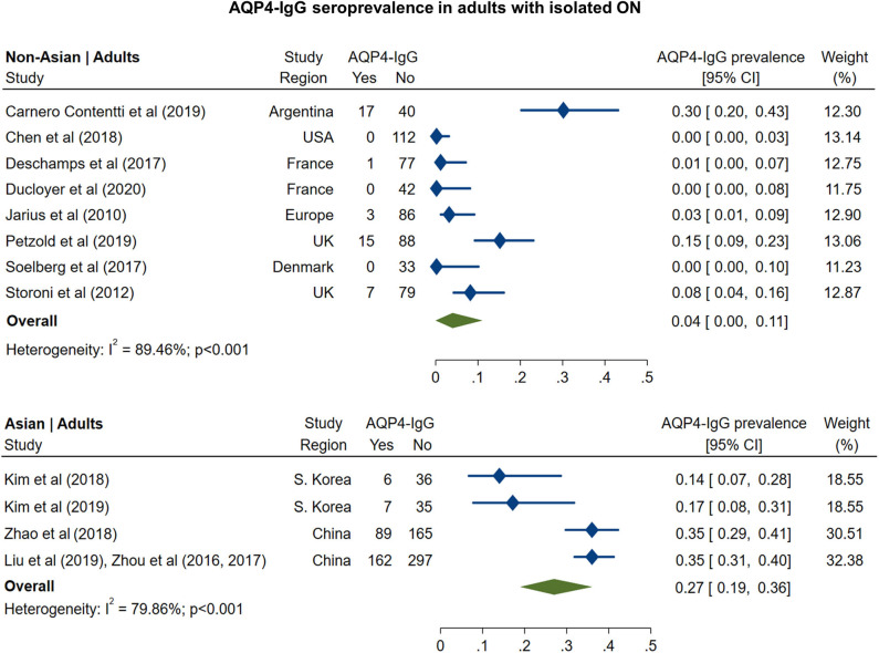
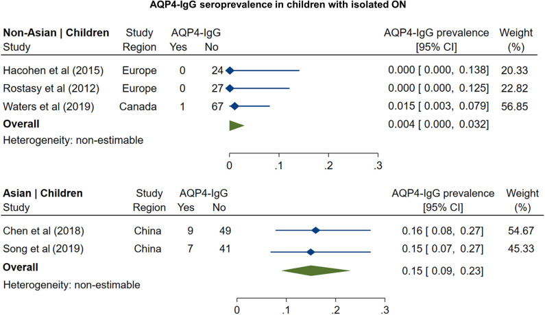
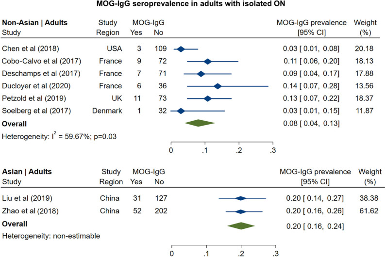
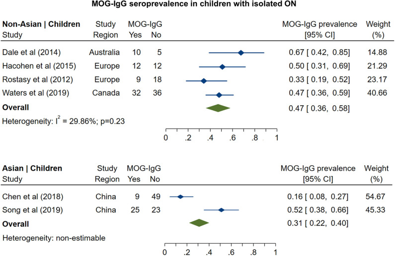
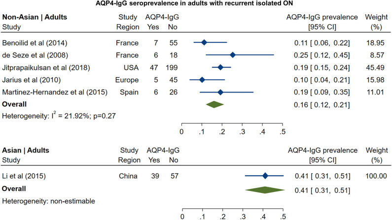
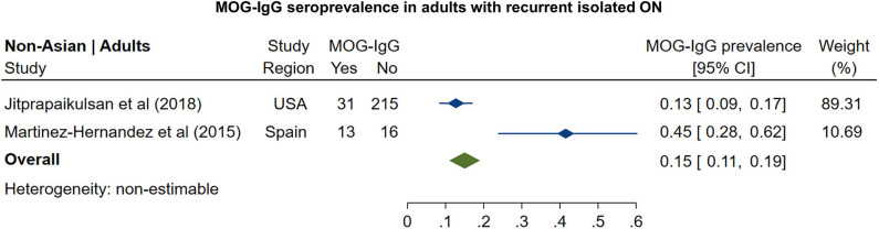
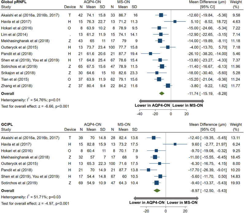
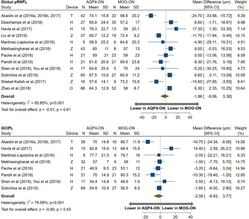
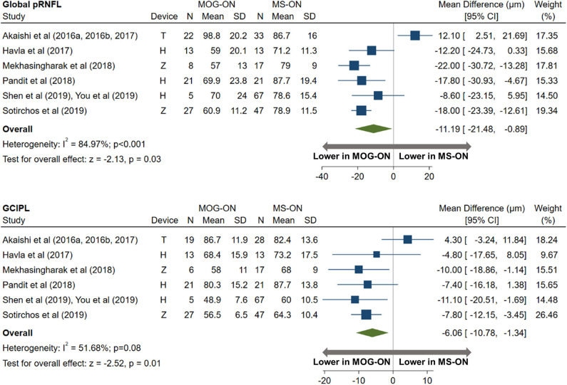
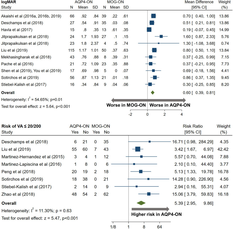
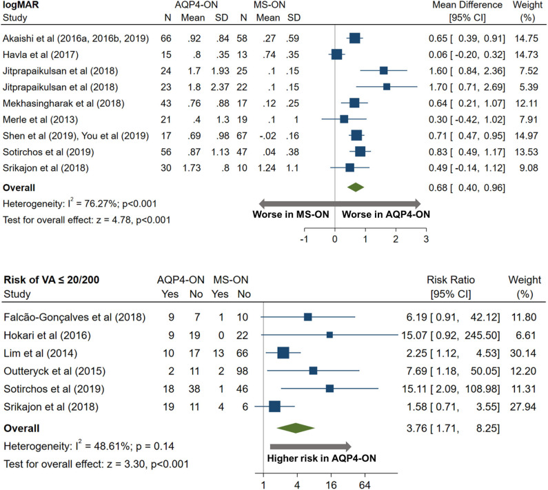
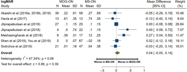
Background: Optic neuritis (ON) is a cardinal manifestation of multiple sclerosis (MS), aquaporin-4 (AQP4)-IgG-, and myelin oligodendrocyte glycoprotein (MOG)-IgG-associated disease. However, the prevalence of AQP4-IgG seropositivity and MOG-IgG seropositivity in isolated ON is unclear, and studies comparing visual outcomes and optical coherence tomography (OCT)-derived structural retinal measures between MS-ON, AQP4-ON, and MOG-ON eyes are limited by small sample sizes. Objectives: (1) To assess the prevalence of AQP4-IgG and MOG-IgG seropositivity among patients presenting with isolated ON; (2) to compare visual outcomes and OCT measures between AQP4-ON, MOG-ON, and MS-ON eyes. Methods: In this systematic review and meta-analysis, a total of 65 eligible studies were identified by PubMed search. Statistical analyses were performed with random effects models. Results: In adults with isolated ON, AQP4-IgG seroprevalence was 4% in non-Asian and 27% in Asian populations, whereas MOG-IgG seroprevalence was 8 and 20%, respectively. In children, AQP4-IgG seroprevalence was 0.4% in non-Asian and 15% in Asian populations, whereas MOG-IgG seroprevalence was 47 and 31%, respectively. AQP4-ON eyes had lower peri-papillary retinal nerve fiber layer (pRNFL; -11.7 μm, 95% CI: -15.2 to -8.3 μm) and macular ganglion cell + inner plexiform layer (GCIPL; -9.0 μm, 95% CI: -12.5 to -5.4 μm) thicknesses compared with MS-ON eyes. Similarly, pRNFL (-11.2 μm, 95% CI: -21.5 to -0.9 μm) and GCIPL (-6.1 μm, 95% CI: -10.8 to -1.3 μm) thicknesses were lower in MOG-ON compared to MS-ON eyes, but did not differ between AQP4-ON and MOG-ON eyes (pRNFL: -1.9 μm, 95% CI: -9.1 to 5.4 μm; GCIPL: -2.6 μm, 95% CI: -8.9 to 3.8 μm). Visual outcomes were worse in AQP4-ON compared to both MOG-ON (mean logMAR difference: 0.60, 95% CI: 0.39 to 0.81) and MS-ON eyes (mean logMAR difference: 0.68, 95% CI: 0.40 to 0.96) but were similar in MOG-ON and MS-ON eyes (mean logMAR difference: 0.04, 95% CI: -0.05 to 0.14). Conclusions: AQP4-IgG- and MOG-IgG-associated disease are important diagnostic considerations in adults presenting with isolated ON, especially in Asian populations. Furthermore, MOG-IgG seroprevalence is especially high in pediatric isolated ON, in both non-Asian and Asian populations. Despite a similar severity of GCIPL and pRNFL thinning in AQP4-ON and MOG-ON, AQP4-ON is associated with markedly worse visual outcomes.

Keywords: aquaporin-4 (AQP4) IgG; myelin oligodendrocyte glycoprotein (MOG) IgG associated disease; neuromyelitis optica (NMO); neuromyelitis optica spectrum disorder (NMOsd); optic neuritis (ON); optical coherence tomography (OCT); retina; visual acuity.


Figures

Figure 1
Figure 1
Forest plot of the prevalence of AQP4-IgG seropositivity in adults with monosymptomatic isolated ON.
Figure 2
Figure 2
Forest plot of the prevalence of AQP4-IgG seropositivity in children with monosymptomatic isolated ON.
Figure 3
Figure 3
Forest plot of the prevalence of MOG-IgG seropositivity in adults with monosymptomatic isolated ON.
Figure 4
Figure 4
Forest plot of the prevalence of MOG-IgG seropositivity in children with monosymptomatic isolated ON.
Figure 5
Figure 5
Forest plot of the prevalence of AQP4-IgG seropositivity in adults with recurrent isolated ON.
Figure 6
Figure 6
Forest plot of the prevalence of MOG-IgG seropositivity in adults with recurrent isolated ON.
Figure 7
Figure 7
Forest plot of the mean difference in global pRNFL and GCIPL thickness between AQP4-ON and MS-ON. The SD-OCT devices used are indicated as H (Spectralis, Heidelberg Engineering; Heidelberg, Germany), O (RTVue, Optovue Inc; Fremont, CA, USA), and T (3D OCT-2000, Topcon Corporation; Tokyo, Japan), Z (Cirrus, Carl Zeiss Meditec; Dublin, CA, USA).
Figure 8
Figure 8
Forest plot of the mean difference in global pRNFL and GCIPL thickness between AQP4-ON and MOG-ON. The SD-OCT devices used are indicated as H (Spectralis, Heidelberg Engineering; Heidelberg, Germany), O (RTVue, Optovue Inc; Fremont, CA, USA), and T (3D OCT-2000, Topcon Corporation; Tokyo, Japan), Z (Cirrus, Carl Zeiss Meditec; Dublin, CA, USA).
Figure 9
Figure 9
Forest plot of the mean difference in global pRNFL and GCIPL thickness between MOG-ON and MS-ON. The SD-OCT devices used are indicated as H (Spectralis, Heidelberg Engineering; Heidelberg, Germany), O (RTVue, Optovue Inc; Fremont, CA, USA), and T (3D OCT-2000, Topcon Corporation; Tokyo, Japan), Z (Cirrus, Carl Zeiss Meditec; Dublin, CA, USA).
Figure 10
Figure 10
Forest plot of the mean difference in logMAR (high-contrast visual acuity) between AQP4-ON and MOG-ON; forest plot of the relative risk of a poor visual outcome (VA worse than 20/200) in AQP4-ON vs MOG-ON.
Figure 11
Figure 11
Forest plot of the mean difference in logMAR (high-contrast visual acuity) between AQP4-ON and MS-ON; forest plot of the relative risk of a poor visual outcome (VA worse than 20/200) in AQP4-ON vs MS-ON.
Figure 12
Figure 12
Forest plot of the mean difference in logMAR (high-contrast visual acuity) between MOG-ON and MS-ON.

Similar articles

Cited by

References

    1. Toosy AT, Mason DF, Miller DH. Optic neuritis. Lancet Neurol. (2014) 13:83–99. 10.1016/S1474-4422(13)70259-X - DOI - PubMed
    1. Petzold A, Wattjes MP, Costello F, Flores-Rivera J, Fraser CL, Fujihara K, et al. . The investigation of acute optic neuritis: a review and proposed protocol. Nat Rev Neurol. (2014) 10:447–58. 10.1038/nrneurol.2014.108 - DOI - PubMed
    1. Reindl M, Waters P. Myelin oligodendrocyte glycoprotein antibodies in neurological disease. Nat Rev Neurol. (2019) 15:89–102. 10.1038/s41582-018-0112-x - DOI - PubMed
    1. Jarius S, Paul F, Aktas O, Asgari N, Dale RC, de Seze J, et al. . MOG encephalomyelitis: international recommendations on diagnosis and antibody testing. J Neuroinflammation. (2018) 15:134. 10.1186/s12974-018-1144-2 - DOI - PMC - PubMed
    1. Borisow N, Mori M, Kuwabara S, Scheel M, Paul F. Diagnosis and Treatment of NMO Spectrum Disorder and MOG-Encephalomyelitis. Front Neurol. (2018) 9:888. 10.3389/fneur.2018.00888 - DOI - PMC - PubMed

Publication types