

Large Sinus of Valsalva Aneurysm Complicated by Thrombus Formation
- PMID: 33133369
- PMCID: PMC7587321
- DOI: 10.14797/mdcj-16-3-e8
Large Sinus of Valsalva Aneurysm Complicated by Thrombus Formation
Abstract
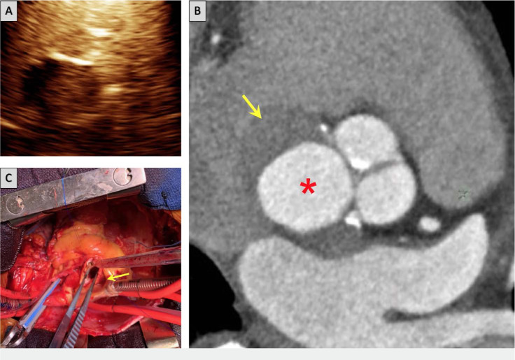
Sinus of Valsalva aneurysm (SOVA) is an unusual cardiac anomaly that is potentially fatal with rupture. It is often asymptomatic but has various presentations. We describe a case of a 67-year-old male who presented with atypical chest pain. Transthoracic echocardiogram and cardiac computed tomography scan confirmed a large SOVA complicated by thrombus formation and compression of the left atrium and left ventricular outflow tract. The patient successfully underwent a Bentall procedure-surgical aortic aneurysm repair with mechanical aortic valve conduit. We discuss several clinical decision-making branch points to highlight the complexity of managing this condition. Even in asymptomatic or minimally symptomatic patients with SOVA, surgery may be indicated if the aneurysm meets the criteria for size or has thrombus formation or compressive effects.
Keywords: SOVA; cardiac anomaly; sinus of Valsalva aneurysm.
© 2020 Houston Methodist Hospital Houston, Texas.
Conflict of interest statement
Conflict of Interest Disclosure: The authors have completed and submitted the Methodist DeBakey Cardiovascular Journal Conflict of Interest Statement and none were reported.
Figures
References
-
- Bricker AO, Avutu B, Mohammed TL, et al. Valsalva sinus aneurysms: Findings at CT and MR imaging. RadioGraphics. 2010 Jan;30(1):99–110. - PubMed
-
- Cheng TO, Yang, Xie M, et al. Echocardiographic diagnosis of sinus of Valsalva aneurysm: a 17-year (1995–2012) experience of 212 surgically treated patients from one single medical center in China. Int J Cardiol. 2014 Apr 15;173(1):33–9. - PubMed
Publication types
MeSH terms
LinkOut - more resources
Full Text Sources
Medical
