

First-trimester rupture of a gravid bicornuate uterus after prior vaginal deliveries, simulating a ruptured ectopic pregnancy: a case report
- PMID: 33133501
- PMCID: PMC7584461
- DOI: 10.1093/jscr/rjaa366
First-trimester rupture of a gravid bicornuate uterus after prior vaginal deliveries, simulating a ruptured ectopic pregnancy: a case report
Abstract
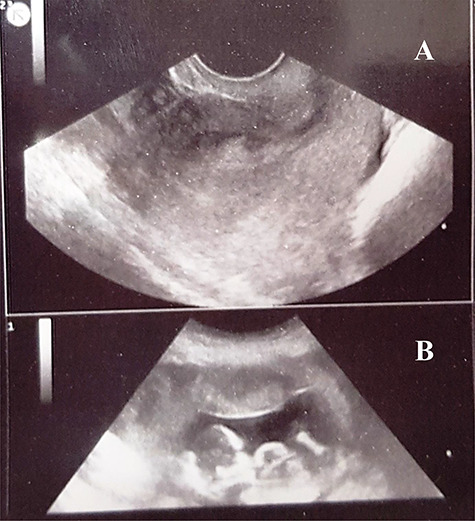
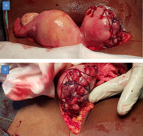
The first-trimester rupture of a bicornuate uterus (BU) is a rare obstetrical emergency, especially following previous normal vaginal deliveries where it is often misdiagnosed. A 24-year-old G3P2002 woman presented at 11 weeks of gestation with sudden onset of severe left iliac fossa pain without other symptoms. On examination, she was fully conscious and hemodynamically unstable with signs of peritoneal irritation, a distended pouch of Douglas and a slightly enlarged uterus and a tender left adnexal mass. The diagnosis of a ruptured ectopic pregnancy was made and a laparotomy was done. Intra-operative findings were hemoperitoneum, a left ruptured BU and a dead fetus. Surgical management entailed hysterorrarphy, left salpingectomy and conservation of both ovaries. Her postoperative course was uneventful and future fertility was preserved. We recommend a high index of suspicion of ruptured BU as a differential diagnosis of acute abdomen in the first trimester in women with previous term vaginal deliveries.
Keywords: gynecology; obstetrics; ruptured bicornuate uterus.
Published by Oxford University Press and JSCR Publishing Ltd. All rights reserved. © The Author(s) 2020.
Figures

Similar articles
-
Spontaneous rupture of a bicornuate uterus at 19 weeks gestation: A case report.Int J Surg Case Rep. 2025 Jun;131:111239. doi: 10.1016/j.ijscr.2025.111239. Epub 2025 Mar 31. Int J Surg Case Rep. 2025. PMID: 40306100 Free PMC article.
-
Mid-trimester spontaneous rupture of a bicornuate uterus: A case report.Case Rep Womens Health. 2023 Jun 30;39:e00524. doi: 10.1016/j.crwh.2023.e00524. eCollection 2023 Sep. Case Rep Womens Health. 2023. PMID: 37954229 Free PMC article.
-
Second Trimester Spontaneous Fundal Rupture of Unscarred Bicornuate Uterus in Primipara: A Case Report and Literature Review; Jigjiga University Sheik Hassen Yabare Comprehensive Specialized Hospital, Jigjiga, Ethiopia.Int Med Case Rep J. 2024 Mar 19;17:181-185. doi: 10.2147/IMCRJ.S446718. eCollection 2024. Int Med Case Rep J. 2024. PMID: 38524802 Free PMC article.
-
Pregnancy after advanced ovarian cancer with spontaneous uterine rupture in second trimester: A case report and review of the literature.Int J Gynaecol Obstet. 2025 Jan;168(1):57-62. doi: 10.1002/ijgo.15837. Epub 2024 Aug 1. Int J Gynaecol Obstet. 2025. PMID: 39087457 Free PMC article. Review.
-
Misdiagnosis of appendicitis in tubally sterilized women.Aust N Z J Surg. 1993 Jan;63(1):68-70. doi: 10.1111/j.1445-2197.1993.tb00037.x. Aust N Z J Surg. 1993. PMID: 8466465 Review.
Cited by
-
Management of Interstitial Ectopic Pregnancy in a Bicornuate Uterus Simulating an Incomplete Abortion.Cureus. 2024 Apr 15;16(4):e58351. doi: 10.7759/cureus.58351. eCollection 2024 Apr. Cureus. 2024. PMID: 38756287 Free PMC article.
References
-
- Singh N, Singh U, Verma ML. Ruptured bicornuate uterus mimicking ectopic pregnancy: a case report. J Obstet Gynaecol Res 2013;39:364–6. - PubMed
-
- Elyan A, Saeed M. Mullerian duct anomalies: clinical concepts. Ain Shams J Obstetr Gynecol 2004;1:11–20.
-
- Dohbit JS, Mbouche L, Ngo Um ME, Tompeen I, Tochie JN, Ayissi G, et al. A fortuitous diagnosis of pyonephrosis complicating pyeloureteral junction syndrome during pregnancy:a case report. Clin Res Obst Gynecol 2018;1:1–4.
Publication types
LinkOut - more resources
Full Text Sources
