. 2020 Sep 10;8(10):5717-5728.
doi: 10.1002/fsn3.1875. eCollection 2020 Oct.

Lysimachia christinae Hance as an anticancer agent against breast cancer cells

Affiliations

Lysimachia christinae Hance as an anticancer agent against breast cancer cells

Hyun A Kim et al. Food Sci Nutr. .

Abstract

Breast cancer is the most common cancer in women, and metastasis is the leading cause of death in breast cancer patients. Although chemoprevention is widely employed to treat breast cancer, anticancer drugs can cause significant adverse effects. Lysimachia christinae Hance (LH) is a traditional Chinese medicinal plant with diverse therapeutic effects. However, its potential anticancer activity has not been fully investigated in breast cancers to date. Using high-performance liquid chromatography-mass spectrometry, we found that the main constituent of LH extract (LHE) was rutin. Our results indicated that LHE or rutin markedly decreased the proliferation and viability of estrogen receptor (ER)-positive MCF-7 and ER-negative HCC38 human breast cancer cells. LHE treatment induced morphological changes in apoptotic nuclei using 4',6-diamidino-2-phenylindole (DAPI) staining. Annexin V-fluorescein isothiocyanate (FITC) propidium iodide (PI) staining assay revealed that apoptosis significantly increased in both breast cancer cell types after LHE treatment. Additionally, the expression of poly (ADP-ribose) polymerase (PARP), Bcl-2, and phospho-Akt decreased, while that of cleaved PARP and p53 increased, in both cell types. Furthermore, LHE treatment inhibited epithelial-mesenchymal transition (EMT). LHE treatment significantly upregulated E-cadherin level in MCF-7 and HCC38 cells, while vimentin level was downregulated in HCC38 cells. In addition, transwell and wound-healing assays revealed that LHE or rutin inhibited breast cancer cell migration. Overall, these findings demonstrate that LHE is a promising therapeutic agent that acts by promoting apoptosis and reducing cell proliferation, EMT, and cell migration in ER-positive and ER-negative breast cancer cells.

Keywords: Lysimachia christinae Hance; apoptosis; breast cancer; epithelial–mesenchymal transition.


Conflict of interest statement

The authors declare no conflicts of interest.

Figures

FIGURE 1
FIGURE 1
HPLC chromatogram of LHE (a) and rutin (b) as a standard compound. LHE, Lysimachia christinae Hance extract; HPLC, high‐performance liquid chromatography
FIGURE 2
FIGURE 2
Effects of LHE on the proliferation or viability of MCF‐7 and HCC38 human breast cancer cells. Breast cancer cell lines were treated with LHE (0.1, 0.2, 0.4, 0.8 mg/ml) for 24 hr and 48 hr by MTS assay (a). Trypan blue exclusion staining assay was performed to determine cell viability using LHE (b) or rutin (c) for 24 hr and 48 hr in breast cancer cells. Data are expressed as the mean ± SD of at least three independent experiments. Significantly different (p < .05) compared with the *DMSO control. LHE, Lysimachia christinae Hance extract; SD, standard deviation
FIGURE 3
FIGURE 3
LHE‐induced apoptosis in breast cancer cells. (a) Apoptosis of MCF‐7 and HCC38 cells was increased by LHE extract. (b) The quantification of apoptotic nuclei. Data are expressed as the mean ± SD of at least three independent experiments. Significantly different (p < .05) compared with the *DMSO control. LHE, Lysimachia christinae Hance extract; DAPI, 4′,6‐diamidino‐2‐phenylindole
FIGURE 4
FIGURE 4
Effects of LHE on apoptosis in breast cancer cells. (a) The apoptotic cells were evaluated after annexin V‐FITC/PI staining using flow cytometry after LHE treatment for 48 hr. Quantification of the total apoptotic cell population (early + late) is shown. Data are expressed as the mean ± SD of at least three independent experiments. Significantly different (p < .05) compared with the *DMSO control. LHE, Lysimachia christinae Hance extract; FITC/PI, fluorescein isothiocyanate/propidium iodide; SD, standard deviation
FIGURE 5
FIGURE 5
Western blotting analysis of PARP, cleaved PARP, and Bcl‐2 proteins in MCF‐7 and HCC38 cells treated with LHE or rutin for 48 hr. β‐Actin was used as the protein loading control. Data are expressed as the mean ± SD of at least three independent experiments. Significantly different (p < .05) compared with the *DMSO control. PARP, poly (ADP‐ribose) polymerase; LHE, Lysimachia christinae Hance extract; SD, standard deviation
FIGURE 6
FIGURE 6
Western blotting analysis of p53, phospho‐Akt, and total Akt proteins in MCF‐7 and HCC38 cells treated with LHE for 48 hr. β‐Actin was used as the protein loading control. Data are expressed as the mean ± SD of at least three independent experiments. Significantly different (p < .05) compared with the *DMSO control. LHE, Lysimachia christinae Hance extract; SD, standard deviation
FIGURE 7
FIGURE 7
Effects of LHE on EMT‐related protein expression in breast cancer cells. Western blotting analysis of E‐cadherin and vimentin proteins in MCF‐7 and HCC38 cells treated with LHE for 24 hr. β‐Actin was used as the protein loading control. Data are expressed as the mean ± SD of at least three independent experiments. Significantly different (p < .05) compared with the *DMSO control. LHE, Lysimachia christinae Hance extract; EMT, epithelial–mesenchymal transition; SD, standard deviation
FIGURE 8
FIGURE 8
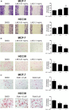
Effects of LHE or rutin on breast cancer cells migration in wound‐healing and transwell migration assay. (a) The confluent monolayers of MCF‐7 and HCC38 cells treated with LHE for 24 hr. Quantification of scratched area was performed using ImageJ software. (b) MCF‐7 and HCC38 cells in serum‐free medium with LHE or rutin were seeded into the transwell upper chamber. The lower chamber was filled with 5% FBS containing media. The cells were allowed to migrate for 24 hr. Migrating cells were stained with H&E solutions and counted. Data are expressed as the mean ± SD of at least three independent experiments. Significantly different (p < .05) compared with the *DMSO control. H&E, hematoxylin and eosin; LHE, Lysimachia christinae Hance extract; SD, standard deviation

References

    1. Al‐Mahmood, S. , Sapiezynski, J. , Garbuzenko, O. B. , & Minko, T. (2018). Metastatic and triple‐negative breast cancer: Challenges and treatment options. Drug Delivery and Translational Research, 8(5), 1483–1507. 10.1007/s13346-018-0551-3 - DOI - PMC - PubMed
    1. Ashaie, M. A. , & Chowdhury, E. H. (2016). Cadherins: The superfamily critically involved in breast cancer. Current Pharmaceutical Design, 22(5), 616–638. 10.2174/138161282205160127095338 - DOI - PubMed
    1. Aung, T. N. , Qu, Z. , Kortschak, R. D. , & Adelson, D. L. (2017). Understanding the effectiveness of natural compound mixtures in cancer through their molecular mode of action. International Journal of Molecular Sciences, 18(3), 656 10.3390/ijms18030656 - DOI - PMC - PubMed
    1. Balunas, M. J. , & Kinghorn, A. D. (2005). Drug discovery from medicinal plants. Life Sciences, 78(5), 431–441. 10.1016/j.lfs.2005.09.012 - DOI - PubMed
    1. Ban, K. A. , Godellas, C. V . (2014). Epidemiology of breast cancer. Surgical Oncology Clinics of North America, 23(3), 409–422. 10.1016/j.soc.2014.03.011 - DOI - PubMed