

Generation of human liver organoids from pluripotent stem cell-derived hepatic endoderms
- PMID: 33133779
- PMCID: PMC7580584
- DOI: 10.7717/peerj.9968
Generation of human liver organoids from pluripotent stem cell-derived hepatic endoderms
Abstract
Background: The use of a personalized liver organoid derived from human-induced pluripotent stem cells (HuiPSCs) is advancing the use of in vitro disease models for the design of specific, effective therapies for individuals. Collecting patient peripheral blood cells for HuiPSC generation is preferable because it is less invasive; however, the capability of blood cell-derived HuiPSCs for hepatic differentiation and liver organoid formation remains uncertain. Moreover, the currently available methods for liver organoid formation require a multistep process of cell differentiation or a combination of hepatic endodermal, endothelial and mesenchymal cells, which is a major hurdle for the application of personalized liver organoids in high-throughput testing of drug toxicity and safety. To demonstrate the capability of blood cell-derived HuiPSCs for liver organoid formation without support from endothelial and mesenchymal cells.
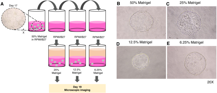
Methods: The peripheral blood-derived HuiPSCs first differentiated into hepatic endoderm (HE) in two-dimensional (2D) culture on Matrigel-coated plates under hypoxia for 10 days. The HE was then collected and cultured in 3D culture using 50% Matrigel under ambient oxygen. The maturation of hepatocytes was further induced by adding hepatocyte growth medium containing HGF and oncostatin M on top of the 3D culture and incubating the culture for an additional 12-17 days. The function of the liver organoids was assessed using expression analysis of hepatocyte-specific gene and proteins. Albumin (ALB) synthesis, glycogen and lipid storage, and metabolism of indocyanine were evaluated. The spatial distribution of albumin was examined using immunofluorescence and confocal microscopy.
Results: CD34+ hematopoietic cell-derived HuiPSCs were capable of differentiating into definitive endoderm expressing SOX17 and FOXA2, hepatic endoderm expressing FOXA2, hepatoblasts expressing AFP and hepatocytes expressing ALB. On day 25 of the 2D culture, cells expressed SOX17, FOXA2, AFP and ALB, indicating the presence of cellular heterogeneity. In contrast, the hepatic endoderm spontaneously formed a spherical, hollow structure in a 3D culture of 50% Matrigel, whereas hepatoblasts and hepatocytes could not form. Microscopic observation showed a single layer of polygonal-shaped cells arranged in a 3D structure. The hepatic endoderm-derived organoid synthesis ALB at a higher level than the 2D culture but did not express definitive endoderm-specific SOX17, indicating the greater maturity of the hepatocytes in the liver organoids. Confocal microscopic images and quantitative ELISA confirmed albumin synthesis in the cytoplasm of the liver organoid and its secretion. Overall, 3D culture of the hepatic endoderm is a relatively fast, simple, and less laborious way to generate liver organoids from HuiPSCs that is more physiologically relevant than 2D culture.
Keywords: Differentiation; Hepatic endoderm; Hepatocyte; Liver organoid; Matrigel; Three-dimensional culture; Albumin; Hepatocyte; Human induced pluripotent stem cell.
©2020 Kulkeaw et al.
Conflict of interest statement
The authors declare there are no competing interests.
Figures


Similar articles
-
Human liver organoids are susceptible to Plasmodium vivax infection.Malar J. 2024 Dec 5;23(1):368. doi: 10.1186/s12936-024-05202-8. Malar J. 2024. PMID: 39639330 Free PMC article.
-
Liver Organoids: Formation Strategies and Biomedical Applications.Tissue Eng Regen Med. 2021 Aug;18(4):573-585. doi: 10.1007/s13770-021-00357-w. Epub 2021 Jun 16. Tissue Eng Regen Med. 2021. PMID: 34132985 Free PMC article. Review.
-
Retaining mTeSR1 Medium during Hepatic Differentiation Facilitates Hepatocyte-Like Cell Survival by Decreasing Apoptosis.Cell Physiol Biochem. 2018;51(4):1533-1543. doi: 10.1159/000495644. Epub 2018 Nov 29. Cell Physiol Biochem. 2018. PMID: 30497075
-
Developing a Cost-Effective and Scalable Production of Human Hepatic Competent Endoderm from Size-Controlled Pluripotent Stem Cell Aggregates.Stem Cells Dev. 2018 Feb 15;27(4):262-274. doi: 10.1089/scd.2017.0074. Epub 2018 Feb 1. Stem Cells Dev. 2018. PMID: 29298619
-
Challenges for the Applications of Human Pluripotent Stem Cell-Derived Liver Organoids.Front Cell Dev Biol. 2021 Oct 1;9:748576. doi: 10.3389/fcell.2021.748576. eCollection 2021. Front Cell Dev Biol. 2021. PMID: 34660606 Free PMC article. Review.
Cited by
-
Female Reproductive Tract Organoids: Applications from Physiology to Pathology.Biomolecules. 2025 Jun 24;15(7):925. doi: 10.3390/biom15070925. Biomolecules. 2025. PMID: 40723797 Free PMC article. Review.
-
Progress and Challenges in the Use of a Liver-on-a-Chip for Hepatotropic Infectious Diseases.Micromachines (Basel). 2021 Jul 19;12(7):842. doi: 10.3390/mi12070842. Micromachines (Basel). 2021. PMID: 34357252 Free PMC article. Review.
-
Human liver organoids are susceptible to Plasmodium vivax infection.Malar J. 2024 Dec 5;23(1):368. doi: 10.1186/s12936-024-05202-8. Malar J. 2024. PMID: 39639330 Free PMC article.
-
Liver Organoids: Formation Strategies and Biomedical Applications.Tissue Eng Regen Med. 2021 Aug;18(4):573-585. doi: 10.1007/s13770-021-00357-w. Epub 2021 Jun 16. Tissue Eng Regen Med. 2021. PMID: 34132985 Free PMC article. Review.
-
Addressing Key Questions in Organoid Models: Who, Where, How, and Why?Int J Mol Sci. 2023 Nov 6;24(21):16014. doi: 10.3390/ijms242116014. Int J Mol Sci. 2023. PMID: 37958996 Free PMC article. Review.
References
-
- Aguila JC, Blak A, Van Arensbergen J, Sousa A, Vazquez N, Aduriz A, Gayosso M, Lopez Mato MP, Lopez de Maturana R, Hedlund E, Sonntag KC, Sanchez-Pernaute R. Selection based on FOXA2 expression is not sufficient to enrich for dopamine neurons from human pluripotent stem cells. Stem Cells Translational Medicine. 2014;3:1032–1042. doi: 10.5966/sctm.2014-0011. - DOI - PMC - PubMed
-
- Boj SF, Hwang CI, Baker LA, Chio II, Engle DD, Corbo V, Jager M, Ponz-Sarvise M, Tiriac H, Spector MS, Gracanin A, Oni T, Yu KH, Van Boxtel R, Huch M, Rivera KD, Wilson JP, Feigin ME, Ohlund D, Handly-Santana A, Ardito-Abraham CM, Ludwig M, Elyada E, Alagesan B, Biffi G, Yordanov GN, Delcuze B, Creighton B, Wright K, Park Y, Morsink FH, Molenaar IQ, BorelRinkes IH, Cuppen E, Hao Y, Jin Y, Nijman IJ, Iacobuzio-Donahue C, Leach SD, Pappin DJ, Hammell M, Klimstra DS, Basturk O, Hruban RH, Offerhaus GJ, Vries RG, Clevers H, Tuveson DA. Organoid models of human and mouse ductal pancreatic cancer. Cell. 2015;160:324–338. doi: 10.1016/j.cell.2014.12.021. - DOI - PMC - PubMed
-
- Bruening J, Lasswitz L, Banse P, Kahl S, Marinach C, Vondran FW, Kaderali L, Silvie O, Pietschmann T, Meissner F, Gerold G. Hepatitis C virus enters liver cells using the CD81 receptor complex proteins calpain-5 and CBLB. PLOS Pathogens. 2018;14:e1007111. doi: 10.1371/journal.ppat.1007111. - DOI - PMC - PubMed
LinkOut - more resources
Full Text Sources
Other Literature Sources
Miscellaneous
