

Mapping of the Motor Cortex
- PMID: 33133815
- PMCID: PMC7586383
- DOI: 10.7759/cureus.10645
Mapping of the Motor Cortex
Abstract
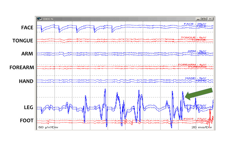
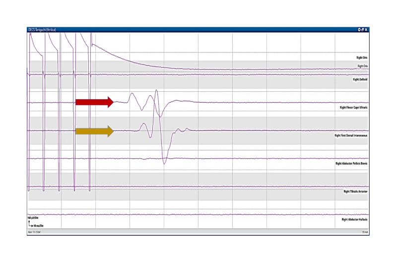
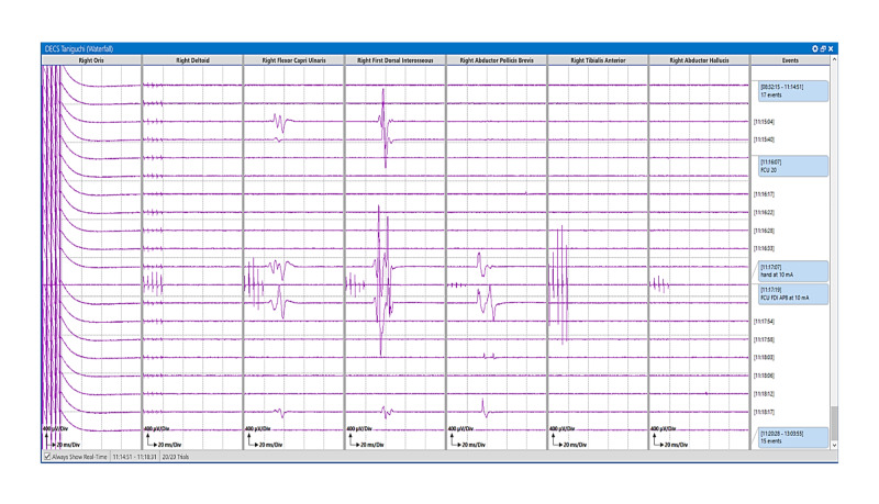
The resection of brain tumors located within or near the eloquent tissue has a higher risk of postoperative neurological deficits. The primary concerns include loss of sensory and motor functions in the contralateral face, upper and lower extremities, as well as speech deficits. Intraoperative neurophysiological monitoring (IONM) techniques are performed routinely for the identification and preservation of the functional integrity of the eloquent brain areas during neurosurgical procedures. The IONM modalities involve sensory, motor, and language mapping, which helps in the identification of the boundaries of these areas during surgical resection. Cortical motor Mapping (CmM) technique is considered as a gold-standard technique for mapping of the brain. We present the intraoperative CmM technique, including anesthesia recommendations, types of electrodes, as well as stimulation and recording parameters for successful monitoring.
Keywords: brain tumor; cortical mapping; eeg; electrocorticography; ionm; motor mapping; neuro-monitoring; neuro-surgery.
Copyright © 2020, Jahangiri et al.
Conflict of interest statement
The authors have declared that no competing interests exist.
Figures


Similar articles
-
Intraoperative neurophysiological monitoring in pediatric neurosurgery: why, when, how?Childs Nerv Syst. 2002 Jul;18(6-7):264-87. doi: 10.1007/s00381-002-0582-3. Epub 2002 Jun 13. Childs Nerv Syst. 2002. PMID: 12172930 Review.
-
Awake craniotomy for brain tumors near eloquent cortex: correlation of intraoperative cortical mapping with neurological outcomes in 309 consecutive patients.Neurosurgery. 2009 May;64(5):836-45; discussion 345-6. doi: 10.1227/01.NEU.0000342405.80881.81. Neurosurgery. 2009. PMID: 19404147 Clinical Trial.
-
[Surgical treatment of eloquent brain area tumors using neurophysiological mapping of the speech and motor areas and conduction tracts].Zh Vopr Neirokhir Im N N Burdenko. 2017;81(1):39-50. doi: 10.17116/neiro201780739-50. Zh Vopr Neirokhir Im N N Burdenko. 2017. PMID: 28291212 Russian.
-
Mapping of the Language Cortex.Cureus. 2021 May 11;13(5):e14960. doi: 10.7759/cureus.14960. Cureus. 2021. PMID: 34123657 Free PMC article.
-
Intraoperative neurophysiological mapping and monitoring in spinal tumor surgery: sirens or indispensable tools?Neurosurg Focus. 2016 Aug;41(2):E18. doi: 10.3171/2016.5.FOCUS16141. Neurosurg Focus. 2016. PMID: 27476842 Review.
Cited by
-
Intraoperative Testing During the Mapping of the Language Cortex.Cureus. 2023 Mar 26;15(3):e36718. doi: 10.7759/cureus.36718. eCollection 2023 Mar. Cureus. 2023. PMID: 37123781 Free PMC article.
-
New frontiers in intraoperative neurophysiologic monitoring: a narrative review.Ann Transl Med. 2023 Oct 25;11(11):388. doi: 10.21037/atm-22-4586. Epub 2023 Jul 26. Ann Transl Med. 2023. PMID: 37970609 Free PMC article. Review.
-
Frontal lobe disconnection: How I do it.Acta Neurochir (Wien). 2024 Oct 29;166(1):429. doi: 10.1007/s00701-024-06319-0. Acta Neurochir (Wien). 2024. PMID: 39470854
-
Optimization of direct cortical stimulation using tibial versus median nerve sensory mapping during midline brain tumor resection: illustrative case.J Neurosurg Case Lessons. 2024 Mar 25;7(13):CASE23704. doi: 10.3171/CASE23704. Print 2024 Mar 25. J Neurosurg Case Lessons. 2024. PMID: 38531084 Free PMC article.
-
Motor Mapping of the Brain: Taniguchi Versus Penfield Method.Cureus. 2022 May 11;14(5):e24901. doi: 10.7759/cureus.24901. eCollection 2022 May. Cureus. 2022. PMID: 35706721 Free PMC article.
References
-
- The first primary brain-tumor operation. Kirkpatrick DB. J Neurosurg. 1984;61:809–813. - PubMed
-
- The excitable cerebral cortex. Fritsch G, Hitzig E. Über die elektrische Erregbarkeit des Grosshirns. Arch Anat Physiol Wissen 1870; 37: 300-32. Carlson C, Devinsky O. Epilepsy Behav. 2009;15:131–132. - PubMed
-
- Somatic motor and sensory representation in the cerebral cortex of man as studied by electrical stimulation. Penfield W, Boldrey E. Brain. 1937;60:389–443.
-
- Modification of cortical stimulation for motor evoked potentials under general anesthesia: technical description. Taniguchi M, Cedzich C, Schramm J, Cedzich C, Schramm J. Neurosurgery. 1993;32:219–226. - PubMed
LinkOut - more resources
Full Text Sources