

Bone-Tendon-Autograft Anterior Cruciate Ligament Reconstruction: A New Anterior Cruciate Ligament Graft Option
- PMID: 33134055
- PMCID: PMC7587499
- DOI: 10.1016/j.eats.2020.06.021
Bone-Tendon-Autograft Anterior Cruciate Ligament Reconstruction: A New Anterior Cruciate Ligament Graft Option
Abstract
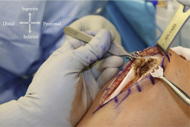
The bone-tendon-bone (BTB) autograft is widely used for anterior cruciate ligament (ACL) reconstruction. However, the primary disadvantages of this technique include postoperative kneeling pain, the risk of perioperative patellar fracture, and graft-tunnel mismatch. Therefore, a single bone plug technique for ACL reconstructions was developed to mitigate the disadvantages of the BTB technique. To differentiate this graft, we have coined the term BTA, for bone-tendon-autograft. The middle third of the patellar tendon is used with a typical width of 10 to 11 mm. A standard tibial tubercle bone plug is harvested. The length of the patellar tendon and graft construct is then measured. If the tendon is >45 mm and the construct at least 70 mm, then we proceed with the BTA technique. At the inferior pole of the patella, electrocautery is used to harvest the tendon from the patella. The advantages of this technique include faster graft harvest and preparation. Obviating the patellar bone plug harvest should eliminate the risk of perioperative patellar fracture and theoretically will mitigate donor site morbidity and kneeling pain, 2 of the most commonly cited complications of the use of BTB autografts for ACL reconstruction. In conclusion, the BTA technique is a reliable technique for ACL reconstruction.
© 2020 by the Arthroscopy Association of North America. Published by Elsevier.
Figures


Similar articles
-
Comparing Bone-Tendon Autograft With Bone-Tendon-Bone Autograft for ACL Reconstruction: A Matched-Cohort Analysis.Orthop J Sports Med. 2020 Dec 4;8(12):2325967120970224. doi: 10.1177/2325967120970224. eCollection 2020 Dec. Orthop J Sports Med. 2020. PMID: 33330739 Free PMC article.
-
Remnant-preserving, selective single-bundle augmentation of the anterior cruciate ligament using a bone-patellar tendon-bone autograft: A technical note.Knee. 2016 Jun;23(3):554-8. doi: 10.1016/j.knee.2015.11.024. Epub 2016 Feb 8. Knee. 2016. PMID: 26869506
-
Anterior Cruciate Ligament Reconstruction Basics: Bone-Patellar Tendon-Bone Autograft Harvest.Arthrosc Tech. 2017 Jul 31;6(4):e1189-e1194. doi: 10.1016/j.eats.2017.04.006. eCollection 2017 Aug. Arthrosc Tech. 2017. PMID: 29354416 Free PMC article.
-
A meta-analysis of bone-patellar tendon-bone autograft versus four-strand hamstring tendon autograft for anterior cruciate ligament reconstruction.Knee. 2015 Mar;22(2):100-10. doi: 10.1016/j.knee.2014.11.014. Epub 2014 Dec 11. Knee. 2015. PMID: 25547048 Review.
-
Anterior Cruciate Ligament Reconstruction: A Systematic Review and Meta-analysis of Outcomes for Quadriceps Tendon Autograft Versus Bone-Patellar Tendon-Bone and Hamstring-Tendon Autografts.Am J Sports Med. 2019 Dec;47(14):3531-3540. doi: 10.1177/0363546518825340. Epub 2019 Feb 21. Am J Sports Med. 2019. PMID: 30790526
Cited by
-
Electrospun, Resorbable, Drug-Eluting, Nanofibrous Membranes Promote Healing of Allograft Tendons.Membranes (Basel). 2022 May 18;12(5):529. doi: 10.3390/membranes12050529. Membranes (Basel). 2022. PMID: 35629855 Free PMC article.
-
Supplementary Tibial Fixation in Anterior Cruciate Ligament Reconstruction With Bone-Tendon-Bone Graft.Arthrosc Tech. 2022 Aug 6;11(9):e1551-e1556. doi: 10.1016/j.eats.2022.04.006. eCollection 2022 Sep. Arthrosc Tech. 2022. PMID: 36185108 Free PMC article.
-
Anterior cruciate ligament reconstruction: Effect of graft tunnel position on early to mid-term clinical outcomes.World J Orthop. 2024 Aug 18;15(8):744-753. doi: 10.5312/wjo.v15.i8.744. eCollection 2024 Aug 18. World J Orthop. 2024. PMID: 39165872 Free PMC article.
References
-
- Gifstad T., Foss O.A., Engebretsen L. Lower risk of revision with patellar tendon autografts compared with hamstring autografts: A registry study based on 45,998 primary ACL reconstructions in Scandinavia. Am J Sports Med. 2014;42:2319–2328. - PubMed
-
- Kartus J., Movin T., Karlsson J. Donor-site morbidity and anterior knee problems after anterior cruciate ligament reconstruction using autografts. Arthroscopy. 2001;17:971–980. - PubMed
-
- Stein D.A., Hunt S.A., Rosen J.E., Sherman O.H. The incidence and outcome of patella fractures after anterior cruciate ligament reconstruction. Arthroscopy. 2002;18:578–583. - PubMed
-
- Busam M.L., Provencher M.T., Bach B.R. Complications of anterior cruciate ligament reconstruction with bone-patellar tendon-bone constructs: Care and prevention. Am J Sports Med. 2008;36:379–394. - PubMed
LinkOut - more resources
Full Text Sources
