. 2020 Oct 1;9(10):e1607-e1611.
doi: 10.1016/j.eats.2020.05.005. eCollection 2020 Oct.

Arthroscopic Transosseous Suture-bridge Fixation for Anterior Cruciate Ligament Tibial Avulsion Fractures

Affiliations

Arthroscopic Transosseous Suture-bridge Fixation for Anterior Cruciate Ligament Tibial Avulsion Fractures

Supamongkol Mutchamee et al. Arthrosc Tech. .

Abstract

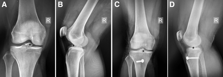
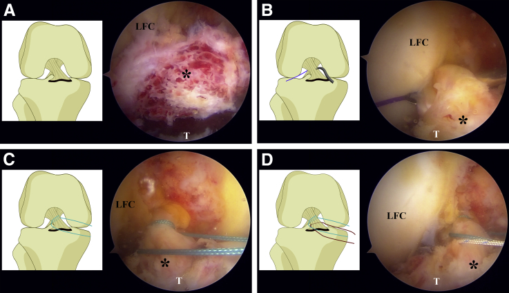
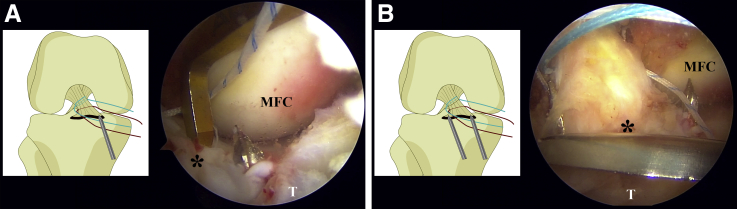
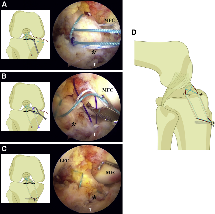
Few cases of tibial spine avulsion injuries occur in adolescents. An open or arthroscopic surgical approach is indicated for displaced and nonreducible fractures, but evidence for a gold standard is insufficient. Various arthroscopic techniques are available. Suture fixation is popular and shows good results. The proposed technique is a modified suture-bridge fixation using 2 high-strength sutures tied through 2 transosseous tunnels. This simple and low-cost technique avoids the potential complications of hardware fixation within a joint. It represents an arthroscopic treatment option for anterior cruciate ligament tibial avulsion injuries.


Figures

Fig 1
Fig 1
(A) Preoperative anteroposterior view of right knee. (B) Lateral view, showing a completely displaced tibial spine fracture, that is, type III according to the Meyers and McKeever classification. (C) Postoperative anteroposterior view. (D) Lateral view of right knee at 6 months postoperatively, showing fracture union with anatomic reduction. The asterisks indicate the tibial spine avulsion fragment. (R, right.)
Fig 2
Fig 2
(A) Arthroscopic photograph and drawing of right knee (in supine position), viewed from standard anterolateral portal. The tibial spine avulsion fragment is shown after debridement and blood clot removal. (B) A 45°, left-curved suture passer loaded with a No. 1 polydioxanone suture is inserted through the anteromedial portal and pierced through the most fibrous portion of the anterior cruciate ligament from the medial to lateral side, as close as possible to the tibial footprint. (C) One limb of the No. 5 Ethibond is shuttled through the anterior cruciate ligament fiber. The 45°, left-curved suture passer is used to shuttle the medial limb of the No. 5 Ethibond suture, making a locking loop. (D) The same technique is repeated: One limb of the Ultrabraid suture is shuttled, making a simple loop. The asterisks indicate the tibial spine avulsion fragment. (LFC, lateral femoral condyle; T, tibia.)
Fig 3
Fig 3
(A) Arthroscopic photograph and drawing of right knee (in supine position), viewed from standard anterolateral portal. Creation of a tibial tunnel (2.5-mm diameter) is shown, using an anterior cruciate ligament tibial guide with the angle set at 45° and aiming at the anteromedial rim through the fracture site. (B) The same technique is performed again—this time aiming at the anterolateral rim through the fracture site from a separate tibial entry point—with at least 1 cm of bone bridge between the 2 tibial tunnels. The asterisks indicate the tibial spine avulsion fragment. (MFC, medial femoral condyle; T, tibia.)
Fig 4
Fig 4
(A) Arthroscopic photograph and drawing of right knee (in supine position), viewed from standard anterolateral portal. Two suture limbs are shown: one limb of the No. 5 Ethibond and the opposite limb of the Ultrabraid. The 2 limbs are retrieved through the polydioxanone suture loop and shuttled to the anterolateral tibial tunnel in a crossing-bridge pattern. (B) The remaining suture limbs are shuttled to the anteromedial tibial tunnel in the same manner. (C) Two limbs of the sutures (one each from the anteromedial and anterolateral tibial tunnels) are tied separately over a screw and post, thereby completing the suture-bridge fixation system. (D) Drawing of right knee in lateral view, illustrating transosseous suture-bridge fixation technique. The asterisks indicate the tibial spine avulsion fragment. (LFC, lateral femoral condyle; MFC, medial femoral condyle; T, tibia.)

Similar articles

Cited by

References

    1. Skak S.V., Jensen T.T., Poulsen T.D., Sturup J. Epidemiology of knee injuries in children. Acta Orthop Scand. 1987;58:78–81. - PubMed
    1. Meyers M.H., McKeever F.M. Fracture of the intercondylar eminence of the tibia. J Bone Joint Surg Am. 1970;52:1677–1684. - PubMed
    1. Zaricznyj B. Avulsion fracture of the tibial eminence: Treatment by open reduction and pinning. J Bone Joint Surg Am. 1977;59:1111–1114. - PubMed
    1. Gans I., Baldwin K.D., Ganley T.J. Treatment and management outcomes of tibial eminence fractures in pediatric patients: A systematic review. Am J Sports Med. 2014;42:1743–1750. - PubMed
    1. Furlan D., Pogorelic Z., Biocic M., Juric I., Mestrovic J. Pediatric tibial eminence fractures: Arthroscopic treatment using K-wire. Scand J Surg. 2010;99:38–44. - PubMed

LinkOut - more resources