. 2020 Oct 1;9(10):e1619-e1626.
doi: 10.1016/j.eats.2020.07.002. eCollection 2020 Oct.

Restoring Horizontal Stability of the Acromioclavicular Joint: Open Acromioclavicular Ligament Reconstruction and Repair With Semitendinosus Allograft

Affiliations

Restoring Horizontal Stability of the Acromioclavicular Joint: Open Acromioclavicular Ligament Reconstruction and Repair With Semitendinosus Allograft

Gianna M Aliberti et al. Arthrosc Tech. .

Abstract

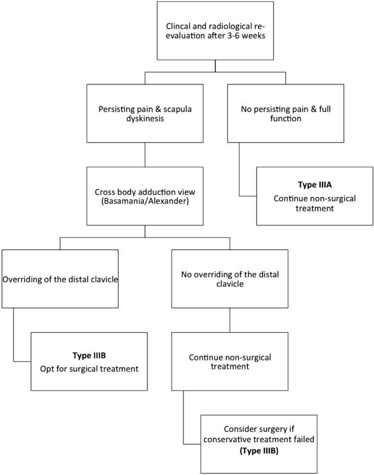
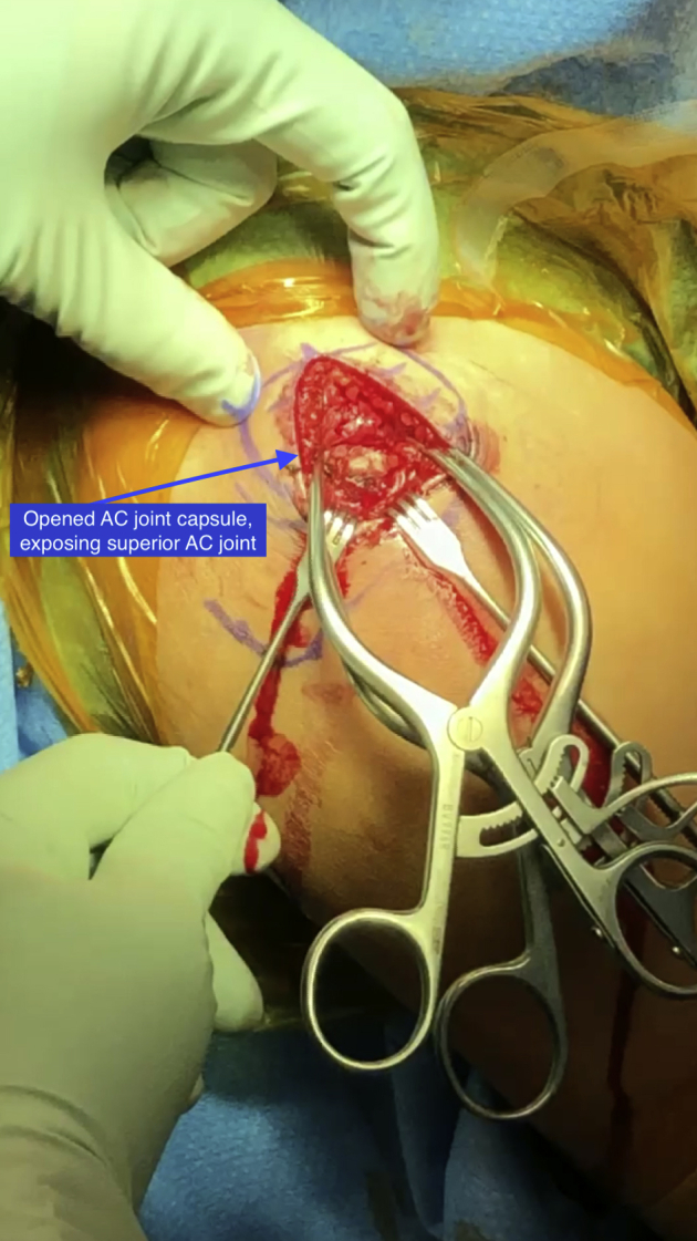
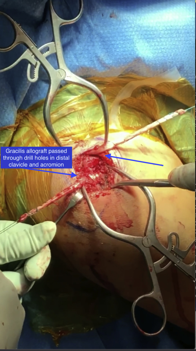
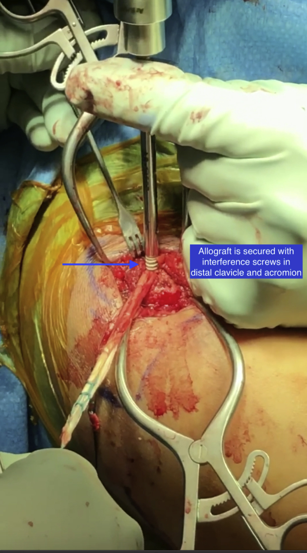
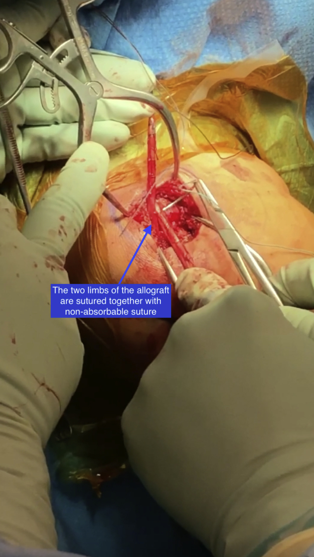
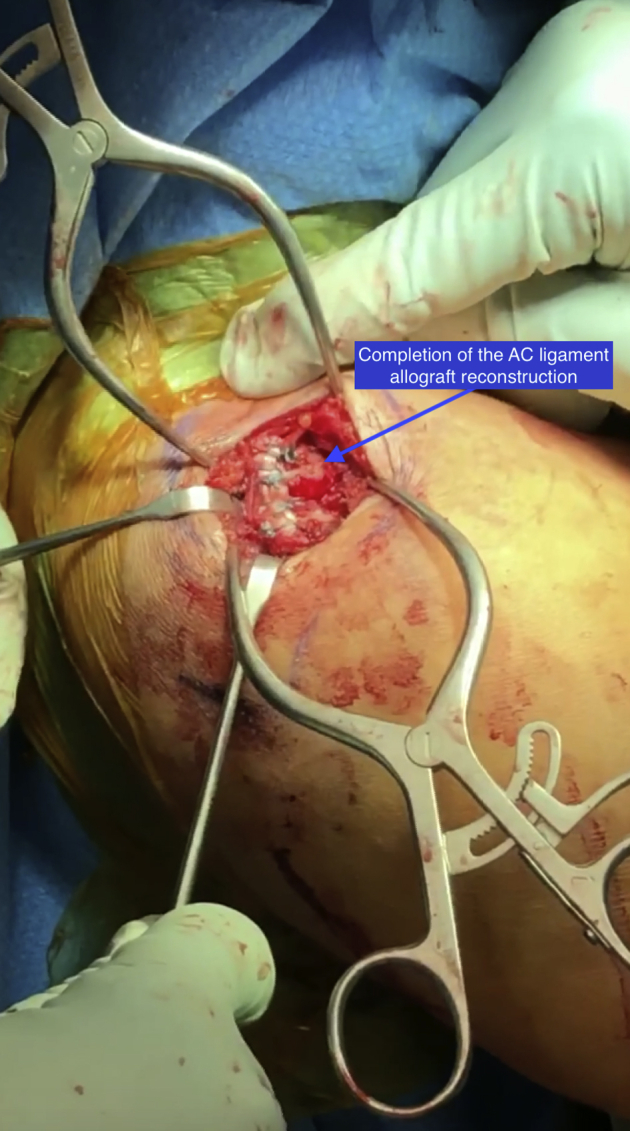
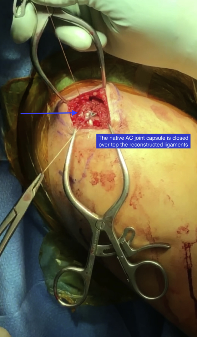
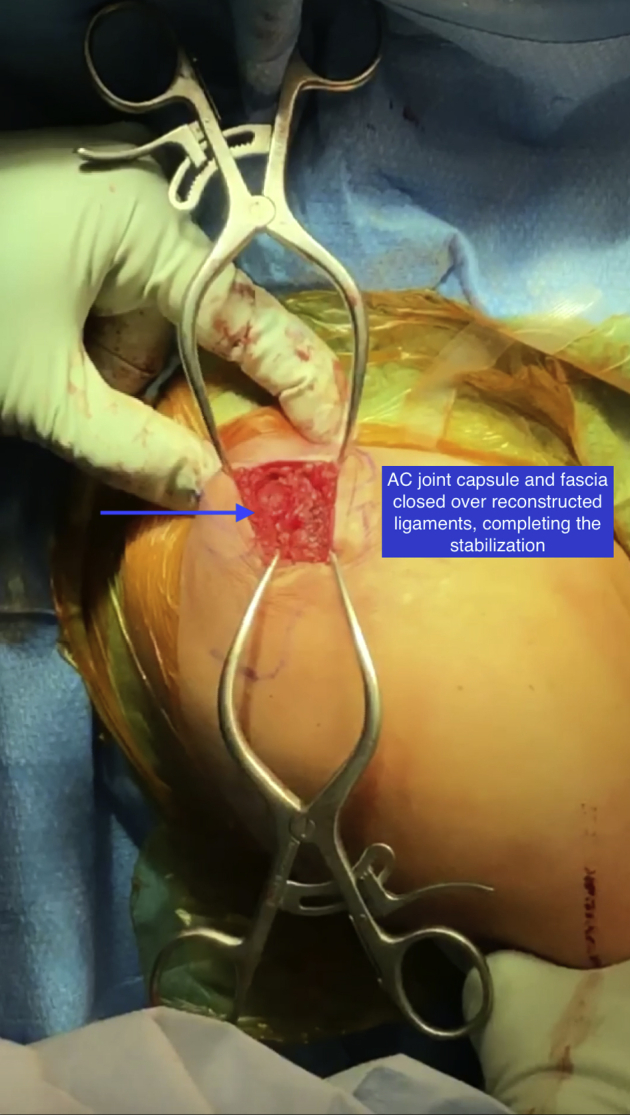
Injuries to the acromioclavicular (AC) joint are common and comprise ∼12% of shoulder injuries. Stability to the AC joint depends on the coracoclavicular (CC) ligaments for vertical stability and AC ligaments and capsular structures for horizontal stability. Injuries to the AC ligaments can lead to horizontal instability of the AC joint. There is no gold standard technique for treating these injuries surgically, and many of the described procedures focus on vertical instability rather than horizontal instability. This article describes an open AC ligament reconstruction with semitendinosus allograft to restore horizontal stability of the AC joint.


Figures

Fig 1
Fig 1
Modified Rockwood classification. The original Rockwood classification describes 6 types of acromioclavicular (AC) joint injuries. The modified Rockwood classification expands on Rockwood's original classification by separating type III injuries into stable (IIIA) and unstable (IIIB). Type IIIA injuries involve a stable AC joint without overriding of the clavicle on cross-body adduction view and without significant scapular dysfunction, whereas type IIIB injuries involve therapy-resistant scapular dysfunction and an overriding clavicle on cross-body adduction view.
Fig 2
Fig 2
The right shoulder of a patient diagnosed with a type II acromioclavicular (AC) joint separation confirmed by magnetic resonance imaging is exposed while the patient is in the beach chair position. The AC joint is exposed through a superior incision in line with the clavicle. Here, the AC joint capsule has been opened, exposing the superior AC joint.
Fig 3
Fig 3
Superior view of the acromioclavicular (AC) joint in a right shoulder, with the patient in the beach chair position. The gracilis allograft has been passed through drill holes in the distal clavicle and acromion.
Fig 4
Fig 4
Superior view of the acromioclavicular (AC) joint in a right shoulder, with the patient in the beach chair position. After reducing the AC joint, the allograft is secured with 2 interference screws, 1 in the distal clavicle and 1 in the acromion.
Fig 5
Fig 5
Superior view of the acromioclavicular (AC) joint in a right shoulder, with the patient in the beach chair position. The 2 limbs of the allograft are sutured together with nonabsorbable suture.
Fig 6
Fig 6
Superior view of the acromioclavicular (AC) joint in a right shoulder, with the patient in the beach chair position, depicting completion of the AC ligament allograft reconstruction. The horizontal stability of the AC joint can be assessed at the completion of the reconstruction.
Fig 7
Fig 7
Superior view of the acromioclavicular (AC) joint in a right shoulder, with the patient in the beach chair position. The native AC joint capsule is closed over the top of the reconstructed ligaments.
Fig 8
Fig 8
Superior view of the acromioclavicular (AC) joint in a right shoulder, with the patient in the beach chair position. The AC joint capsule and fascia have been closed over the reconstructed ligaments, completing the stabilization. The wound is thoroughly irrigated, closed, and dressed, and the patient is placed into an abduction pillow sling at the completion of the case.

Similar articles

Cited by

References

    1. Braun S., Imhoff A.B., Martetschlaeger F. Primary fixation of acromioclavicular joint disruption. Oper Tech Sports Med. 2014;22:221–226.
    1. Li X., Ma R., Bedi A., Dines D.M., Altchek D.W., Dines J.S. Management of acromioclavicular joint injuries. J Bone Joint Surg Am. 2014;96:73–84. - PubMed
    1. Martetschläger F., Tauber M., Habermeyer P., Hawi N. Arthroscopically assisted acromioclavicular and coracoclavicular ligament reconstruction for chronic acromioclavicular joint instability. Arthrosc Tech. 2016;5:e1239–e1246. - PMC - PubMed
    1. Dyrna F.G.E., Imhoff F.B., Voss A. The integrity of the acromioclavicular capsule ensures physiological centering of the acromioclavicular joint under rotational loading. Am J Sports Med. 2018;46:1432–1440. - PubMed
    1. Saccomanno M.F., De Ieso C., Milano G. Acromioclavicular joint instability: Anatomy, biomechanics and evaluation. Joints. 2014;2:87–92. - PMC - PubMed

LinkOut - more resources