Review
. 2020 Sep 10:8:818.
doi: 10.3389/fchem.2020.00818. eCollection 2020.

Nano-Sized Iron Sulfide: Structure, Synthesis, Properties, and Biomedical Applications

Affiliations
Review

Nano-Sized Iron Sulfide: Structure, Synthesis, Properties, and Biomedical Applications

Ye Yuan et al. Front Chem. .

Abstract

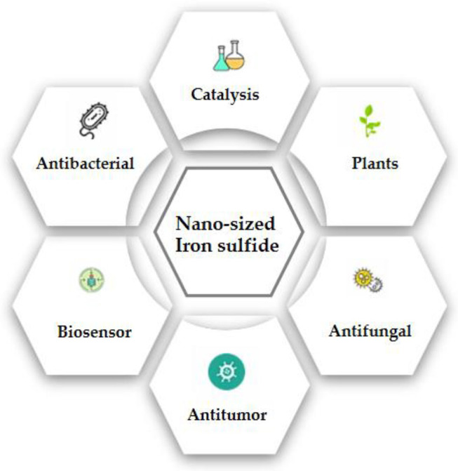
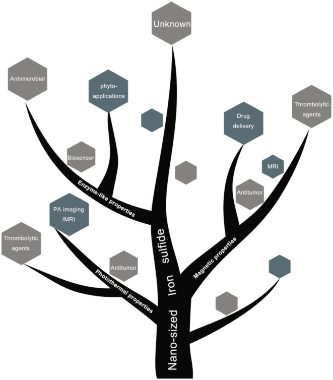
Nano-sized iron sulfides have attracted intense research interest due to the variety of their types, structures, and physicochemical properties. In particular, nano-sized iron sulfides exhibit enzyme-like activity by mimicking natural enzymes that depend on an iron-sulfur cluster as cofactor, extending their potential for applications in biomedicine. The present review principally summarizes the synthesis, properties and applications in biomedical fields, demonstrating that nano-sized iron sulfides have considerable potential for improving human health and quality of life.

Keywords: biomedical applications; enzyme-like activities; nano-sized iron sulfide; structure; synthesis.


Figures

Figure 1
Figure 1
Crystal structure of iron sulfide. (A) FeS. (B) FeS2. (C) Fe2S2. (D) Fe3S4. Reproduced with permission from Rickard and Luther (2007). Copyright 2007, American Chemical Society.
Figure 2
Figure 2
Representative images of nano-sized iron sulfide. (A) TEM image of the spherical FeS. Reproduced with permission from Dai et al. (2009). Copyright 2009, Wiley-VCH Verlag GmbH & Co. KGaA, Weinheim. (B) TEM and HRTEM of FeS2 nanodots. Reproduced with permission from Jin et al. (2018). Copyright 2017, American Chemical Society. (C) TEM images of platelet-like Fe3S4. Reproduced with permission from Paolella et al. (2011). Copyright 2011, American Chemical Society. (D) SEM and HRTEM of Fe7S8 nanowires. Reproduced with permission from Yao et al. (2013). Copyright 2013, Wiley-VCH Verlag GmbH & Co. KGaA, Weinheim.
Figure 3
Figure 3
Magnetocaloric conversion performance (a–d) and photothermal property of iron sulfides. (a) Temperature change of the Fe3S4 nanoparticles in water at varied concentrations of Fe2+ (i.e., 0, 0.5, and 1.0 mg/mL) as a function of magnetic field action time. (b) Plot of temperature change over 300s vs. the concentration of Fe3S4 nanoparticles. Thermal imaging of (c) pure water and (d) Fe3S4 nanoparticles (1.0 mg/mL) under the action of an AMF for 5 min. Reproduced with permission from Fu et al. (2019). Copyright 2019, Frontiers in Materials. (e) Synthetic route and applications of FeS2 nanodots in the base of its photothermal activity. (f) Scheme showing photothermal enhanced cellular uptake of FeS2@BSA-Ce6 nanoparticles. Reproduced with permission from Jin et al. (2018). Copyright 2017, American Chemical Society.
Scheme 1
Scheme 1
Schematic diagram of biomedical applications of nano-sized iron sulfide.
Figure 4
Figure 4
Schematic diagram of catalysis of iron sulfide as a nanozyme. (A) The peroxidase-like activity of FeS. Images of the suspension of sheet-like FeS nanostructure (1), mixture of TMB and H2O2 after catalytic reaction by sheet-like FeS nanostructure (2), mixture of TMB and H2O2 after adding H2SO4 to quench the catalytic reaction by sheet-like FeS nanostructure (3). Reproduced with permission from Dai et al. (2009). Copyright 2009, Wiley-VCH Verlag GmbH & Co. KGaA, Weinheim. (B) The catalase-like activity of Cys-nFeS. The trend of KM and the ratio of Vmax/KM in the kinetics assay with varied cysteine. Reproduced with permission from Xu et al. (2018). Copyright 2018, Nature Publishing Group.
Figure 5
Figure 5
(A) Morphology of P. aeruginosa before (control) and after Cys-nFeS treatment. The red triangles indicate flagella. Scale bars: 1 μm. (B) Photographs of P. aeruginosa infected wounds treated with buffer (control), Cys-nFeS, H2O2, and Cys-nFeS + H2O2 at different times (five mice in each group). (C) Confocal 3D image of a S. mutans UA159 biofilm treated by Cys-nFeS. Scale bars: 100 μm. (D) SEM image of a S. mutans biofilm treated by Cys-nFeS. The red arrows indicate EPS. Left scale bars: 100 μm. Right scale bars: 3 μm. (E) Scheme of polysulfane release from nFeS. Reproduced with permission from Xu et al. (2018). Copyright 2018, Nature Publishing Group.
Figure 6
Figure 6
(A) In vivo fluorescence images of 4T1 tumor-bearing nude mice taken at different time points post iv injection of FeS2@BSA-Ce6 nanodots (3.5 mg/kg Ce6 and 12 mg/kg FeS2). (B) Photoacoustic images of tumors in mice taken at different time points post iv injection of FeS2@BSA-Ce6 nanodots. (C) MR images of 4T1 tumor-bearing nude mice before and 8 h after iv injection of FeS2@BSA-Ce6 nanodots. (3.5 mg/kg Ce6 and 12 mg/kg FeS2). (D) IR thermal images of tumors in mice iv injected with FeS2@BSA-Ce6 under 808 nm laser irradiation (0.8W/cm2, 15 min). (E) Surface temperature changes of tumors monitored by the IR thermal camera during laser irradiation. (F) Representative immunofluorescence images of tumor slices after hypoxia staining. The cell nucleus, hypoxia areas, and blood vessels were stained with DAPI (blue), antipimonidazole antibody (green), and anti-CD31 antibody (red), respectively. Reproduced with permission from Jin et al. (2018). Copyright 2017, American Chemical Society.
Figure 7
Figure 7
(A) Schematical illustration of the greigite-containing magnetosome and the chemical synthesized magnetosome-like GMNCs. (B) Growth inhibition effect in murine S180 sarcoma model of the sample. The photos and weight ratios of tumor tissue from mice treated with normal saline (control group), GMNCs, Dox at low concentration, and GMNCs loading with Dox (without and with the guidance of external magnetic field), respectively. Reproduced with permission from Feng et al. (2013). Copyright 2013, Nature Publishing Group.
Figure 8
Figure 8
(A) Thrombolytic capacity in vitro of Fe3S4 nanoparticles under the indicated conditions. Fe3S4 nanoparticles: 0.5 mg/mL; NIR: 808 nm, 0.33 W cm−2; AMF: 4.2 × 10⁹ A m−1 s−1. (B) T2-weighted MR imaging in vivo of a mouse with celiac vein thrombosis before (left) and after (right) an intravenous injection of a solution of the Fe3S4 nanoparticles followed by the simulation of AMF. Reproduced with permission from Fu et al. (2019). Copyright 2019, Frontiers in Materials.
Figure 9
Figure 9
Plant growth parameters: control vs. test (FeS2). (a) Leaf area/Plant, showing significant increase in leaf area/test plants (52.4 ± 0.3) as compared to control (25.6 ± 0.2). (b) Leaf area index signifying total photosynthetic area available to plant and high values for test samples (1.5 ± 0.07) can be correlated with high biomass content of pro-fertilized spinach plants in comparison with (0.9 ± 0.02). (c) Comparative photograph of leaves showing larger leaf area in test plants as compared to control plants. Plant growth parameters: control vs. test (FeS2). (d) Number of leaves/Plant: control: 13 ± 1.0; test: 19 ± 1.0. (e) Specific leaf area signifies leaf thickness and was found similar for both test and control samples. (f) Field photograph taken at day 50 (just before harvesting the crop) depicting that the test group plants have comparatively more foliage as compared to the control plants. (g) Proposed outline of the mechanism of action of FeS2 on spinach seed in enhancing germination and plant growth. Reproduced with permission from Srivastava al. (2014). Copyright 2014, The Royal Society of Chemistry.
Scheme 2
Scheme 2
Schematic diagram of biomedical applications based on physiochemical properties of nano-sized iron sulfide.

Similar articles

Cited by

References

    1. Agnihotri S., Mohan T., Jha D., Gautam H., Roy I. (2020). Dual Modality FeS Nanoparticles with Reactive Oxygen Species-Induced and Photothermal Toxicity toward Pathogenic Bacteria. ACS OMEGA 5, 597–602. 10.1021/acsomega.9b03177 - DOI - PMC - PubMed
    1. Ahuja R., Sidhu A., Bala A. (2019). Synthesis and evaluation of iron(ii) sulfide aqua nanoparticles (FeS-NPs) against Fusarium verticillioides causing sheath rot and seed discoloration of rice. Eur. J. Plant Pathol. 155, 163–171. 10.1007/s10658-019-01758-3 - DOI
    1. Argueta-Figueroa L., Martinez-Alvarez O., Santos-Cruz J., Garcia-Contreras R., Acosta-Torres L. S., Fuente-Hernández J., et al. . (2017). Nanomaterials made of non-toxic metallic sulfides: a systematic review of their potential biomedical applications. Mater. Sci. Eng. C 76, 1305–1315. 10.1016/j.msec.2017.02.120 - DOI - PubMed
    1. Argueta-Figueroa L., Torres-Gómez N., García-Contreras R., Vilchis-Nestor A. R., Martínez-Alvarez O., Acosta-Torres L. S., et al. (2018). Hydrothermal synthesis of pyrrhotite (Fex−1S) nanoplates and their antibacterial, cytotoxic activity study. Progress Nat. Sci. 28, 447–455. 10.1016/j.pnsc.2018.06.003 - DOI
    1. Barand A., Russo S. (2009). Modeling the environmental stability of FeS2 nanorods, using lessons from biomineralization. Nanotechnology 20:115702. 10.1088/0957-4484/20/11/115702 - DOI - PubMed

LinkOut - more resources