. 2020 Oct 6:7:569415.
doi: 10.3389/fmolb.2020.569415. eCollection 2020.

Shortwave Infrared-Emitting Theranostics for Breast Cancer Therapy Response Monitoring

Affiliations

Shortwave Infrared-Emitting Theranostics for Breast Cancer Therapy Response Monitoring

Jay V Shah et al. Front Mol Biosci. .

Abstract

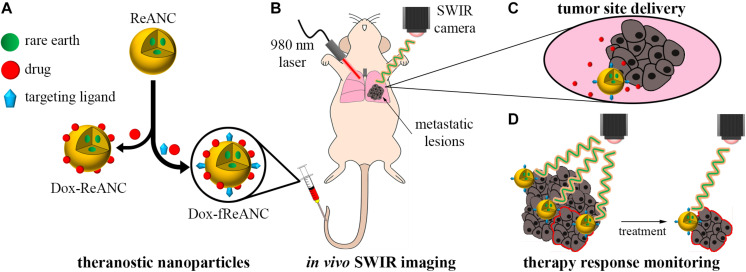
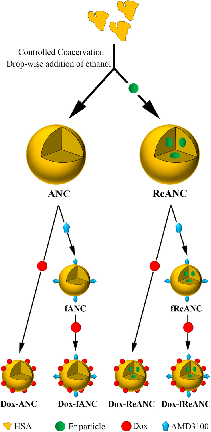
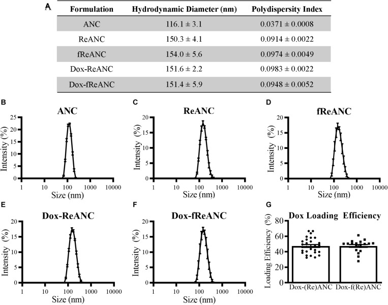
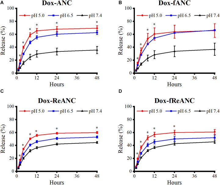
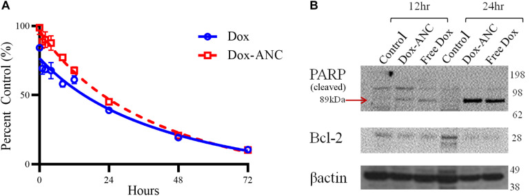
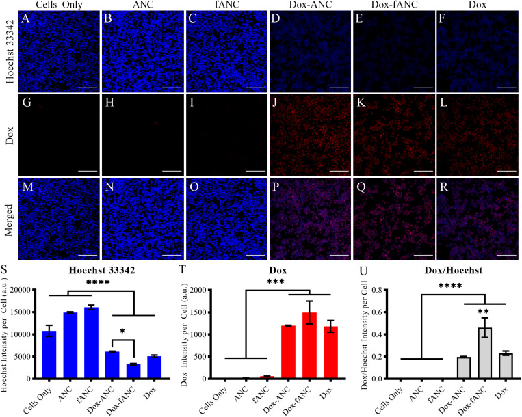
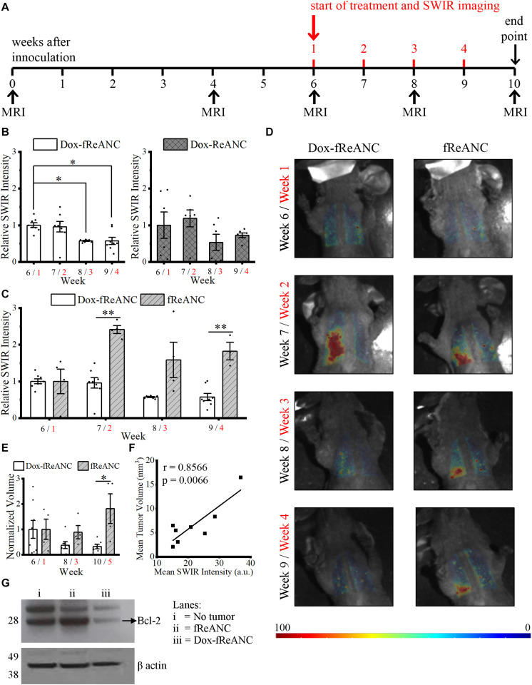
Therapeutic drug monitoring (TDM) in cancer, while imperative, has been challenging due to inter-patient variability in drug pharmacokinetics. Additionally, most pharmacokinetic monitoring is done by assessments of the drugs in plasma, which is not an accurate gauge for drug concentrations in target tumor tissue. There exists a critical need for therapy monitoring tools that can provide real-time feedback on drug efficacy at target site to enable alteration in treatment regimens early during cancer therapy. Here, we report on theranostic optical imaging probes based on shortwave infrared (SWIR)-emitting rare earth-doped nanoparticles encapsulated with human serum albumin (abbreviated as ReANCs) that have demonstrated superior surveillance capability for detecting micro-lesions at depths of 1 cm in a mouse model of breast cancer metastasis. Most notably, ReANCs previously deployed for detection of multi-organ metastases resolved bone lesions earlier than contrast-enhanced magnetic resonance imaging (MRI). We engineered tumor-targeted ReANCs carrying a therapeutic payload as a potential theranostic for evaluating drug efficacy at the tumor site. In vitro results demonstrated efficacy of ReANCs carrying doxorubicin (Dox), providing sustained release of Dox while maintaining cytotoxic effects comparable to free Dox. Significantly, in a murine model of breast cancer lung metastasis, we demonstrated the ability for therapy monitoring based on measurements of SWIR fluorescence from tumor-targeted ReANCs. These findings correlated with a reduction in lung metastatic burden as quantified via MRI-based volumetric analysis over the course of four weeks. Future studies will address the potential of this novel class of theranostics as a preclinical pharmacological screening tool.

Keywords: NIR-II bioimaging; nanotechnology; rare earths; shortwave infrared imaging; theranostic; tumor therapy monitoring.


Figures

FIGURE 1
FIGURE 1
Therapeutic drug monitoring by SWIR-emitting rare-earth albumin nanocomposites. Human serum albumin encapsulated SWIR-emitting rare earth-doped nanoparticles (ReANCs) were synthesized by controlled coacervation. The targeting ligand (AMD3100) and/or the drug (doxorubicin) were physically adsorbed sequentially onto the surface of ReANCs (A). SWIR imaging was used to detect lung metastatic lesions in a mouse model of triple negative breast cancer metastasis (B). Tumor-targeting fReANCs facilitated delivery of doxorubicin to the lesion site (C). SWIR emissions from the theranostic particles retained in the lungs were used as a metric of therapy response that correlated with tumor progression (D).
FIGURE 2
FIGURE 2
Design and fabrication of albumin-based nanocarriers and theranostic nanoparticles. Albumin nanocomposites (ANCs) were synthesized via controlled coacervation by adding pure ethanol drop-wise to a solution of human serum albumin. Dissolving Er-doped nanoparticles in the ethanol prior to synthesis resulted in encapsulation of the nanoprobes in albumin, producing diagnostic ReANCs. Tumor-targeting particles (fANCs and fReANCs) were synthesized following physical adsorption of the CXCR4 antagonist AMD3100, which improved uptake of the particles by CXCR4-expressing cancer cells. Doxorubicin (Dox) was then loaded to both tumor-targeting and non-targeting albumin particles by physical adsorption, producing therapeutic particles. ReANCs loaded with Dox (Dox-ReANCs and Dox-fReANCs), possessing both diagnostic and therapeutic components, served as theranostic agents.
FIGURE 3
FIGURE 3
Characterization of ANC and ReANC formulations. The table (A) displays the hydrodynamic diameters and polydispersity indices (PDI) of ANCs (B), ReANCs (C), fReANCs (D), Dox-ReANCs (E), and Dox-fReANCs (F) were measured using dynamic light scattering. Data is presented as the mean ± SEM. All particle formulations had low polydispersity indices in solution. Functionalization with AMD3100 and Dox loading did not affect the size of ReANCs (p = 0.9535 assessed by an ordinary one-way ANOVA) or PDI (p = 0.3877). The loading efficiency of Dox adsorption onto the albumin shells of (Re)ANCs and f(Re)ANCs was calculated based on the fluorescence (Ex: 470 nm, Em: 595 nm) of unloaded Dox (G). Loading efficiency was approximately 47% for both tumor-targeted and untargeted formulations, indicating that adsorption of AMD3100 for precision targeting prior to Dox loading did not affect the loading efficiency (p = 0.9833 assessed by a two-tailed student’s t test).
FIGURE 4
FIGURE 4
Pharmacokinetic release profiles of Dox from ANCs in vitro. Dox release from Dox-ANCs (A), Dox-fANCs (B), Dox-ReANCs (C), and Dox-fReANCs (D) was evaluated at 37°C in PBS at varying pH (5.0, 6.5, and 7.4) via microdialysis. Particles were loaded into microdialysis devices, which were then placed in 1 mL of release buffer consisting of PBS of varying pH in 5 mL centrifuge tubes (n = 3). At each time point, the microdialysis device was moved into a new tube containing fresh release buffer. The amount of released Dox was a measure of fluorescence (Ex: 470 nm, Em: 595 nm) from the release buffer at each indicated time point. Data is expressed as mean ± SEM. Dox was released from nanoparticles at a sustained rate over the course of 48 h in a pH-dependent manner, as assessed by a one-way ANOVA. Addition of rare earth nanoparticle cores or AMD3100 did not affect the release profiles, as assessed by a two-tailed student’s t test.
FIGURE 5
FIGURE 5
Dox-ANCs induce cytotoxicity through activation of cell-death pathways. MDA-MB-231 cells (104 cells/well) were treated with an equivalent dose of 0.02 mg/mL Dox-ANCs or free Dox over 72 h. Viability was assessed via an MTT assay at times 0, 1, 2, 4, 8, 12, 24, 48, and 72 h of treatment, and the absorbance at 570 nm was normalized to untreated controls (A). Data is expressed as mean ± SEM for n = 3. The cytotoxic effect of Dox-ANC was delayed in comparison to free Dox during the first 24 h. To assess the effect of Dox and Dox-ANCs on cellular pathways of cell death, an immunoblot was run from samples of MDA-MB-231 cells lysed after 12 and 24 h of treatment with Dox (Dox-ANCs or free Dox) or untreated. Qualitative analysis of the blots revealed elevated levels of cleaved PARP and decreased expression of Bcl2 in cells treated with Dox-ANC and free Dox compared to untreated control (B). This indicates an active cell death process in response to the delivery of Dox.
FIGURE 6
FIGURE 6
Increased Dox uptake in vitro by tumor-targeted nanoparticles. MDA-MB-231 cells were seeded into 96 well plates and treated with either culture media (cells only), ANCs, fANCs, Dox-ANCs, Dox-fANCs, or free Dox at an equivalent concentration of 0.02 mg/mL Dox and a 1x concentration of particles for 8 h. Cells were then fixed and stained with Hoechst 33342 to image the cell nuclei (A–F). The natural fluorescence of Dox was used to image the uptake of Dox by cells (G–L). Dox and Hoechst 33342 compete for binding spots on DNA; therefore, the Hoechst and Dox images were merged to observe colocalization in the cell nuclei (M–R). The signal from the Hoechst stain (S) and Dox (T) was quantified from three distinct fields of view for each sample and normalized to the cell count. The effects of treatment and functionalization were assessed via a two-way ANOVA with a Tukey’s post hoc test to correct for multiple comparisons. The intensity of the Hoechst stain was significantly diminished in response to treatment. Inversely, the Dox signal was significantly greater in cells that received treatment. Since the decrease in Hoechst signal is due to intercalation of Dox to the DNA, the Dox/Hoechst ratio was assessed (U). The Dox/Hoechst ratio was greatest in cells treated with Dox-fANCs, indicating increased uptake of Dox when a targeted nanocarrier is used for administration in vitro. Data representative of three independent experiments. Scale bar = 200 μm. Data in S-U are expressed as mean +SEM from triplicate samples. p < 0.05, ∗∗p < 0.01, ∗∗∗p < 0.001, ****p < 0.0001.
FIGURE 7
FIGURE 7
Theranostic rare earth-based nanoparticle mediated therapeutic drug monitoring in a mouse model of lung metastases. Mice were inoculated with human triple negative breast cancer cells (MDA-MB-231) via intravenous tail vein injection. Biweekly MRI was performed from inoculation until the end of the experiment. The first week of the four-week treatment started six weeks after inoculation, when tumors in the lungs were discernible by MRI (A). Weeks after inoculation are presented in black text, and weeks after initiating treatment are presented in red text. Animals in the Dox treatment groups received weekly 150 μL injections of Dox-ReANCs (n = 6) or Dox-fReANCs (n = 8) at an equivalent dose of 2–2.5 mg/kg Dox. Tumor-bearing control mice received weekly injections of ReANCs (n = 5) or fReANCs (n = 4). Whole body SWIR imaging of the mice was performed at 24 h post-injection. To compare the effects of treatment among groups, data was normalized to the SWIR signal from week 1, and a two-way ANOVA was performed, followed by a post hoc Tukey’s test to correct for multiple comparisons. The SWIR signal was similar between mice that received Dox-ReANCs and Dox-fReANCs (B). However, there was a significant decrease in SWIR emissions from the lungs in mice injected with Dox-fReANCs during the third (p = 0.0204) and fourth (p = 0.0176) week of treatment, as determined by a one-way ANOVA followed by a Tukey’s post hoc test, indicating that functionalization led to better therapeutic outcomes. There was a significant difference in the SWIR signals in response to treatment, as seen by the differences in quantified SWIR signal (C) and representative SWIR images (D) of the lungs of mice that received Dox-fReANCs and non-therapeutic fReANCs. There was an overall increase in the SWIR signal in mice that received fReANCs. There was also a significant decrease in SWIR signal over the course of treatment in mice treated with Dox-fReANCs compared to mice injected with fReANCs during week 2 (p = 0.0011) and week 4 (p = 0.0010). Changes in tumor burden in fReANC and Dox-fReANC treated mice were validated by volumetric analysis from MRI (E), with a significant reduction in tumor volume following treatment compared to the untreated control (p = 0.0380). Mean SWIR signal from weeks 1 and 3 were found to correlate with the respective tumor volumes assessed by MRI (F), with a Pearson’s r of 0.8566 (p = 0.0066). At endpoint, the lungs were harvested, homogenized, and lysed. An immunoblot was performed from the lung lysates and probed for Bcl-2 expression (G). Protein levels of Bcl-2 were decreased in the lungs of mice treated with Dox-fReANCs when compared to untreated mice or non-tumor-bearing healthy controls. All data is expressed as the mean ± SEM. p < 0.05, ∗∗p < 0.01.

Similar articles

Cited by

References

    1. Abbasi S., Paul A., Shao W., Prakash S. (2012). Cationic albumin nanoparticles for enhanced drug delivery to treat breast cancer: preparation and in vitro assessment. J. Drug Deliv. 2012:686108. 10.1155/2012/686108 - DOI - PMC - PubMed
    1. Abou-Zied O. K. (2015). Understanding the physical and chemical nature of the warfarin drug binding site in human serum albumin: experimental and theoretical studies. Curr. Pharm. Des. 21 1800–1816. 10.2174/1381612821666150304163447 - DOI - PubMed
    1. Alnaim L. (2007). Therapeutic drug monitoring of cancer chemotherapy. J. Oncol. Pharm. Pract. 13 207–221. 10.1177/1078155207081133 - DOI - PubMed
    1. An F. F., Zhang X. H. (2017). Strategies for preparing albumin-based nanoparticles for multifunctional bioimaging and drug delivery. Theranostics 7 3667–3689. 10.7150/thno.19365 - DOI - PMC - PubMed
    1. Andhariya N., Chudasama B., Mehta R. V., Upadhyay R. V. (2010). Biodegradable thermoresponsive polymeric magnetic nanoparticles: a new drug delivery platform for doxorubicin. J. Nanopart. Res. 13 1677–1688. 10.1007/s11051-010-9921-9926 - DOI