Review
. 2020 Sep 29:7:578433.
doi: 10.3389/fmolb.2020.578433. eCollection 2020.

The Early Phase of β2-Microglobulin Aggregation: Perspectives From Molecular Simulations

Affiliations
Review

The Early Phase of β2-Microglobulin Aggregation: Perspectives From Molecular Simulations

Rui J S Loureiro et al. Front Mol Biosci. .

Abstract

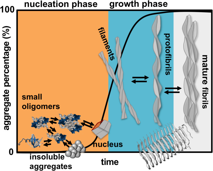
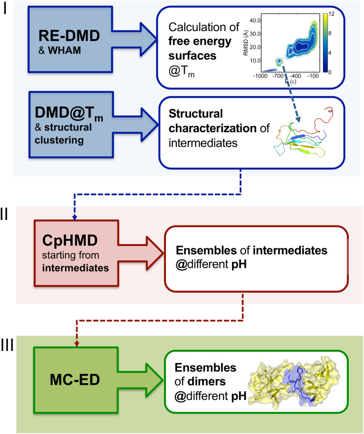
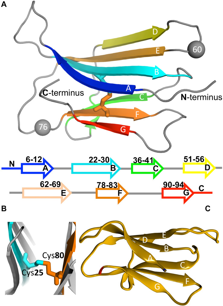
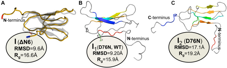
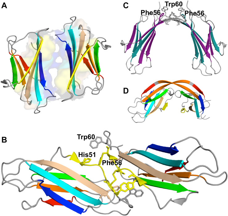
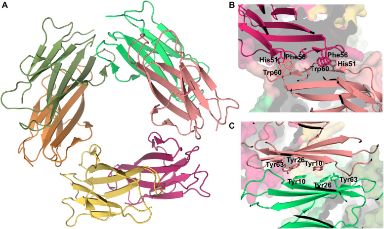
Protein β2-microglobulin is the causing agent of two amyloidosis, dialysis related amyloidosis (DRA), affecting the bones and cartilages of individuals with chronic renal failure undergoing long-term hemodialysis, and a systemic amyloidosis, found in one French family, which impairs visceral organs. The protein's small size and its biomedical significance attracted the attention of theoretical scientists, and there are now several studies addressing its aggregation mechanism in the context of molecular simulations. Here, we review the early phase of β2-microglobulin aggregation, by focusing on the identification and structural characterization of monomers with the ability to trigger aggregation, and initial small oligomers (dimers, tetramers, hexamers etc.) formed in the so-called nucleation phase. We focus our analysis on results from molecular simulations and integrate our views with those coming from in vitro experiments to provide a broader perspective of this interesting field of research. We also outline directions for future computer simulation studies.

Keywords: dimer; docking; intermediates; molecular dynamics; native-centric simulations; protein aggregation.


Figures

FIGURE 1
FIGURE 1
Schematics of protein aggregation. Representation of the process of protein aggregation as a function of time, starting from aggregation prone monomers till the formation of mature amyloid fibrils. This article focus on the nucleation phase, whereby small oligomers (dimers, trimers, tetramers etc.) form prior to the establishment of an aggregation nucleus that triggers the growth phase leading to fibrils.
FIGURE 2
FIGURE 2
Three-stage methodology to investigate the early phase of protein aggregation. The conformational space is explored with RE-DMD of a full atomistic Gō model to compute equilibrium folding thermodynamics (including the melting temperature, Tm, and free energy surfaces) to detect the formation of intermediate states). Large ensembles of intermediates are collected from DMD simulations at constant temperature, and structurally characterized to select those with aggregation potential. Representative conformations are then subjected to a CpHMD protocol with explicit titration to obtain ensembles of intermediates representative of different pH conditions. The latter are finally used in a protein-protein docking protocol that creates ensembles of dimers whose statistical analysis allows determining the most likely regions and residues involved in the onset of aggregation.
FIGURE 3
FIGURE 3
Protein beta-2-microglobulin (β2m). Three-dimensional structure of β2m highlighting the 7 beta-strands, organized in two beta-sheets (comprising strands A-B-E-D, and C-F-G, respectively), and the position of Trp60 (which is a key residue in aggregation) and Asp76, which is mutated to Asn in the D76N mutant (PDB ID: 4FXL) (A). A cysteine bond links strands B and F (B). Native structure of the ΔN6 structural variant lacking the N-terminal hexapeptide (PDB ID: 2XKU) (C).
FIGURE 4
FIGURE 4
Simulated monomers of β2m. Three dimensional structure of three aggregation prone monomeric states predicted by a full atomistic Gō model, used to explore the folding space of ΔN6 (A) and D76N mutant (B,C). The intermediate populated by ΔN6 features an unstructured N terminal region (A), while those populated by D76N display the C-terminus (B), or both termini unstructured (C).
FIGURE 5
FIGURE 5
Dimers of β2m. P32A dimer featuring an anti-parallel eight-stranded beta-sheet ABED-DEBA (PDB ID: 2F8O), which is mediated by antiparallel interactions of two complete D strands (A), DIMC33 formed by two S33C monomers linked by a disulfide bond at position 33 (PDB ID: 4R9H), which exhibits the DD strand interface (B), domain-swapped dimer of the wt formed under reducing conditions, where strands E-F-G are exchanged between monomers (PDB ID: 3LOW); the residues forming the hinge loop, which contains amyloidogenic peptide segments, is highlighted (C). Domain-swapped dimer of the ΔN6 variant, which exchanges strand G (PDB ID: 2 × 89) (D).
FIGURE 6
FIGURE 6
Tetramer of β2m. Three-dimensional structure of the tetramer (A), formed by two disulfide linked homodimers at Cys50 (PDB ID: 3TM6). In this representation the homodimers are colored green and pink, respectively. The DD strand interfacial region in (B) is the largest found in this tetramer being stabilized by hydrogen bonds between His31 and Asp34, and hydrophobic interactions involving Trp60, Leu54, Leu64, and Tyr66.
FIGURE 7
FIGURE 7
Hexamer of β2m. Three dimensional structure of the hexamer formed by the H13F mutant in the presence of Cu2+ (PDB ID: 3CIQ) (A). The hexamer results from the association of dimers that form an interface mediated by D-strands from adjacent chains stabilized by hydrogen bonds between Leu54, Asp34, and His31 (not shown) and hydrophobic interactions involving Phe56 and Trp60 from one chain and His51 and Asp34 from the other (B). The other interface is mediated by the stacking of the ABED sheet from one chain onto the ABED sheet of an adjacent monomer in an antiparallel arrangement and stabilized by successive interactions involving Tyr residues (C).

Similar articles

Cited by

References

    1. Abeln S., Vendruscolo M., Dobson C. M., Frenkel D. (2014). A simple lattice model that captures protein folding, aggregation and amyloid formation. PLoS One 9:e85185. 10.1371/journal.pone.0085185 - DOI - PMC - PubMed
    1. Antwi K., Mahar M., Srikanth R., Olbris M. R., Tyson J. F., Vachet R. W. (2008). Cu(II) organizes beta-2-microglobulin oligomers but is released upon amyloid formation. Protein Sci. 17 748–759. 10.1110/ps.073249008 - DOI - PMC - PubMed
    1. Armen R. S., Daggett V. (2005). Characterization of two distinct beta2-microglobulin unfolding intermediates that may lead to amyloid fibrils of different morphology. Biochemistry 44 16098–16107. 10.1021/bi050731h - DOI - PubMed
    1. Armen R. S., DeMarco M. L., Alonso D. O. V., Daggett V. (2004). Pauling and Corey’s alpha-pleated sheet structure may define the prefibrillar amyloidogenic intermediate in amyloid disease. Proc. Natl. Acad. Sci. U.S.A. 101 11622–11627. 10.1073/pnas.0401781101 - DOI - PMC - PubMed
    1. Baias M., Smith P. E., Shen K., Joachimiak L. A., Zerko S., Kozminski W., et al. (2017). Structure and dynamics of the huntingtin exon-1 N-terminus: a solution NMR perspective. J. Am. Chem. Soc. 139 1168–1176. 10.1021/jacs.6b10893 - DOI - PubMed

LinkOut - more resources