. 2020 Oct 2;6(6):e522.
doi: 10.1212/NXG.0000000000000522. eCollection 2020 Dec.

Blended phenotype of adult-onset Alexander disease and spinocerebellar ataxia type 6

Affiliations

Blended phenotype of adult-onset Alexander disease and spinocerebellar ataxia type 6

Takashi Odo et al. Neurol Genet. .
No abstract available


Figures

Figure
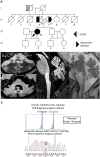
Figure. Genetic data of the proband and brain MRI of the proband and his mother
(A) Pedigree chart of the family. The proband is shown with an arrow. Those who underwent genetic tests are indicated as E+ and not as E−. Patients whose diagnosis was established as corresponding diseases are shown in black and suggestive but not established in gray. Her paternal grandfather developed a progressive gait abnormality suggestive of SCA6. Her mother was diagnosed as Alexander disease based on typical brain MRI findings, although she did not undergo genetic tests. (B–E) Brain MRIs. T2-weighted images of the proband (B) and her mother (E) and fluid-attenuated inversion recovery (FLAIR) images of the proband (C, D) showed atrophy of the midbrain, cerebellum, medulla, and upper cervical spinal cord, the latter 2 designated as tadpole appearance characteristic for Alexander disease. High-intensity signals were observed in the dentate nucleus (C) and medulla oblongata (B, D) in the proband. Cerebellar atrophy was more severe in the proband than in her mother (D, E), presumably due to concomitant SCA6. (F) Mutational analysis of the proband. The upper row: PCR fragment analysis for the triplet repeat expansion in the SCA6 locus. The lower row: an electropherogram of a GFAP mutation c.827G>T (p.R276L). An arrow shows the heterozygous mutation in GFAP. GFAP = glial fibrillary acidic protein.

References

    1. Yoshida T, Sasaki M, Yoshida M, et al. . Nationwide survey of Alexander disease in Japan and proposed new guidelines for diagnosis. J Neurol 2011;258:1998–2008. - PubMed
    1. Tsuji S, Onodera O, Goto J, Nishizawa M; Diseases OBotSGoA. Sporadic ataxias in Japan—a population-based epidemiological study. Cerebellum 2008;7:189–197. - PubMed
    1. Namekawa M, Takiyama Y, Aoki Y, et al. . Identification of GFAP gene mutation in hereditary adult-onset Alexander's disease. Ann Neurol 2002;52:779–785. - PubMed
    1. Namekawa M, Takiyama Y, Honda J, Shimazaki H, Sakoe K, Nakano I. Adult-onset Alexander disease with typical "tadpole" brainstem atrophy and unusual bilateral basal ganglia involvement: a case report and review of the literature. BMC Neurol 2010;10:21. - PMC - PubMed
    1. Cvetanovic M, Ingram M, Orr H, Opal P. Early activation of microglia and astrocytes in mouse models of spinocerebellar ataxia type 1. Neuroscience 2015;289:289–299. - PMC - PubMed