

Total Body PET: Why, How, What for?
- PMID: 33134653
- PMCID: PMC7595297
- DOI: 10.1109/trpms.2020.2985403
Total Body PET: Why, How, What for?
Abstract
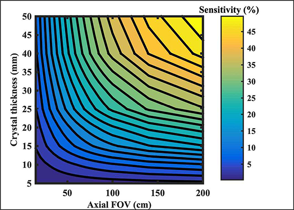
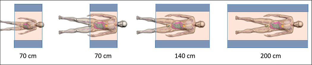
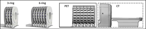
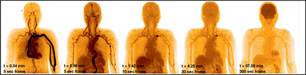
PET instruments are now available with a long axial field-of-view (LAFOV) to enable imaging the total-body, or at least head and torso, simultaneously and without bed translation. This has two major benefits, a dramatic increase in system sensitivity and the ability to measure kinetics with wider axial coverage so as to include multiple organs. This manuscript presents a review of the technology leading up to the introduction of these new instruments, and explains the benefits of a LAFOV PET-CT instrument. To date there are two platforms developed for TB-PET, an outcome of the EXPLORER Consortium of the University of California at Davis (UC Davis) and the University of Pennsylvania (Penn). The uEXPLORER at UC Davis has an AFOV of 194 cm and was developed by United Imaging Healthcare. The PennPET EXPLORER was developed at Penn and is based on the digital detector from Philips Healthcare. This multi-ring system is scalable and has been tested with 3 rings but is now being expanded to 6 rings for 140 cm. Initial human studies with both EXPLORER systems have demonstrated the successful implementation and benefits of LAFOV scanners for both clinical and research applications. Examples of such studies are described in this manuscript.
Keywords: (TB-PET); Long axial-field-of-view (LAFOV); PET-CT; PennPET EXPLORER; Total-Body PET; uEXPLORER.
Figures
Similar articles
-
Total Body PET/CT: Future Aspects.Semin Nucl Med. 2025 Jan;55(1):107-115. doi: 10.1053/j.semnuclmed.2024.10.011. Epub 2024 Nov 13. Semin Nucl Med. 2025. PMID: 39542814 Review.
-
Performance Characteristics of Long Axial Field-of-View PET Scanners with Axial Gaps.IEEE Trans Radiat Plasma Med Sci. 2021 May;5(3):322-330. doi: 10.1109/trpms.2020.3027257. Epub 2020 Sep 28. IEEE Trans Radiat Plasma Med Sci. 2021. PMID: 34179595 Free PMC article.
-
State of the art in total body PET.EJNMMI Phys. 2020 May 25;7(1):35. doi: 10.1186/s40658-020-00290-2. EJNMMI Phys. 2020. PMID: 32451783 Free PMC article. Review.
-
Walk-through flat panel total-body PET: a patient-centered design for high throughput imaging at lower cost using DOI-capable high-resolution monolithic detectors.Eur J Nucl Med Mol Imaging. 2023 Oct;50(12):3558-3571. doi: 10.1007/s00259-023-06341-x. Epub 2023 Jul 19. Eur J Nucl Med Mol Imaging. 2023. PMID: 37466650 Free PMC article.
-
Performance evaluation of the PennPET explorer with expanded axial coverage.Phys Med Biol. 2023 Apr 19;68(9):095007. doi: 10.1088/1361-6560/acc722. Phys Med Biol. 2023. PMID: 36958051 Free PMC article.
Cited by
-
A simulation study of the system characteristics for a long axial FOV PET design based on monolithic BGO flat panels compared with a pixelated LSO cylindrical design.EJNMMI Phys. 2023 Dec 1;10(1):75. doi: 10.1186/s40658-023-00593-0. EJNMMI Phys. 2023. PMID: 38036794 Free PMC article.
-
Total Body PET/CT: Future Aspects.Semin Nucl Med. 2025 Jan;55(1):107-115. doi: 10.1053/j.semnuclmed.2024.10.011. Epub 2024 Nov 13. Semin Nucl Med. 2025. PMID: 39542814 Review.
-
Multi-angle acquisition and 3D composite reconstruction for organ-targeted PET using planar detectors.Med Phys. 2025 Apr;52(4):2507-2519. doi: 10.1002/mp.17606. Epub 2024 Dec 27. Med Phys. 2025. PMID: 39729623 Free PMC article.
-
Reinventing Molecular Imaging with Total-Body PET, Part II: Clinical Applications.PET Clin. 2020 Oct;15(4):463-475. doi: 10.1016/j.cpet.2020.06.013. PET Clin. 2020. PMID: 32888545 Free PMC article. Review.
-
Principles of Tracer Kinetic Analysis in Oncology, Part II: Examples and Future Directions.J Nucl Med. 2022 Apr;63(4):514-521. doi: 10.2967/jnumed.121.263519. J Nucl Med. 2022. PMID: 35361713 Free PMC article.
References
-
- Budinger TF, “PET instrumentation: What are the limits?,” JCAT, vol. 28, no. 3, pp. 247–267, 1998. - PubMed
-
- Casey ME and Nutt R, “A multicrystal two dimensional BGO detector system for positron emission tomography,” IEEE Trans. Nucl. Sci, vol. 33, no. 1, pp. 460–463, 1986.
Grants and funding
LinkOut - more resources
Full Text Sources