. 2020;1(1):tgaa070.
doi: 10.1093/texcom/tgaa070. Epub 2020 Sep 29.

Impression Formation in the Human Infant Brain

Affiliations

Impression Formation in the Human Infant Brain

Kathleen M Krol et al. Cereb Cortex Commun. 2020.

Abstract

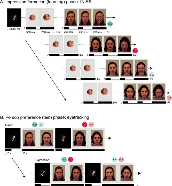
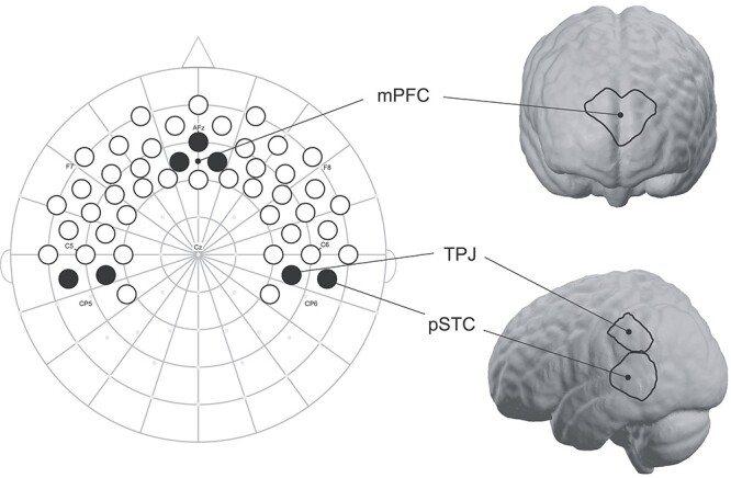
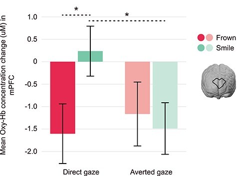
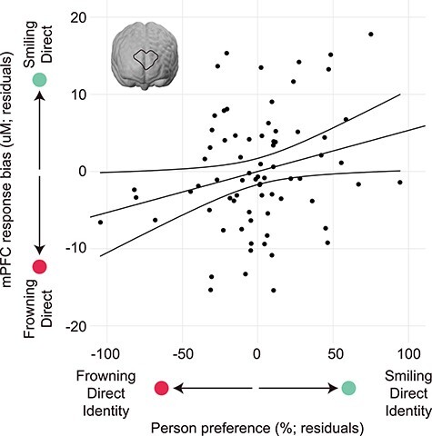
Forming an impression of another person is an essential aspect of human social cognition linked to medial prefrontal cortex (mPFC) function in adults. The current study examined the neurodevelopmental origins of impression formation by testing the hypothesis that infants rely on processes localized in mPFC when forming impressions about individuals who appear friendly or threatening. Infants' brain responses were measured using functional near-infrared spectroscopy while watching 4 different face identities displaying either smiles or frowns directed toward or away from them (N = 77). This was followed by a looking preference test for these face identities (now displaying a neutral expression) using eyetracking. Our results show that infants' mPFC responses distinguish between smiling and frowning faces when directed at them and that these responses predicted their subsequent person preferences. This suggests that the mPFC is involved in impression formation in human infants, attesting to the early ontogenetic emergence of brain systems supporting person perception and adaptive behavior.

Keywords: emotion; fNIRS; impression formation; infancy; mPFC.


Figures

Figure 1
Figure 1
Full experimental paradigm. (A) “Impression formation (learning) phase.” Infants first viewed blocks of dynamic presentation of 4 identities shifting their gaze (from averted [A] to direct [D], or direct to averted) and subsequently presenting a facial expression (smile [S] or frown [F]), for a total of 4 possible expression-gaze combinations per block while fNIRS was recorded. Each trial began with a dynamic baseline consisting of nonsocial (vegetable) stimuli for at least 6 s, followed by a facial stimulus presentation for 6 s. Both baseline and facial stimulus presentation were preceded by a bell tone to maintain infant attention. Blocks were preceded by a 3 s audiovisual attention-getter in the center of the screen. (B) “Person preference (test) phase.” After fNIRS recording, infants immediately underwent a looking preference paradigm using eyetracking. Infants viewed static pairings of the 4 identities from the fNIRS paradigm; now presenting neutral, direct-gaze expressions for 15 s each. Each trial began with a 3 s audiovisual attention-getter in the center of the screen. Pairings were created to directly compare preferences for gaze (i.e., direct vs. averted-gaze within the 2 individuals who smiled [S-D vs. S-A]) and expression (i.e., smile vs. frown within the 2 individuals who exhibited direct gaze [S-D vs. F-D]) for a total of 4 pairings, each presented twice. Note that infants viewed photographs from the FACES database (35), but due to copyright restrictions we have recreated the stimuli using the publicly available Karolinska Directed Emotional Faces (KDEF) database (www.kdef.se); (ms = milliseconds, s = seconds, x = times).
Figure 2
Figure 2
fNIRS cap template and ROIs. Shown is our fNIRS cap template mapped onto 10–20 space. International 10-20 coordinates are indicated by small gray dots, and relevant coordinates are labeled. Forty-nine channels (source-detector pairs) are presented as circles; those shaded in black are the channels used to create ROIs for mPFC, bilateral TPJ, and bilateral posterior superior temporal cortices (pSTC). Channels have been projected onto MNI brain space using NIRSite and nirsLAB software (NIRx) for readers’ reference.
Figure 3
Figure 3
Infant mPFC is sensitive to frowning and smiling faces with direct and averted-gaze. Displayed is the interaction between expression (smile vs. frown) and gaze (direct vs. averted) in the mPFC, F(1, 76) = 4.68, P = 0.034, ηp2 = 0.058, as indexed by the concentration change of oxygenated hemoglobin (oxy-Hb). The mPFC discriminates between frowning and smiling faces of direct gaze, as well as between direct and averted-gaze within smiling faces, displaying the highest response to smiling, direct-gaze faces; error bars represent standard error of the mean, *P < 0.05 (uM = microMolar).
Figure 4
Figure 4
Infants’ mPFC response during impression formation predicts person preference. Plotted are the residuals from a regression demonstrating that heightened mPFC response bias for the smiling, direct-gaze face during impression formation predicts an increased looking preference for that identity in a looking preference paradigm (now displaying a neutral face). In contrast, a heightened mPFC response bias for the frowning, direct-gaze face predicts an increased looking preference for that identity (now displaying a neutral face), β = 2.50, t = 2.142, P = 0.036 (uM = microMolar).
Figure 5
Figure 5
Infant TPJ is sensitive to direct and averted-gaze. Displayed is the interaction between hemisphere (left vs. right) and gaze (direct vs. averted) in the TPJ, F(1, 76) = 6.32, P = 0.014, ηp2 = 0.077, as indexed by the concentration change of oxygenated hemoglobin (oxy-Hb). The TPJ better discriminates between direct and averted-gaze in the left hemisphere, displaying a heightened response to direct-gaze versus averted-gaze faces; error bars represent standard error of the mean (uM = microMolar).

Similar articles

Cited by

References

    1. Ames DL, Fiske ST, Todorov AT. 2011. Impression formation: a focus on others’ intents. In: Decety J, Cacioppo JT. The Oxford Handbook of Social Neuroscience. Oxford: Oxford University Press.
    1. Amodio DM, Frith CD. 2006. Meeting of minds: the medial frontal cortex and social cognition. Nat Rev Neurosci. 7(4):268–277. - PubMed
    1. Argyle M, Dean J. 1965. Eye-contact, distance and affiliation. Sociometry. 28(3):289–304. - PubMed
    1. Baillargeon R, Scott RM, He Z. 2010. False-belief understanding in infants. Trends Cogn Sci. 14(3):110–118. - PMC - PubMed
    1. Baron-Cohen S, Wheelwright S. 2004. The empathy quotient: an investigation of adults with Asperger syndrome or high functioning autism, and normal sex differences. J Autism Dev Disord. 34(2):163–175. - PubMed

LinkOut - more resources