Review
. 2021 May 25;117(6):1450-1488.
doi: 10.1093/cvr/cvaa324.

Cardiac fibrosis

Affiliations
Review

Cardiac fibrosis

Nikolaos G Frangogiannis. Cardiovasc Res. .

Abstract

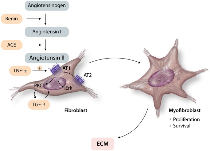
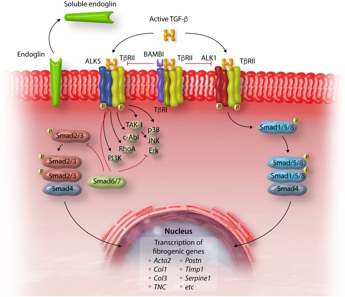
Myocardial fibrosis, the expansion of the cardiac interstitium through deposition of extracellular matrix proteins, is a common pathophysiologic companion of many different myocardial conditions. Fibrosis may reflect activation of reparative or maladaptive processes. Activated fibroblasts and myofibroblasts are the central cellular effectors in cardiac fibrosis, serving as the main source of matrix proteins. Immune cells, vascular cells and cardiomyocytes may also acquire a fibrogenic phenotype under conditions of stress, activating fibroblast populations. Fibrogenic growth factors (such as transforming growth factor-β and platelet-derived growth factors), cytokines [including tumour necrosis factor-α, interleukin (IL)-1, IL-6, IL-10, and IL-4], and neurohumoral pathways trigger fibrogenic signalling cascades through binding to surface receptors, and activation of downstream signalling cascades. In addition, matricellular macromolecules are deposited in the remodelling myocardium and regulate matrix assembly, while modulating signal transduction cascades and protease or growth factor activity. Cardiac fibroblasts can also sense mechanical stress through mechanosensitive receptors, ion channels and integrins, activating intracellular fibrogenic cascades that contribute to fibrosis in response to pressure overload. Although subpopulations of fibroblast-like cells may exert important protective actions in both reparative and interstitial/perivascular fibrosis, ultimately fibrotic changes perturb systolic and diastolic function, and may play an important role in the pathogenesis of arrhythmias. This review article discusses the molecular mechanisms involved in the pathogenesis of cardiac fibrosis in various myocardial diseases, including myocardial infarction, heart failure with reduced or preserved ejection fraction, genetic cardiomyopathies, and diabetic heart disease. Development of fibrosis-targeting therapies for patients with myocardial diseases will require not only understanding of the functional pluralism of cardiac fibroblasts and dissection of the molecular basis for fibrotic remodelling, but also appreciation of the pathophysiologic heterogeneity of fibrosis-associated myocardial disease.

Keywords: Extracellular matrix; Fibrosis; Growth factor; Heart failure; Myofibroblast.


Figures

Figure 1
Figure 1
Histological types of cardiac fibrosis. Images show sections of adult mouse hearts stained with picrosirius red in order to label collagen fibres. (A) The normal adult mammalian heart contains an intricate network of collagenous matrix. Every cardiomyocyte is surrounded by a thin network of endomysial collagen. Thicker perimysial collagen fibres (arrows) enwrap bundles of cardiomyocytes. (B) Following myocardial infarction, dead cardiomyocytes are replaced by collagen-based scar. Replacement fibrosis is shown in a mouse model of reperfused myocardial infarction (arrows, 1 h ischaemia–7 days reperfusion). Infarction is midmyocardial; interstitial fibrosis is noted in remodelling subendocardial and subepicardial areas (arrowheads). (C) Interstitial fibrosis (arrows) is noted in a model of left ventricular pressure overload, induced through transverse aortic constriction (7 days). (D) Perivascular fibrosis (arrows) is also noted in the pressure-overloaded myocardium, with large amounts of collagen surrounding an intracoronary vessel (*). Scalebar = 80 µm.
Figure 2
Figure 2
The myofibroblasts are the main cellular effectors of fibrosis in injured and remodelling hearts. Myofibroblasts are activated fibroblasts that express contractile proteins, such as α-SMA and secrete large amounts of ECM proteins. Images show abundant myofibroblasts in infarcted hearts in both large animal and rodent models. (A) Immunohistochemical staining identifies non-vascular α-SMA+ myofibroblasts in the border zone of a reperfused canine infarct (arrows, 1 h ischaemia/7 days reperfusion). (BD) Serial staining of frozen sections from the infarcted canine heart show that border zone infarct myofibroblasts express α-SMA (D) and the embryonal isoform of smooth muscle myosin (SMemb) (C), but not the SM2 isoform, a marker of mature smooth muscle cells (B). Thus, these cells (arrows) are myofibroblasts and not smooth muscle cells. Arrowhead shows an arteriole, exhibiting expression of α-SMA (D) and SM2 (B). (E) Abundant α-SMA+ myofibroblasts are found in the border zone of a mouse infarct (7 days ischaemia). The short arrow points to the vascular smooth muscle cells on the arteriolar (*) media, which are also strongly positive for α-SMA. Scalebar = 80 µm. Original data published in ref.
Figure 3
Figure 3
Fibrogenic effects of angiotensin II. Angiotensin II generated in injured or remodelling myocardium exerts potent fibrogenic actions on cardiac fibroblasts through interactions with the AT1 receptor, promoting myofibroblast conversion, fibroblast proliferation and survival and stimulating ECM synthesis. In contrast, signalling through the AT2 receptor inhibits the pro-fibrotic effects of AT1. Pro-inflammatory cytokines, such as TNF-α, induce AT1 in cardiac fibroblasts and may accentuate the activating effects of angiotensin II. Fibrogenic effects of angiotensin II are mediated through activation of PKC and Erk signalling. Some of the effects of angiotensin II may involve downstream induction and activation of TGF-β.
Figure 4
Figure 4
Regulation of TGF-β signalling in cardiac fibrosis. Active TGF-β binds to type II and type I receptors, activating downstream Smad-dependent signalling cascades and Smad-independent pathways. TGF-β binding to the ALK5 type 1 receptor and downstream activation of Smad3 signalling, induces a matrix-preserving programme in cardiac fibroblasts and plays an important role in their activation following cardiac injury. In contrast, the role of ALK1/Smad1/5 signalling in regulation of fibroblast phenotype is poorly understood. Activation of Smad-independent pathways, including RhoA and MAPK signalling, mediates some of the effects of TGF-β in cardiac fibroblasts. Endogenous pathways for negative regulation of TGF-β cascades may protect from excessive or unrestrained fibrotic responses. The inhibitory Smads (Smad6/7), pseudoreceptors such as BAMBI, and soluble endoglin may serve as endogenous inhibitors of TGF-β signalling, limiting pro-fibrotic responses.
Figure 5
Figure 5
The Wnt/β-catenin pathway plays an important role in cardiac fibrosis. In the absence of Wnt, cytoplasmic β-catenin is continuously degraded by the Axin complex, which involves casein kinase 1 (CK1), adenomatous polyposis coli (APC) and glycogen synthase kinase 3 (GSK3). Wnts are released in injured and remodelling myocardium and bind to Frizzled transmembrane receptors and their co-receptors LRP5/6. This complex recruits the scaffolding protein Dishevelled (Dvl) and inhibits axin-mediated β-catenin phosphorylation, which is required for ubiquitination and proteasomal degradation. Thus, through the effects of Wnts, β-catenin is stabilized and translocates to the nucleus, where it interacts with T cell Factor (TCF), regulating gene expression. Activation of β-catenin in cardiac fibroblasts has been implicated in fibrogenic signalling and in paracrine hypertrophic effects on cardiomyocytes. Secreted frizzled-related proteins (sFRP) bind to Wnts and inhibit their actions.
Figure 6
Figure 6
Mechanosensitive pathways play a critical role in fibroblast activation in many cardiac pathologic conditions. Integrins, mechanosenstive ion channels, growth factor receptor activation, and activation of G-protein coupled receptors stimulate FAK, MAPK, RhoA/ROCK, and PI-3K signalling, mediating responses of fibroblasts to mechanical stress. Cytoskeletal changes are also sensed through the MRTF/SRF axis or the YAP/TAZ system and regulate transcription of fibrogenic genes. TF, transcription factors.
Figure 7
Figure 7
Fibrotic changes in patients with ischaemic cardiomyopathy in the absence of myocardial infarction. Patients with ischaemic cardiomyopathy undergoing aortocoronary bypass surgery underwent transesophageal echocardiography (TEE)-guided biopsy to sample non-infarcted segments perfused by partially occluded coronaries. (A) Prominent interstitial fibrosis (arrows), and occasional small foci of replacement fibrosis (arrowheads) were typically noted in these segments. (B) Immunohistochemical staining shows interstitial deposition of the matricellular protein tenascin-C (arrows), an indicator of active remodelling. C. α-SMA staining identifies myofibroblasts in the cardiac interstitium (arrows). The findings of the study (originally published in ref. and ref.27) suggested that segments with an active interstitum, characterized by more inflammatory cells, more myofibroblasts, but less mature collagen had a higher chance of recovery following revascularization. These observations may support a role for interstitial cells in resolution and regression of fibrosis. Scalebar = 50 µm.

Similar articles

Cited by

References

    1. Wynn TA. Cellular and molecular mechanisms of fibrosis. J Pathol 2008;214:199–210. - PMC - PubMed
    1. Frangogiannis NG. Cardiac fibrosis: cell biological mechanisms, molecular pathways and therapeutic opportunities. Mol Aspects Med 2019;65:70–99. - PubMed
    1. Berk BC, Fujiwara K, Lehoux S.. ECM remodeling in hypertensive heart disease. J Clin Invest 2007;117:568–575. - PMC - PubMed
    1. Gao XM, White DA, Dart AM, Du XJ.. Post-infarct cardiac rupture: recent insights on pathogenesis and therapeutic interventions. Pharmacol Ther 2012;134:156–179. - PubMed
    1. Borg TK, Caulfield JB.. The collagen matrix of the heart. Fed Proc 1981;40:2037–2041. - PubMed

Publication types

MeSH terms

Substances