. 2022 Jan 7;118(1):141-155.
doi: 10.1093/cvr/cvaa311.

Neutralization of S100A4 induces stabilization of atherosclerotic plaques: role of smooth muscle cells

Affiliations

Neutralization of S100A4 induces stabilization of atherosclerotic plaques: role of smooth muscle cells

Antonija Sakic et al. Cardiovasc Res. .

Abstract

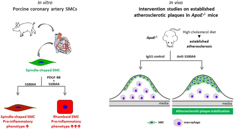
Aims: During atherosclerosis, smooth muscle cells (SMCs) accumulate in the intima where they switch from a contractile to a synthetic phenotype. From porcine coronary artery, we isolated spindle-shaped (S) SMCs exhibiting features of the contractile phenotype and rhomboid (R) SMCs typical of the synthetic phenotype. S100A4 was identified as a marker of R-SMCs in vitro and intimal SMCs, in pig and man. S100A4 exhibits intra- and extracellular functions. In this study, we investigated the role of extracellular S100A4 in SMC phenotypic transition.

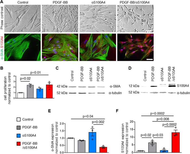
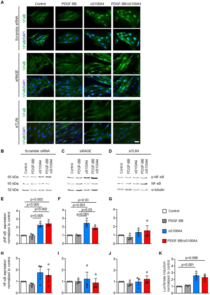
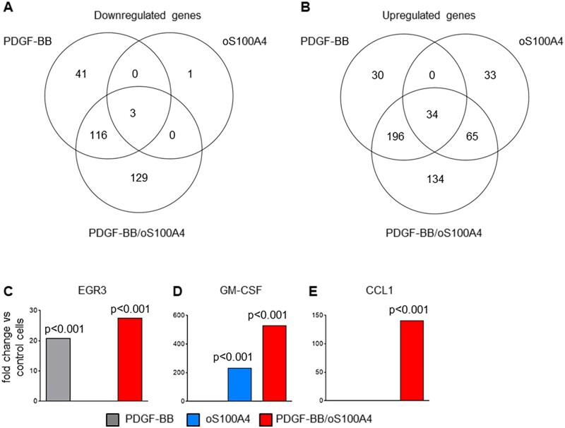
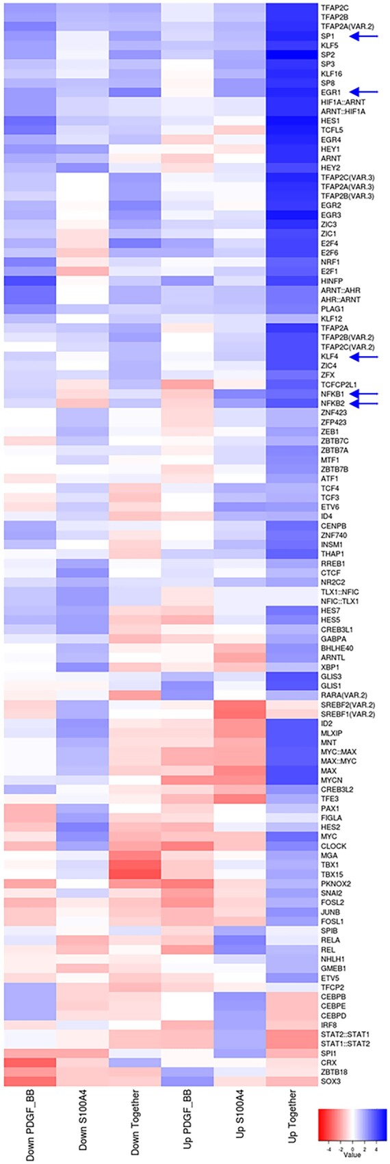
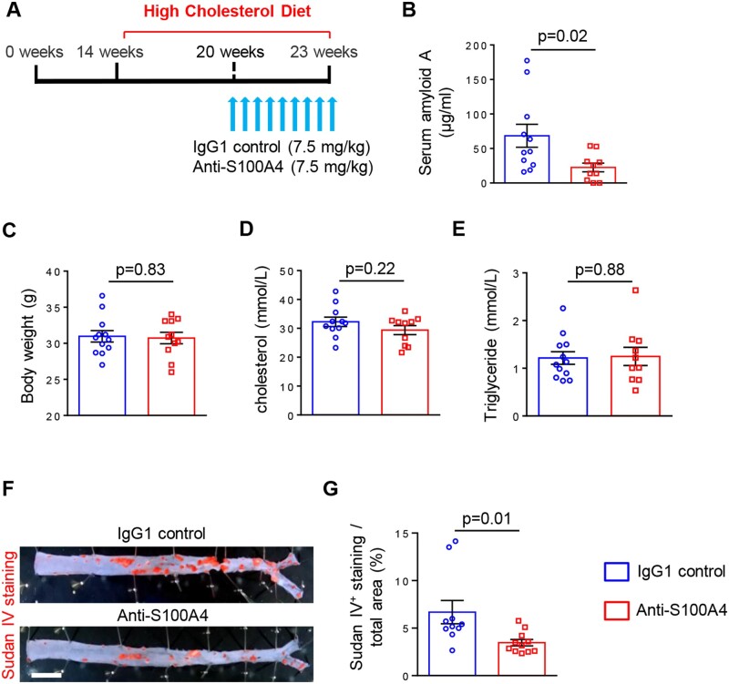
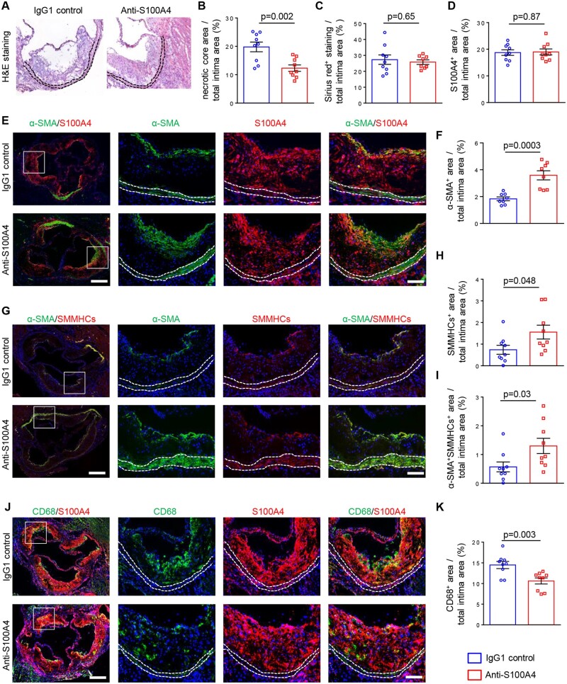
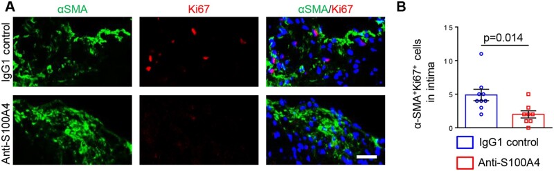
Methods and results: S-SMCs were treated with oligomeric recombinant S100A4 (oS100A4), which induced nuclear factor (NF)-κB activation. Treatment of S-SMCs with oS100A4 in combination with platelet-derived growth factor (PDGF)-BB induced a complete SMC transition towards a pro-inflammatory R-phenotype associated with NF-κB activation, through toll-like receptor-4. RNA sequencing of cells treated with oS100A4/PDGF-BB revealed a strong up-regulation of pro-inflammatory genes and enrichment of transcription factor binding sites essential for SMC phenotypic transition. In a mouse model of established atherosclerosis, neutralization of extracellular S100A4 decreased area of atherosclerotic lesions, necrotic core, and CD68 expression and increased α-smooth muscle actin and smooth muscle myosin heavy chain expression.

Conclusion: We suggest that the neutralization of extracellular S100A4 promotes the stabilization of atherosclerotic plaques. Extracellular S100A4 could be a new target to influence the evolution of atherosclerotic plaques.

Keywords: ApoE−/−; CD68; Extracellular S100A4; NF-κB; RAGE; Smooth muscle myosin heavy chains; TLR4; α-Smooth muscle actin.


Figures

None
Graphical abstract
Figure 1
Figure 1
Oligomeric S100A4 in combination with PDGF-BB promotes SMC phenotypic transition. Phase-contrast photomicrographs and double-immunofluorescence staining for α-SMA and S100A4 (A), quantification of proliferating cells normalized to control condition (B), representative Western blots for α-SMA (C) and corresponding quantification of α-SMA (E), representative Western blots for S100A4 (D) and corresponding quantification of S100A4 (F) in S-SMCs treated with PDGF-BB, oS100A4, or PDGF-BB/oS100A4 for 4 days. Note that the full phenotypic transition is acquired only in PDGF-BB/oS100A4 condition (A). In (D), the band detected above S100A4 corresponds to the exogenous His-tagged S100A4. In (A), nuclei are stained in blue by DAPI. In (C and D), α-tubulin is used as a housekeeping protein. Bar = 75  μm for phase contrast, bar = 25 μm for immunofluorescence, n = 3 biological replicates for each experiment. Comparisons were performed by using one-way ANOVA [B, F = 6.951, F(DFn, DFd) = 0.9611 (3,8); E, F = 10.68, F(DFn, DFd) = 2 (3, 8); F, F = 29.86 F(DFn, DFd) = 0.8042 (3, 8)].
Figure 2
Figure 2
Oligomeric S100A4 activates SMCs through TLR4-dependent NF-κB pathway. Immunofluorescence staining (A) and representative Western blots for NF-κB and pNF-κB (B–D) followed by quantification (E–J) after transfection of S-SMCs with scramble siRNA, siRAGE, or siTLR4 followed by treatments with PDGF-BB, oS100A4, or PDGF-BB/oS100A4 for 1 h. Note that only by silencing of TLR4, oS100A4 and PDGF-BB/oS100A4-induced NF-κB activation is prevented (A, D, G, and J). In (B–D), α-tubulin is used as a housekeeping protein. In (A), nuclei are stained in blue by DAPI. Luciferase promoter reporter assay (K) after transfection of S-SMCs with pNFκB Tluc16-DD vector followed by treatments with PDGF-BB, oS100A4, or PDGF-BB/oS100A4 for 1 h. Note that promoter is activated after the treatment with oS100A4 and PDGF-BB/oS100A4. Bar = 25 µm, n = 3 biological replicates for each experiment. Comparisons were performed by using one-way ANOVA [E, F = 18.36, F(DFn, DFd) = 0.4488 (3,8); F, F = 16.63, F(DFn, DFd) = 1.201 (3,8); G, F = 1.021, F(DFn, DFd) = 0.6769 (3,8); H, F = 1.504, F(DFn, DFd) = 1.012 (3,8); I, F = 0.1510, F(DFn, DFd) = 0.7588 (3,8); J, F = 0.5399, F(DFn, DFd) = 0.5658 (3,8); K, F = 11.66, F(DFn, DFd) = 1.994 (3,8)].
Figure 3
Figure 3
PDGF-BB amplifies the pro-inflammatory properties induced by oligomeric S100A4. EdgeR analysis of RNA sequencing data represented with Venn diagrams showing the total number and relations of down-regulated (A) or up-regulated (B) genes in S-SMCs after treatments with PDGF-BB, oS100A4, or PDGF-BB/oS100A4 for 4 h and histograms showing the fold-changes vs. the control conditions for EGR3 (C), GM-CSF (D), and CCL1 (E). Note that PDGF-BB amplifies oS100A4-induced up-regulation of GM-CSF (D) and CCL1 (E) is exclusively expressed after PDGF-BB/oS100A4 treatment. The fold-change and the Benjamini–Hochberg corrected P-value thresholds were set to 2 and 0.05, respectively. Experiment was performed with biological triplicates.
Figure 4
Figure 4
Oligomeric S100A4 in synergy with PDGF-BB induces enrichment in TFBSs essential for SMC phenotypic transition. Heat map representing TFBS enrichment in promoter regions of genes that were down- or up-regulated after treatments of SMCs with PDGF-BB, oS100A4, or PDGF-BB/oS100A4 for 4 h. TFBSs were defined according to JASPAR. The heat map shows the relative enrichment (z-score) of TFBSs that are significantly overrepresented (P < 0.05). Blue colour represents up-regulated and red colour down-regulated TFBSs. Arrows indicate important TFBSs in SMC phenotypic transition. Note that KLF4, SP1, EGR1, and NF-kB are significantly enriched only after PDGF-BB/oS100A4 treatment. Experiment was performed with biological triplicates.
Figure 5
Figure 5
Neutralization of S100A4 decreases systemic inflammation and atherosclerotic lesion area without affecting lipid metabolism. Scheme representing the experimental setup, in which mice were fed with HCD for 9 weeks to induce the development of atherosclerosis; the IgG1 control or anti-S100A4 were injected during the last 3 weeks of HCD (A). Quantification of SAA levels (B; n = 11 for IgG1 control and n = 10 for anti-S100A4), body weight (C; n = 12 for IgG1 control and n = 11 for anti-S100A4), cholesterol (D; n = 11 for IgG1 control and n = 10 for anti-S100A4), and triglyceride (E; n = 12 for IgG1 control and n = 10 for anti-S100A4) levels in mouse serum samples. En face Sudan IV staining (F) and quantification of Sudan IV positive area of thoracic-abdominal aorta (G) in ApoE−/− mice treated with IgG1 control (n = 10) and anti-S100A4 (n = 11). Bar = 0.2 cm. Comparisons were performed by using two-tailed unpaired Student’s t-test (B, t = 2.476, df = 19, C: t = 0.2139, df = 21; D, t = 1.267, df = 19; E, t = 0.1440, df = 20; G, t = 2.636, df = 19).
Figure 6
Figure 6
Neutralization of S100A4 increases the amount and differentiation of SMCs and decreases the size of the necrotic core and the burden of inflammatory cells within the lesions. Haematoxylin and eosin staining (A), quantification of necrotic core (B; n = 9 for IgG1 control and anti-S100A4), Sirius red staining (C; n = 9 for IgG1 control and n = 8 for anti-S100A4), and quantification of S100A4+ area (D; n = 9 for IgG1 control and anti-S100A4). Representative double-immunofluorescence staining for α-SMA and S100A4 (E) followed by the quantification of α-SMA+ area (F; n = 8 for IgG1 control and anti-S100A4). Representative double-immunofluorescence staining for α-SMA and SMMHCs (G) followed by the quantification of SMMHCs+ (H; n = 9 for IgG1 control and anti-S100A4) or α-SMA+SMMHCs+ area (I; n = 9 for IgG1 control and anti-S100A4). Representative double-immunofluorescence staining for CD68 and S100A4 (J) followed by the quantification of CD68+ area (K; n = 9 for IgG1 control and anti-S100A4). All analyses were performed on aortic root sections. Dashed lines highlight the media. The box indicates the region magnified in the right panels. Bar = 250 μm for low magnification, and bar = 100 μm for high magnification. Comparisons were performed by using two-tailed unpaired Student’s t-tests (B, t = 3.660, df = 16, C: t = 0.4565, df = 15; D, t = 0.1552, df = 16; F, t = 4.731, df = 14; H, t = 2.141, df = 16; I, t = 2.321, df = 16; K, t = 3.409, df = 16).
Figure 7
Figure 7
Neutralization of S100A4 decreases intimal SMC proliferation. Representative double-immunofluorescence staining for α-SMA and Ki67 within intima (A) followed by the quantification of Ki67 positive SMCs (B; n = 9 for IgG1 control and n = 8 for anti-S100A4). Bar = 50 µm. Comparisons were performed by using two-tailed unpaired Student’s t-tests (t = 2.774, df = 15).

Comment in

Similar articles

Cited by

References

    1. Tabas I, García-Cardeña G, Owens GK. Recent insights into the cellular biology of atherosclerosis. J Cell Biol 2015;209:13–22. - PMC - PubMed
    1. Allahverdian S, Chaabane C, Boukais K, Francis GA, Bochaton-Piallat ML. Smooth muscle cell fate and plasticity in atherosclerosis. Cardiovasc Res 2018;114:540–550. - PMC - PubMed
    1. Bennett MR, Sinha S, Owens GK. Vascular smooth muscle cells in atherosclerosis. Circ Res 2016;118:692–702. - PMC - PubMed
    1. Basatemur GL, Jorgensen HF, Clarke MCH, Bennett MR, Mallat Z. Vascular smooth muscle cells in atherosclerosis. Nat Rev Cardiol 2019;16:727–744. - PubMed
    1. Allahverdian S, Chehroudi AC, McManus BM, Abraham T, Francis GA. Contribution of intimal smooth muscle cells to cholesterol accumulation and macrophage-like cells in human atherosclerosis. Circulation 2014;129:1551–1559. - PubMed

Publication types

MeSH terms