. 2021 Dec 17;117(14):2794-2806.
doi: 10.1093/cvr/cvaa323.

Off-target effects of sodium-glucose co-transporter 2 blockers: empagliflozin does not inhibit Na+/H+ exchanger-1 or lower [Na+]i in the heart

Affiliations

Off-target effects of sodium-glucose co-transporter 2 blockers: empagliflozin does not inhibit Na+/H+ exchanger-1 or lower [Na+]i in the heart

Yu Jin Chung et al. Cardiovasc Res. .

Abstract

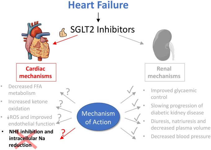
Aims: Emipagliflozin (EMPA) is a potent inhibitor of the renal sodium-glucose co-transporter 2 (SGLT2) and an effective treatment for type-2 diabetes. In patients with diabetes and heart failure, EMPA has cardioprotective effects independent of improved glycaemic control, despite SGLT2 not being expressed in the heart. A number of non-canonical mechanisms have been proposed to explain these cardiac effects, most notably an inhibitory action on cardiac Na+/H+ exchanger 1 (NHE1), causing a reduction in intracellular [Na+] ([Na+]i). However, at resting intracellular pH (pHi), NHE1 activity is very low and its pharmacological inhibition is not expected to meaningfully alter steady-state [Na+]i. We re-evaluate this putative EMPA target by measuring cardiac NHE1 activity.

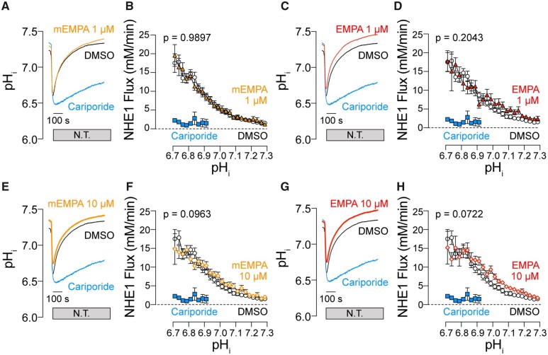
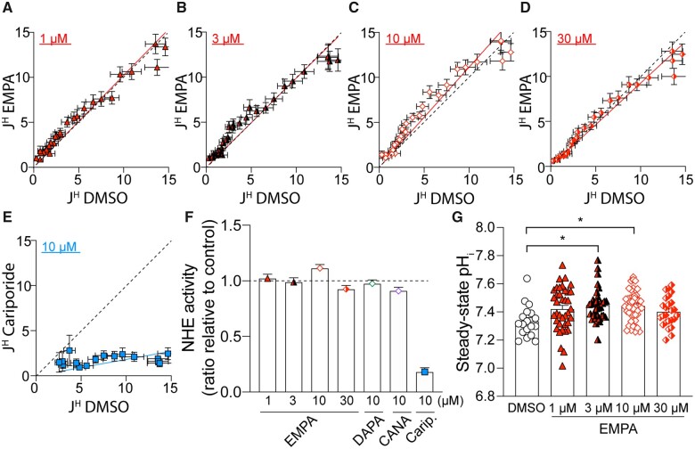
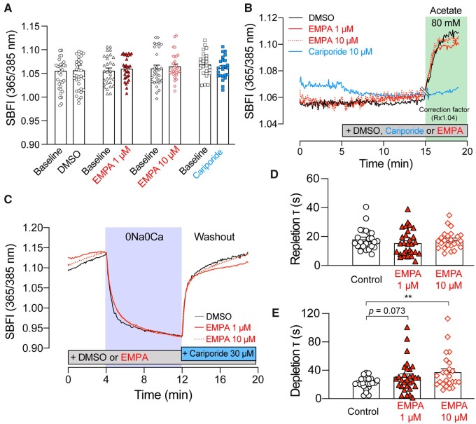
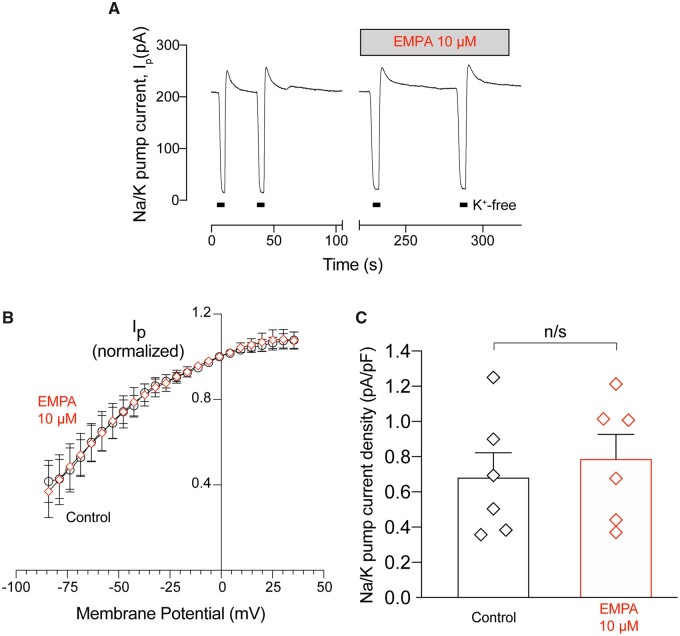
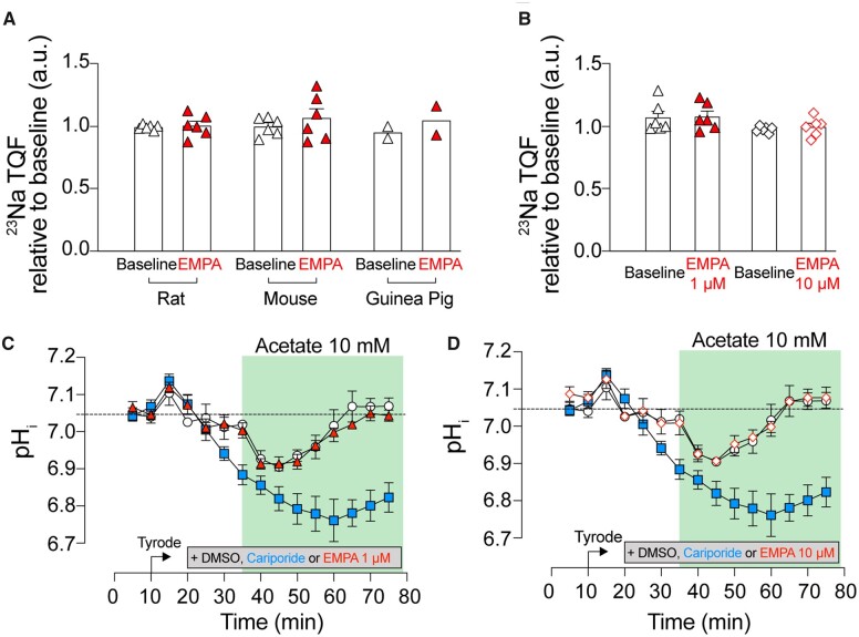
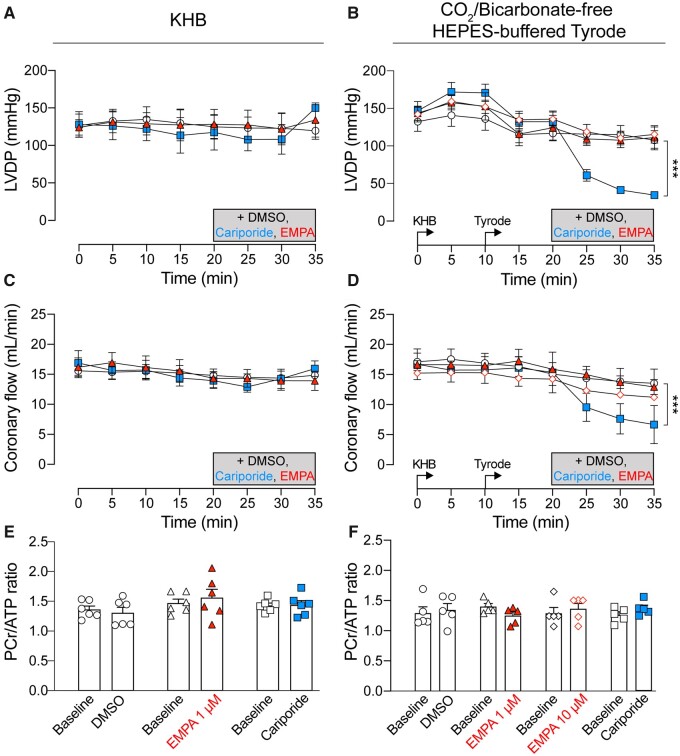
Methods and results: The effect of EMPA on NHE1 activity was tested in isolated rat ventricular cardiomyocytes from measurements of pHi recovery following an ammonium pre-pulse manoeuvre, using cSNARF1 fluorescence imaging. Whereas 10 µM cariporide produced near-complete inhibition, there was no evidence for NHE1 inhibition with EMPA treatment (1, 3, 10, or 30 µM). Intracellular acidification by acetate-superfusion evoked NHE1 activity and raised [Na+]i, reported by sodium binding benzofuran isophthalate (SBFI) fluorescence, but EMPA did not ablate this rise. EMPA (10 µM) also had no significant effect on the rate of cytoplasmic [Na+]i rise upon superfusion of Na+-depleted cells with Na+-containing buffers. In Langendorff-perfused mouse, rat and guinea pig hearts, EMPA did not affect [Na+]i at baseline nor pHi recovery following acute acidosis, as measured by 23Na triple quantum filtered NMR and 31P NMR, respectively.

Conclusions: Our findings indicate that cardiac NHE1 activity is not inhibited by EMPA (or other SGLT2i's) and EMPA has no effect on [Na+]i over a wide range of concentrations, including the therapeutic dose. Thus, the beneficial effects of SGLT2i's in failing hearts should not be interpreted in terms of actions on myocardial NHE1 or intracellular [Na+].

Keywords: Heart failure; Intracellular Na; NMR spectroscopy; Na/H exchanger-1; SGLT2 inhibitor.


Figures

None
Graphical abstract
Figure 1
Figure 1
EMPA does not inhibit NHE1 activity in isolated cardiomyocytes. pHi recovery time course (A), (C), (E), (G) and NHE1 flux as a function of pHi (B), (D), (F), (H) measured in rat ventricular cardiac myocytes exposed to acute NH4+ acid-load and wash out and pre-treated with DMSO, 10 µM cariporide or 1 or 10 µM EMPA obtained from MedChemExpress (mEMPA; yellow) or Boehringer (EMPA; red). Flux is calculated from rate of pHi change (from time courses) multiplied by buffering capacity (obtained in previous studies). Note: in the presence of cariporide, pHi recovery is not complete in 12 min, and the flux is therefore only plotted for pH < 6.9. N.T., Normal Tyrode’s buffer. All data means ± SEM [error bar not included in (A), (C), (E), and (G) for clarity]. n = 14–20 cells from three rats per condition.
Figure 2
Figure 2
EMPA does not inhibit NHE1 flux in isolated cardiomyocytes. (A–E) Acid extrusion flux (JH; mM/min) plotted at matching pHi for control (DMSO) and in presence of drug (EMPA or cariporide) at the concentrations indicated. Red or blue lines show best fit through origin, and the slope describes NHE1 activity in the presence of drug (where 1.0 is no inhibition, indicated in broken black line). (F) Slope of flux–flux relationships for EMPA, DAPA, CANA, and cariporide (Carip.). (G) Steady-state pHi attained at the end of pH recovery. All data are means ± SEM; means ± 95% confidence interval for (F). Data from EMPA and mEMPA were combined. For EMPA and cariporide, n = 30–49 cells from three rats per condition. For DAPA and CANA, n = 31–43 cells from two rats per condition. *P <0.05 by one-way ANOVA.
Figure 3
Figure 3
EMPA does not affect intracellular Na+ handling in isolated cardiomyocytes. Intracellular [Na+] measured using SBFI fluorescence in myocytes treated with DMSO, 1 or 10 µM EMPA or 10 µM cariporide (A) at baseline or (B) in 80 mM Na-acetate. Ratio corrected for pH-artefact (Rx1.04), marked by double line at 15 min. See Supplementary material online, Figure S2 for original time course. n = 24–33 cells from three rats per condition. (C) Interrogation of NHE1-independent Na+ transport pathways using 0Na0Ca protocol. Time constant of (D) Na+ re-uptake and (E) Na+ efflux. All data are means ± SEM [error bar not included in (B) and (C) for clarity]. n = 24–30 cells from three rats per condition. **P <0.01 by two-tail, unpaired student’s t-test.
Figure 4
Figure 4
EMPA does not affect Na+/K+ ATPase current in isolated cardiomyocytes. Whole-cell pump current was measured using the perforated patch technique in rat ventricular myocytes. (A) Representative trace showing pump current (Ip) recorded under control conditions and after the application of EMPA (10 µM). Pump current was recorded at 0 mV. Potassium-free bath solution was applied at the points shown (K+-free). (B) Current–voltage relationships recorded during the application of a negative-going voltage ramp (13 mV/s) under control conditions and after application of EMPA. Data were averaged (factor 800) to obtain ∼1 reading every 5 mV and normalized to the current reading at 0 mV. (C) Average Na+/K+ ATPase current density recorded after 300 s in control and EMPA. All data are means ± SEM. n = 6 cells from six rats.
Figure 5
Figure 5
EMPA does not affect intracellular [Na+] and pHi in Langendorff-perfused hearts. Intracellular [Na+] measurement using 23Na NMR spectroscopy in (A) rat, mouse and guinea pig hearts perfused in CO2/bicarbonate-buffered KHB and treated with 1 µM EMPA and (B) rat hearts perfused with CO2/bicarbonate-free Tyrode’s buffer and treated with either 1 or 10 µM EMPA. n = 6 rats (A and B), 6 mice (A and B), and 2 guinea pigs (A) per group; 23Na signal normalized to baseline. Rat hearts pre-treated with DMSO (open circle), 10 µM cariporide (blue square), (C) 1 µM or (red triangle) (D) 10 µM EMPA (red, open diamond), and perfused in Tyrode’s buffer supplemented with 10 mM Na-acetate. Intracellular pH measured using 31P NMR spectroscopy. All data mean ± SEM. n = 6 rats per group per assay.
Figure 6
Figure 6
EMPA does not affect cardiac contractile function and energetics in Langendorff-perfused rat hearts. (A, B) LVDP measured using balloon inserted into left ventricle of rat heart. (C, D) Coronary flow required to maintain perfusion pressure at 80 mmHg. DMSO control (open circle), 10 µM cariporide (blue square), 1 µM EMPA (red triangle), or 10 µM EMPA (red, open diamond). (E, F) PCr/ATP ratio measured using 31P NMR spectroscopy. Hearts perfused in Kreb’s buffer (KHB) (A), (C), and (E) and in CO2/bicarbonate-free HEPES-buffered Tyrode’s (Tyrode) (B), (D), and (F). All data mean ± SEM. n = 6 rats (KHB) or 5 rats (CO2/bicarbonate-free Tyrode) per group. ***P <0.001 by two-way ANOVA.

Comment in

Similar articles

Cited by

References

    1. Grempler R, Thomas L, Eckhardt M, Himmelsbach F, Sauer A, Sharp DE, Bakker RA, Mark M, Klein T, Eickelmann P.. Empagliflozin, a novel selective sodium glucose cotransporter-2 (SGLT-2) inhibitor: characterisation and comparison with other SGLT-2 inhibitors. Diabetes Obes Metab 2012;14:83–90. - PubMed
    1. Zinman B, Wanner C, Lachin JM, Fitchett D, Bluhmki E, Hantel S, Mattheus M, Devins T, Johansen OE, Woerle HJ, Broedl UC, Inzucchi SE, EMPA-REG OUTCOME Investigators. Empagliflozin, cardiovascular outcomes, and mortality in type 2 diabetes. N Engl J Med 2015;373:2117–2128. - PubMed
    1. Yurista SR, Silljé HHW, Oberdorf-Maass SU, Schouten E-M, Pavez Giani MG, Hillebrands J-L, van Goor H, van Veldhuisen DJ, de Boer RA, Westenbrink BD.. Sodium-glucose co-transporter 2 inhibition with empagliflozin improves cardiac function in non-diabetic rats with left ventricular dysfunction after myocardial infarction. Eur J Heart Fail 2019;21:862–873. - PubMed
    1. Adingupu DD, Göpel SO, Grönros J, Behrendt M, Sotak M, Miliotis T, Dahlqvist U, Gan L-M, Jönsson-Rylander A-C.. SGLT2 inhibition with empagliflozin improves coronary microvascular function and cardiac contractility in prediabetic ob/ob−/− mice. Cardiovasc Diabetol 2019;18:16. - PMC - PubMed
    1. Li C, Zhang J, Xue M, Li X, Han F, Liu X, Xu L, Lu Y, Cheng Y, Li T, Yu X, Sun B, Chen L.. SGLT2 inhibition with empagliflozin attenuates myocardial oxidative stress and fibrosis in diabetic mice heart. Cardiovasc Diabetol 2019;18:https://doi.org/10.1186/s12933-019-0816-2 - PMC - PubMed

Publication types

MeSH terms