Review
. 2021 Mar;595(6):675-706.
doi: 10.1002/1873-3468.13984. Epub 2020 Dec 4.

ABC-F translation factors: from antibiotic resistance to immune response

Affiliations
Review

ABC-F translation factors: from antibiotic resistance to immune response

Corentin R Fostier et al. FEBS Lett. 2021 Mar.

Abstract

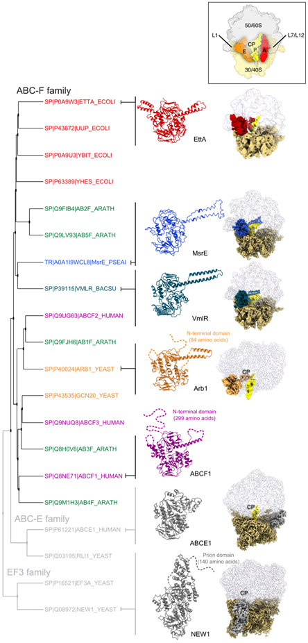
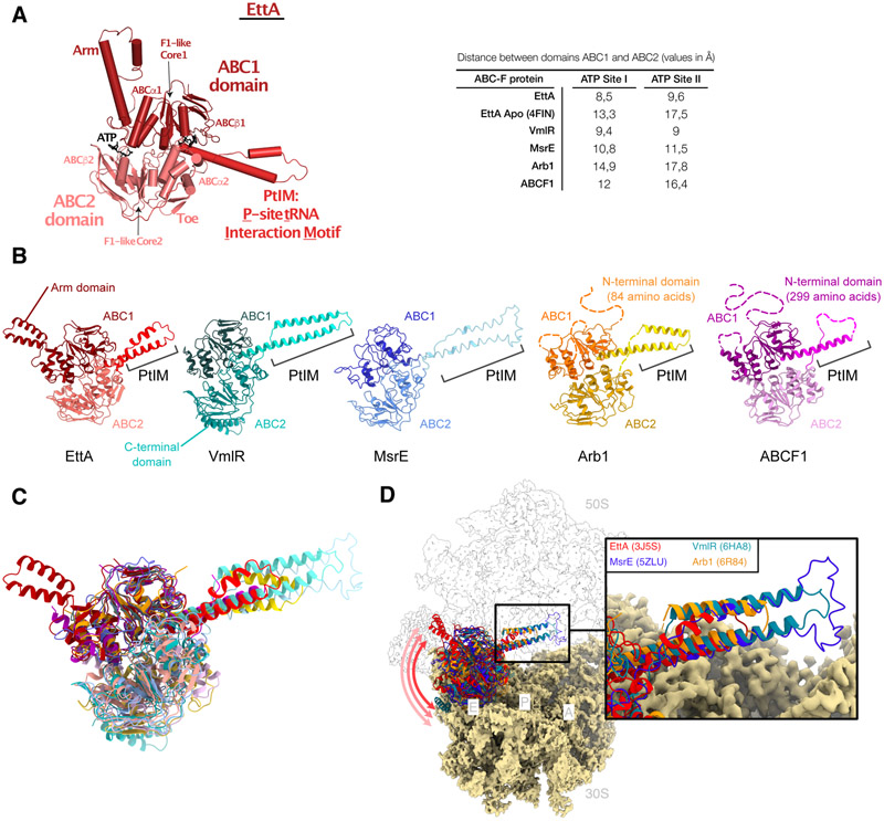
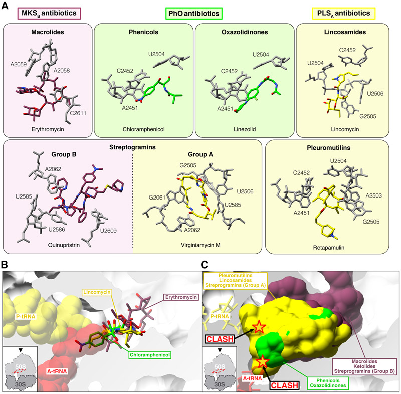
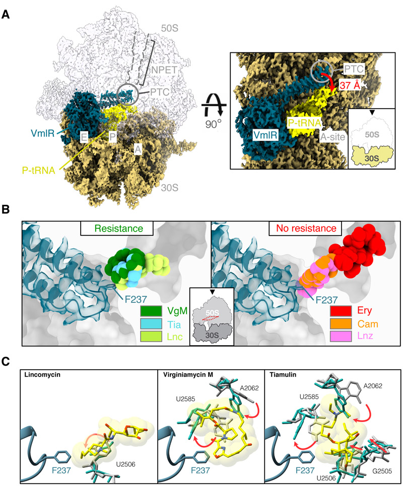
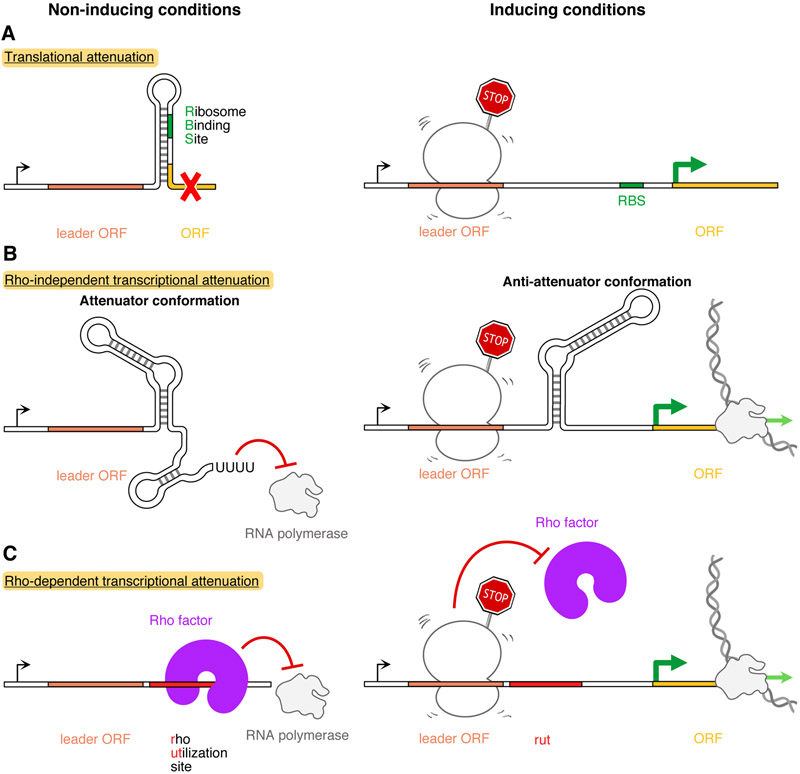
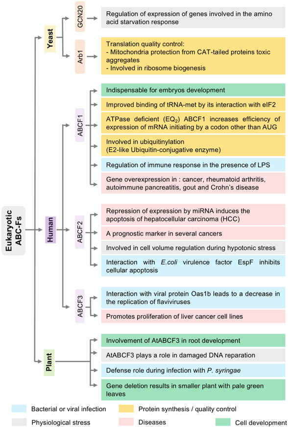
Energy-dependent translational throttle A (EttA) from Escherichia coli is a paradigmatic ABC-F protein that controls the first step in polypeptide elongation on the ribosome according to the cellular energy status. Biochemical and structural studies have established that ABC-F proteins generally function as translation factors that modulate the conformation of the peptidyl transferase center upon binding to the ribosomal tRNA exit site. These factors, present in both prokaryotes and eukaryotes but not in archaea, use related molecular mechanisms to modulate protein synthesis for heterogenous purposes, ranging from antibiotic resistance and rescue of stalled ribosomes to modulation of the mammalian immune response. Here, we review the canonical studies characterizing the phylogeny, regulation, ribosome interactions, and mechanisms of action of the bacterial ABC-F proteins, and discuss the implications of these studies for the molecular function of eukaryotic ABC-F proteins, including the three human family members.

Keywords: ABC ATPase; ABC-F protein family; antibiotic resistance; immune response; infection; mRNA translation; protein synthesis.


Conflict of interest statement

The authors declare that there is no conflict of interest regarding the publication of this article.

Figures

Figure 1:
Figure 1:. Phylogeny and structure of ABC-F factors.
Cladogram labeled with Swiss-Prot (SP) or TrEMBL (TR) accession codes for representative ABC-F proteins as well as proteins from two non-ABC-F families containing ABC domains (the eEF3 and ABCE families), and their corresponding structures. Identifiers correspond to the following species: Escherichia coli (ECOLI), Bacillus subtilis (BACSU), Pseudomonas aeruginosa (PSEAI), Saccharomyces cerevisiae (YEAST), Arabidopsis thaliana (ARATH) and Homo sapiens (HUMAN). Structure of factors were aligned based on least-squared superposition of ABC1, and missing residues were added as dotted lines. The corresponding cryo-EM structures were aligned based on least-squared superposition the 23S (prokaryotes) 25S/28S (eukaryotes) rRNA in the large ribosomal subunit. Orientation and major landmarks are indicated on the schematic ribosome on the top of the figure (A, A tRNA; P, P tRNA; E, E tRNA; CP, central protuberance). Cryo-EM density segments are colored as follows: ribosomal small subunit in sand, ribosomal large subunit in transparent grey, and P tRNA in yellow. Note that the ribosomal small subunit is absent in the Arb1 structure, and no cryo-EM structure has been solved yet for ABCF1. ABC-F factors exclusively bind to the ribosomal E site, while ABCE factors bind near the A site, and EF3-related factors bind near the central protuberance at the junction of small and large ribosomal subunits. PDB accession codes are as follows: EttA (3J5S - Cryo-EM, resolution: 7.5 Å ), MsrE (5ZLU - Cryo-EM, resolution: 3.6 Å), VmlR (6HA8 - Cryo-EM, resolution: 3.5 Å), Arb1 (6R84 - Cryo-EM, resolution: 3.6 Å), ABCF1 (5ZXD - X-ray diffraction, resolution: 2.29 Å), ABCE1 (5LZV - Cryo-EM, resolution: 3.35 Å), NEW1 (6S47 - Cryo-EM, resolution: 3.28 Å) [7,15-17,21,46,159].
Figure 2:
Figure 2:. General structure of ABC-F factors and their binding site on the ribosome.
(A) Architecture of ribosome-bound EttA, showing its domains and subdomains. Structural features are colored as follows: ABC1 in dark red shade, PtIM in bright red shade and ABC2 in light red shade. Table with the distances in Å between serine Cα of the signature motif of the ABC1 domain and the Lysine Cα of the walker A of the ABC2 domain (ATP binding site I) and the serine Cα of the signature motif of the ABC2 domain and the Lysine Cα of the walker A of the ABC1 domain (ATP binding site II). (B, C) Structural alignment of several ABC-F factors. Factors were aligned based on least-squared superposition of ABC1, and their domains are shown in different shades of the same color: ABC1 in a dark shade, the PtIM in a bright shade, and ABC2 in a light shade. Missing residues are represented as dotted lines, and additional sequence motifs and domains are labeled (e.g., the Arm in EttA and the C-terminal domain in VmlR. Structural superposition shows strong structural conservation of ABC1 and ABC2 varying alignment of the PtIM. (D) Structural alignment of ribosome-bound ABC-Fs factors. Cryo-EM structures were aligned on the large subunit 23S rRNA (or the 25S rRNA for Arb1), and the corresponding cryo-EM densities were extracted and colored as in Figure 1. Alignment of ribosome-bound ABC-Fs proteins based on the rRNA in the large subunit shows that PtIMs superimpose well and point in the same direction, while the ABC cassettes are less closely aligned and tending to oscillate in the ribosomal E site as indicated by the arrows. PDB accession codes are the same as in Figure 1
Figure 3:
Figure 3:. ABC-Fs factors and resistance to ribosomally-directed antibiotics.
(A) Binding site of some major representatives of ribosomally-directed antibiotic families to which ARE ABC-Fs provide resistance. Antibiotic families are grouped according to ARE ABC-Fs resistance phenotypes: MKSB antibiotics (Macrolides, Ketolides, group B Streprogramins in purple), PLSA antibiotics (Pleuromutilins, Lincosamides, group A Streptogramins in yellow) and PhO antibiotics (Phenicols and Oxazolidinones in green). (B, C) Resistance phenotypes are correlated but not strictly specified by the location of the antibiotic binding sites in the PTC/NPET. A transverse section of the PTC/NPET indicates the relative position of some antibiotics shown on Figure 3A, namely chloramphenicol, lincomycin and erythromycin as well as A tRNA in red and P tRNA in yellow. Antibiotics belonging to MKSB, PLSA and PhO resistance phenotypes are also depicted as surface to show their relative occupancy and binding promiscuity. Note that PhO antibiotics would sterically clash A tRNA while PLSA antibiotics would clash both A and P tRNAs. Structures were aligned on the 23S rRNA domain V of a high-resolution structure of E. coli ribosome (PDB 4YBB) [160]. PDB accession codes are as follows: Azithromycin (4V7Y) [89], Carbomycin A (1K8A) [88], Chloramphenicol (6ND5) [161], Clindamycin (4V7V) [77], Dalfopristrin (4U24) [162], Erythromycin (4V7U) [77], Lincomycin (5HKV), [163], Linezolid (3CPW) [80], Quinupristrin (4U1U) [162], Retapamulin (2OGO) [107], piramycin (1KD1) [88], Telithromycin (4V7S) [77], Tiamulin (3G4S) [164], Tylosin (1K9M) [88], Virginiamycin M (4U25) [162], Pre-catalysis A and P tRNAs (1VY4), [165].
Figure 4:
Figure 4:. VmlR structure and possible allosteric mechanism.
(A) Cryo-EM density of ribosome-bound VmlR (PDB 6HA8) [16]. Density segments were extracted and colored as follows: ribosomal small subunit in sand, ribosomal large subunit in transparent grey, P site tRNA in yellow, VmlR in dark cyan. The labels indicate functional sites on the ribosome (A, A site; P, P site; E, E site; PTC, peptidyl transferase center; NPET, nascent polypeptide exit tunnel). VmlR binds in the ribosomal E site and displaces the CCA acceptor stem of the P site tRNA CCA by 37 Å in the direction of the A site. The Tip of its PtIM penetrates directly into the PTC. Equivalent structural effects are observed in the cryo-EM structure of ribosome-bound MsrE from Pseudomonas aeruginosa MsrE (PDB 5ZLU) [15]. (B) Direct steric drug displacement by the PtIM in ARE ABC-Fs cannot fully explain the resistance mechanism. Cryo-EM structures of VmlR-bound and antibiotic-bound ribosomes were aligned based on least-squares superposition of the 23S rRNA in the large subunit, demonstrating that the residue F237 in the Tip of the PtIM in VmlR (dark cyan) can interact with several antibiotics to which it provides resistance, namely virginiamycin M (VgM), tiamulin (Tia) and lincomycin (Lnc). However, VmlR does not provide resistance to chloramphenicol (Cam) nor linezolid (Lnz) even though F237 would also sterically clash those two antibiotics. VmlR furthermore does not provide resistance to erythromycin (Ery). (C) 23S rRNA nucleotides rearrangement at the PTC upon VmlR binding would suggest an allosteric mechanism. Nucleotides with lincomycin-, virginiamycin M- and tiamulin-bound in absence of VmlR are shown in grey while they are shown in cyan in presence of VmlR. Relative rearrangements are indicated by red arrows. PDB accession codes are the same as in Figure 3.
Figure 5:
Figure 5:. Ligand-induced translational stalling regulates gene expression.
Top, translational attenuators sequester the ribosome binding site (RBS) of a regulated ORF in a structured stem-loop that blocks ribosome binding and initiation of translation. Drug-induced ribosomal stalling on an upstream ORF encoding a short “leader peptide” induces formation of an alternative stem-loop that opens RBS to promote efficient translation initiation and thereby higher protein expression. This mechanism is exemplified by the erythromycin-sensing ErmBL and ErmCL leader peptides [94,95]. Middle, rho-independent transcriptional attenuators fold into their transcription-attenuating conformation in absence of an inducer compound, leading RNA polymerase terminate transcription prematurely and produce a truncated mRNA. Drug-induced ribosomal stalling on the leader peptide encoded in an upstream ORF stabilizes an alternative “anti-attenuator” conformation that prevents RNA polymerase drop-off and allows transcription of a full-length mRNA. This mechanism is exemplified by the erythromycin-sensing MefAL leader peptide [132]. Bottom, in Rho-dependent transcriptional attenuators, the Rho helicase enzyme is able to terminate transcription by binding to a Rho utilization site (rut), leading RNA polymerase to drop off and produce a truncated mRNA. Drug-induced ribosomal stalling on an ORF encoding a leader peptide could directly mask the rut site or stabilize an alternative mRNA conformation that prevents Rho binding and transcription termination. This mechanism is exemplified by the L-tryptophan-sensing TnaC and the L-ornithine-sensing SpeFL leader peptides [124,125]. Note that, to date, no mechanism involving antibiotic-dependent regulation of a Rho-dependent transcriptional attenuator has been described, making the mechanism illustrated in the bottom panel hypothetical.
Figure 6:
Figure 6:. Known functions of eukaryotic ABC-F factors.
The different roles of ABC-F factors in yeast, human, and plants are presented and distinguished by different colors: protein synthesis and quality control in yellow, physiological stress response in grey, bacterial or viral infection response in light blue, cellular development in green, and diseases processes in pink.

Similar articles

Cited by

References

    1. Jaciuk M, Nowak E, Skowronek K, Tanska A & Nowotny M (2011) Structure of UvrA nucleotide excision repair protein in complex with modified DNA. Nature structural & molecular biology 18, 191–7. - PMC - PubMed
    1. Pakotiprapha D, Samuels M, Shen K, Hu JH & Jeruzalmi D (2012) Structure and mechanism of the UvrA-UvrB DNA damage sensor. Nature structural & molecular biology 19, 291–8. - PubMed
    1. Lammens K, Bemeleit DJ, Mockel C, Clausing E, Schele A, Hartung S, Schiller CB, Lucas M, Angermuller C, Soding J, Strasser K & Hopfner KP (2011) The Mre11:Rad50 structure shows an ATP-dependent molecular clamp in DNA double-strand break repair. Cell 145, 54–66. - PMC - PubMed
    1. Hopfner K-P, Karcher A, Shin DS, Craig L, Arthur LM, Carney JP & Tainer JA (2000) Structural Biology of Rad50 ATPase. Cell 101, 789–800. - PubMed
    1. Goosen N & Moolenaar GF (2001) Role of ATP hydrolysis by UvrA and UvrB during nucleotide excision repair. Research in Microbiology 152, 401–409. - PubMed

Publication types

MeSH terms

Substances

LinkOut - more resources