. 2021 Jan 1;320(1):F31-F46.
doi: 10.1152/ajprenal.00431.2020. Epub 2020 Nov 2.

A uropathogenic E. coli UTI89 model of prostatic inflammation and collagen accumulation for use in studying aberrant collagen production in the prostate

Affiliations

A uropathogenic E. coli UTI89 model of prostatic inflammation and collagen accumulation for use in studying aberrant collagen production in the prostate

Hannah Ruetten et al. Am J Physiol Renal Physiol. .

Abstract

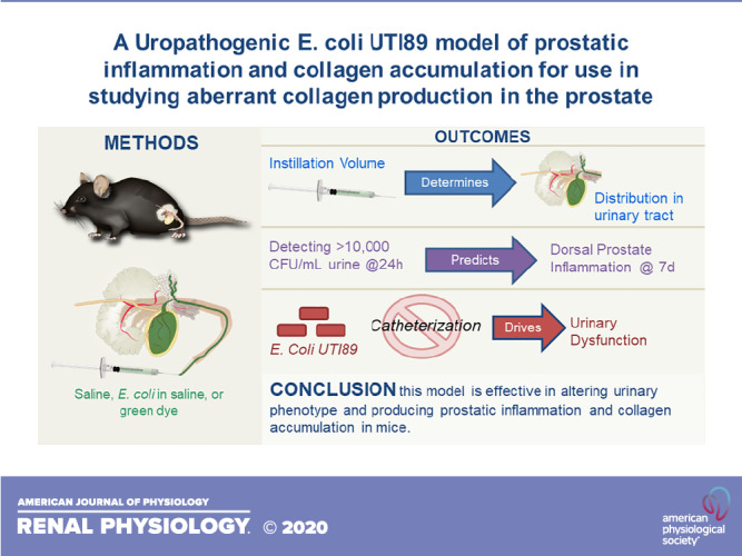
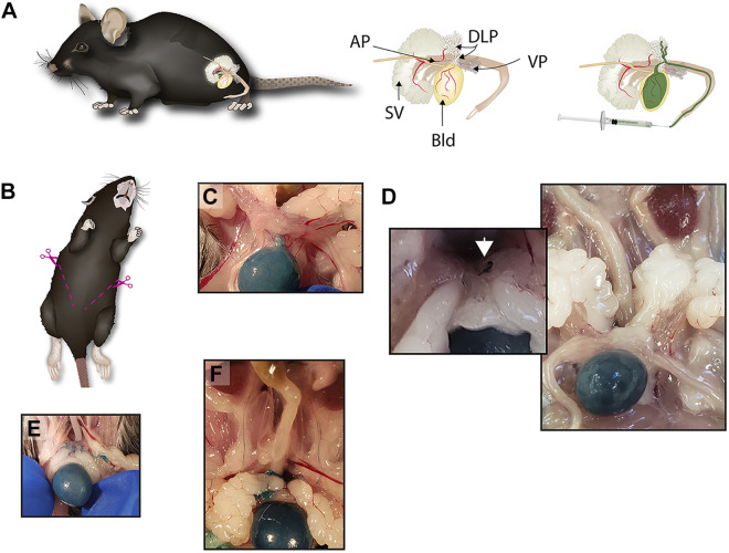
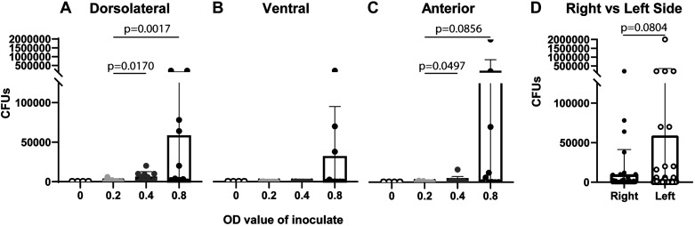
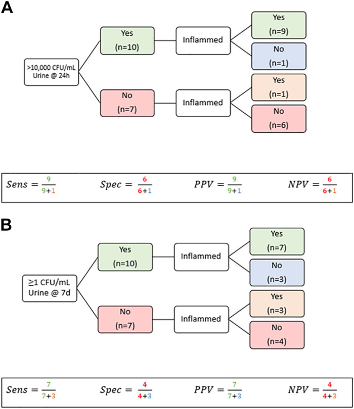
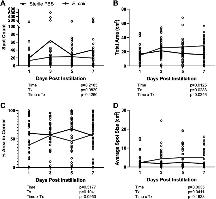
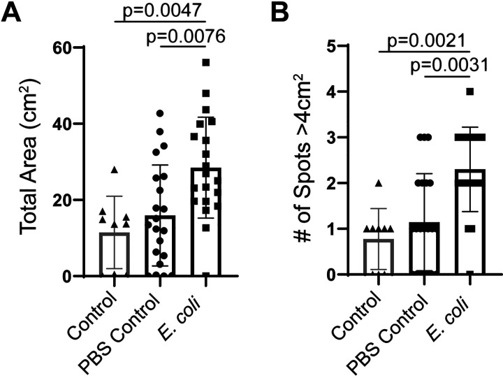
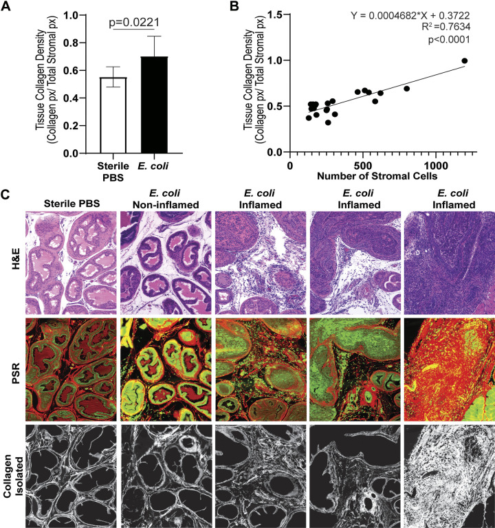
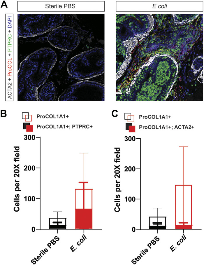
Bacterial infection is one known etiology of prostatic inflammation. Prostatic inflammation is associated with prostatic collagen accumulation and both are linked to progressive lower urinary tract symptoms in men. We characterized a model of prostatic inflammation using transurethral instillations of Escherichia coli UTI89 in C57BL/6J male mice with the goal of determining the optimal instillation conditions, understanding the impact of instillation conditions on urinary physiology, and identifying ideal prostatic lobes and collagen 1a1 prostatic cell types for further analysis. The smallest instillation volume tested (50 µL) distributed exclusively to the bladder, 100- and 200-µL volumes distributed to the bladder and prostate, and a 500-µL volume distributed to the bladder, prostate, and ureter. A threshold optical density of 0.4 E. coli UTI89 in the instillation fluid was necessary for significant (P < 0.05) prostate colonization. E. coli UTI89 infection resulted in a low frequency, high volume spontaneous voiding pattern. This phenotype was due to exposure to E. coli UTI89, not catheterization alone, and was minimally altered by a 50-µL increase in instillation volume and doubling of E. coli concentration. Prostate inflammation was isolated to the dorsal prostate and was accompanied by increased collagen density. This was partnered with increased density of protein tyrosine phosphatase receptor type C+, procollagen type I-α1+ copositive cells and decreased density of α2-smooth muscle actin+, procollagen type I-α1+ copositive cells. Overall, we determined that this model is effective in altering urinary phenotype and producing prostatic inflammation and collagen accumulation in mice.

Keywords: Escherichia coli; benign prostatic hyperplasia; collagen type I-α1; lower urinary tract symptoms; prostatitis.


Conflict of interest statement

No conflicts of interest, financial or otherwise, are declared by the author(s).

Figures

None
Graphical abstract
Figure. 1.
Figure. 1.
The volume of transurethral instillation fluid determines the anatomic distribution of instilled fluid across the male mouse lower urinary tract. A: mouse with lower urinary tract schematic and labeled lower urinary tract anatomic components, including anterior (AP), ventral (VP), dorsolateral (DLP), bladder (Bld), and seminal vesicle (SV). Mice were euthanized, a transurethral catheter (2.5 cm, PE-10) was placed into mice of various genetic backgrounds and strains (see materials and methods), and green Davidson Tissue Dye was instilled. A loose suture was placed around the tip of the penis, the catheter was removed, and the suture was tightened to prevent dye leakage. B: two cuts extending from the pubis to lateral ribs were used to open the abdomen and visualize the urinary tract. Ureters, bladder, seminal vesicles, prostatic lobes, and ductus deferens were visually inspected for green dye. C: instillation of 50 µL of fluid distributed dye to the bladder. D: instillation of 100 µL of fluid distributed dye to the bladder and dorsal prostate. E: instillation of 200 µL of fluid distributed dye to the bladder and multiple prostate lobes. F: instillation of 500 µL distributed dye to the bladder all prostate lobes, seminal vesicle, and ureters.
Fig. 2.
Fig. 2.
One hundred microliters of E. coli UTI89 [optical density (OD) 0.80] contains 1–8.2 × 10⁸ colony-forming units (CFU) of E. coli. To determine the range of CFU for E. coli UTI89 culture and transurethral instillation, and to establish biologically significant cutoffs for CFU values in tissue or urine, we measured the concentration of 12 (6 per tube) instillation aliquots originating from two separate culture tubes [tube 1 (lane A) and tube 2 (lane B)]. Culture tubes of inoculation solution were prepared at an OD of 0.80 and were prepared by the same person on the same day and both colonies were taken from a single streaked plate. An image of the culture plate of serial dilutes from two aliquots are depicted to the left of the graph. The graph on the right shows means ± SD. A Shapiro-Wilk test was used to test for normality, and transformation was applied to normalize data. Data could not be normalized using transformation, so the Mann-Whitney test was applied to compare tubes. P < 0.05 was considered statistically significant.
Fig. 3.
Fig. 3.
Concentration of E. coli in instillation fluid determines acute prostatic colony-forming unit (CFU) load. C57BL/6J male mice were instilled with sterile PBS [optical density (OD) 0] or PBS containing graded concentrations of E. coli (OD 0.2, 0.4, or 0.8). Mice were euthanized within 1 min of instillation. Prostate lobes were collected, homogenized, and plated in serial dilution to determine CFU per mL tissue homogenate. Graphs show means ± SD. The mouse prostate contains bilaterally symmetrical prostate lobes, and for the purposes of this figure, half of one lobe (hemi prostate) was used as the statistical unit. Results are representative of 4–10 hemi prostate lobes from 2−5 mice/group and 2 hemi prostate lobes/mouse. For A−C, the Shapiro-Wilk test was used to test for normality, and transformation was applied to normalize data. Bartlett’s test was used to test for homogeneity of variance. Welsh’s ANOVA was applied when variance was unequal followed by Dunnett's T3 multiple-comparisons test. When variance was equal, comparisons between groups were made using ordinary one-way ANOVA followed by Tukey’s multiple-comparisons test. For D, a Wilcoxon matched-pairs signed rank test was performed to compare right to left in each lobe (dorsolateral, ventral, and anterior) in each mouse instilled with inoculate (OD 0.2–0.8). P < 0.05 was considered statistically significant. P < 0.10 is shown.
Fig. 4.
Fig. 4.
Free catch urine culture can be used to predict prostatic inflammation. Urine was collected and cultured at 1 and 7 days postinstillation and assessed for dorsal prostate inflammation. A: detection of >10,000 colony-forming units (CFU) of E. coli UTI89 in free catch urine culture at 24 h (the clinical cutoff for bacterial infection) had a sensitivity (Sens; 90%), specificity (Spec; 86%), positive predictive value (PPV; 90%), and negative predictive value (NPV; 86%) for dorsal prostate inflammation. B: detection of any CFU of E. coli UTI89 in free catch urine culture at 7 days had a sensitivity (70%), specificity (57%), positive predictive value (70%), and negative predictive value (57%) for dorsal prostate inflammation. Data shown in A and B are from a single cohort of n = 17 mice with urine culture performed at both 24 h and 7 days. Inflammation was assessed on hematoxylin and eosin-stained sections from dorsal prostate lobes collected 7–8 days postinfection.
Fig. 5.
Fig. 5.
E. coli UTI89 infection results in a low frequency, high volume voiding phenotype. All control and E. coli UTI89-infected C57BL/6J male mice were grouped, regardless of instillation concentration or volume, to maximize sample size compared spontaneous void spot assays end points over time and between E. coli UTI89-infected mice and PBS controls. Included are selected end points that summarize the detailed void spot assay data shown in Tables 1–5 [spot count (A), total area (B), percent area in the corners of the paper (C), and average spot size (D)]. Lines are placed at the mean. A mixed-effects model REML was fit to the data, and Geisser-Greenhouse correction was applied. Results are representative of n = 20–22 per group. P < 0.05 was considered statistically significant. Tx, treatment.
Fig. 6.
Fig. 6.
Catheterization alone does not impact voiding. We compared void spot assay results obtained 7 days after sterile PBS instillation with results from historical control C57BL/6J male mice of the same age and strain that were not catheterized (42). A: total area. B: number of spots of >4 cm2. Graphs are means ± SD and representative of n = 9–21 per group. A Shapiro-Wilk test was used to test for normality, and transformation was applied to normalize data. Bartlett’s test was used to test for homogeneity of variance. When variance was equal, comparisons between groups were made using ordinary one-way ANOVA followed by Tukey’s multiple-comparisons test. If data could not be normalized through transformation, a Kruskal-Wallis test was applied. P < 0.05 was considered statistically significant.
Fig. 7.
Fig. 7.
Transurethral instillation of E. coli UTI89 into C57BL/6J male mice results in increased dorsal prostate lobe stromal cell density and collagen accumulation. Histological inflammation was confined to the dorsal prostate, and collagen density was quantified exclusively in this lobe. A: picrosirius red (PSR) staining was conducted to assess dorsal prostate collagen content. Graph shows means ± SD, and results are representative of 6–7 mice/group. A Mann-Whitney test was used to compare groups. B: linear regression analysis was performed to compare dorsal prostate stromal cell number and collagen density. Results are representative of assessment of 21 regions of interest. C: histological inflammation was variable between mice with some E. coli UTI89 infected displaying no inflammation and others with expansive leukocyte recruitment and collagen accumulation. H&E, hematoxylin and eosin.
Fig. 8.
Fig. 8.
Transurethral instillation of UTI89 increases the density of procollagen type I-α1 (proCOL1A1)+ cells in the C57BL/6J male mouse prostate, and transurethral instillation of UTI89 increases the density of protein tyrosine phosphatase receptor type C (PTPRC)+ proCOL1A1+ cells and decreases the percentage of α2-smooth muscle actin (ACTA2)+ proCol1A1+ cells in the C57BL/6J male mouse prostate. A: immunohistochemical staining for ACTA2, proCol1A1, and PTPRC (also known as CD45) was performed on dorsal prostate sections from E. coli UTI89-infected mice and PBS controls. B and C: infection increased the density of total collagen-producing cells (proCol1A1+) compared with the sterile PBS control (P = 0.0369). B: infection also increased the density of collagen-producing bone marrow--derived cells (P = 0.0420). However, the proportion of collagen-producing immune cells (dual PTPRC+; proCOL1A1+/total proCOL1A1+) was similar between groups (P = 0.1531). C: infection did not alter the density of proCOL1A1+ ACTA2+ cells (P = 0.7233). However, the proportion of collagen-producing ACTA2+ cells (dual ACTA2+; proCOL1A1+/total proCOL1A1+) was significantly lower compared with control (P = 0.0326). Graphs show means ± SD and are representative of 6−7 mice/group. A Shapiro-Wilk test was used to test for normality, and transformation was applied to normalize data. Bartlett’s test was used to test for homogeneity of variance. When variance was equal, comparisons between groups were made using ordinary one-way ANOVA followed by Tukey’s multiple-comparisons test. If data could not be normalized through transformation, a Kruskal-Wallis test was applied. P < 0.05 was considered statistically significant.

Similar articles

Cited by

References

    1. Aiello SE, Moses MA, Allen DG. The Merck Veterinary Manual. Kenilworth, NJ: Merck, 2016.
    1. Ashok A, Keener R, Rubenstein M, Stookey S, Bajpai S, Hicks J, Alme AK, Drake CG, Zheng Q, Trabzonlu L, Yegnasubramanian S, De Marzo AM, Bieberich CJ. Consequences of interleukin 1β-triggered chronic inflammation in the mouse prostate gland: Altered architecture associated with prolonged CD4+ infiltration mimics human proliferative inflammatory atrophy. Prostate 79: 732–745, 2019. doi:10.1002/pros.23784. - DOI - PubMed
    1. Bauman TM, Nicholson TM, Abler LL, Eliceiri KW, Huang W, Vezina CM, Ricke WA. Characterization of fibrillar collagens and extracellular matrix of glandular benign prostatic hyperplasia nodules. PLoS One 9: e109102, 2014. doi:10.1371/journal.pone.0109102. - DOI - PMC - PubMed
    1. Bell-Cohn A, Mazur DJ, Hall C, Schaeffer AJ, Thumbikat P. Uropathogenic Escherichia coli-induced fibrosis, leading to lower urinary tract symptoms, is associated with type 2 cytokine signaling. Am J Physiol Renal Physiol 316: F682–F692, 2019. doi:10.1152/ajprenal.00222.2018. - DOI - PMC - PubMed
    1. Bjorling DE, Wang Z, Vezina CM, Ricke WA, Keil KP, Yu W, Guo L, Zeidel ML, Hill WG. Evaluation of voiding assays in mice: impact of genetic strains and sex. Am J Physiol Renal Physiol 308: F1369–F1378, 2015. doi:10.1152/ajprenal.00072.2015. - DOI - PMC - PubMed

Publication types

MeSH terms