. 2020 Dec;14(1):214-226.
doi: 10.1080/19336918.2020.1842131.

Short-term ex-vivo exposure to hydrogen sulfide enhances murine hematopoietic stem and progenitor cell migration, homing, and proliferation

Affiliations

Short-term ex-vivo exposure to hydrogen sulfide enhances murine hematopoietic stem and progenitor cell migration, homing, and proliferation

Anoushka Khanna et al. Cell Adh Migr. 2020 Dec.

Abstract

For successful transplantation of Hematopoietic Stem cells (HSCs), it is quite necessary that efficient homing, engraftment and retention of HSC self-renewal capacity takes place, which is often restricted due to inadequate number of adult HSCs. Here, we report that short-term ex-vivo treatment of mouse bone marrow mononuclear cells (BMMNCs) to Sodium Hydrogen Sulfide (NaHS, hydrogen sulfide-H2S donor) can be used as a possible strategy to overcome such hurdle. H2S increases the expression of CXCR4 on HSPCs, enhancing their migration toward SDF-1α in-vitro and thus homing to BM niche. . Additionally, in-vitro studies revealed that H2S has a role in activating mitochondria, thus, pushing quiescent HSCs into division. These results suggest a readily available and cost-effective method to facilitate efficient HSC transplantation.

Keywords: CXCR4 expression; Stem cell migration; bone marrow transplantation; homing; mitochondrial function.


Conflict of interest statement

No potential conflict of interest was reported by the authors.

Figures
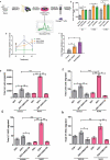
Figure 1.
Figure 1.
Enhanced CXCR4 receptor expression on NaHS treated BMMNCs and chemotaxis of HSPCs toward SDF-1α. (a) Representative experimental setup for evaluating CXCR4 expression and other parameters. Total BMMNCs were isolated and treated with either vehicle or NaHS for 20 min, washed and cultured in IMDM+10% HI-FBS for 24 h. (b) mRNA amplification plots of CXCR4 receptor on BMMNCs. Relative mRNA expression was calculated using Livak method and β-Actin as a house keeping gene. Time course plot for CXCR4 expression after 12, 24 and 36 h of treatment with vehicle or NaHS to optimize the time point for maximum CXCR4 expression after treatment. (c) Changes in the CXCR4 expression in vehicle/NaHS (100 and 300 μM) treated BMMNCs after 24 h using real time PCR. Data are expressed as mean ± SEM fold change in expression with respect to control (n = 4) and the comparisons were done using one-way ANOVA. Linneg cells were sorted from freshly isolated BMMNCs, exposed to NaHS/vehicle, washed and cultured in media for 24 h (37ºC, 5%CO2, 95% humidity). Thereafter, the cells were washed and resuspended in IMDM+0.5%BSA and were allowed to migrate for 4 h. The number of different subsets of HSPCs migrated to the bottom chamber in the presence or absence of SDF-1α were quantified using flow cytometry; (d)Total Linneg cells migrated (e), Migration of gated LSK population (f), Migration of LT-HSCs (CD34) and (g) total ST-HSCs (CD34+) migrated. Data are expressed as mean ± SEM total number of cells migrated. (n = 3, ns = non-significant *p ≤ 0.05, **p ≤ 0.01 and ***p ≤ 0.001) and analyzed using one-way ANOVA
Figure 2.
Figure 2.
H2S enhances the homing potential of HSPCs. (a) Graphical representation of experimental setup for evaluating homing of different subsets of HSPCs. 3 × 106 murine BMMNCs were treated with vehicle/NaHS, labeled with CFSE dye, washed and transplanted into lethally irradiated host mice. After 16 hrs, the host mice were sacrificed, BMMNCs isolated, lineage depleted and stained with surface markers and subsequently analyzed through flow cytometry. (b) This panel shows an increase in the homed CFSE+ LSK cells after NaHS treatment (n = 4 mice/group). (c) LSK population was analyzed further by staining with CD34 surface marker and CFSE+ LSK cells which were CD34+ (ST-HSCs) did not show a significant increase in the homed cells after treatment whereas, (d) a significant increase in the number of CD34 (LT-HSCs) homed events were observed after NaHS treatment. Data are expressed as mean ± SEM for n = 4 mice, each assayed individually
Figure 3.
Figure 3.
H2S enhances proliferation of HSPCs and differentiation of BMMNCs in-vitro. (a) BMMNCs were pulsed with NaHS/vehicle, washed and cultured in Methocult (3X10⁴/plate) for 14 days in a CO2 incubator at 37ºC. On the 14th day, the colonies were identified as shown and scored as (b) mean ± SEM number of CFUs/3X104 cells. Treated cells displayed a significant increase in the CFU-GM and CFU-GEMM. The comparisons were made using student’s t-test. (n = 3, each assayed individually). (c) BMMNCs were treated with NaHS/vehicle, washed and plated in α-MEM+10% HI-FBS for 24 h. Thereafter, the cells were sorted, stained, fixed and permeabilized. Finally, the cells were stained with cell proliferation marker and the proportion of Ki67+ linneg and LSK cells were analyzed through flow cytometry. (d) The mean fluorescence intensity for Ki67 was also noted. Data are expressed as mean ± SEM and analyzed through two-way ANOVA. (e) Similarly, the treated cells were incubated for 6 and 24 h, thereafter sorted stained and fixed and the proportion of LSK population in cell cycle was quantified using 7-AAD viability dye through flow cytometry and represented as fold change in the S+ G2M phase after NaHS exposure. Data are expressed as mean ± SEM and analyzed through two-way ANOVA. (n = 3,4 biological replicates, ns = non-significant *p ≤ 0.05, **p ≤ 0.01 and ***p ≤ 0.001)
Figure 4.
Figure 4.
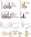
Modulation of intracellular calcium and mitochondrial activity by H2S and the effect of NaHS mediated Calcium influx on CXCR4 expression. (a) Graphs depict the MFI of Fluo-3AM indicative of intracellular calcium measured in LSK cells. The experiment was done in PBS with or without CaCl2 in the presence or absence of NaHS and Nifedipine (NF, L-type calcium channel blocker). (b) intracellular calcium levels before and after NaHS/NF treatment in LSK population when the cells were incubated in the IMDM media (containing pre-added CaCl2, phenol red free) (c) the effect of H2S on intracellular calcium levels in HEK cell line in the presence and absence of CaCl2 indicating the effect of H2S not only in LSK fraction of mice but in the other cells also. (d) Fold change in CXCR4 expression in BMMNCs in vehicle/NaHS/BAPTA-AM treated cells demonstrating the role of calcium in NaHS mediated increase in CXCR4 expression. (e) Line graphs indicating the gradual rise in MFI of Rh123, a mitochondrial membrane potential dye after 6,12 and 24 h in NaHS/vehicle treated cells (LSK fraction). (f) mitochondrial mass in LSK proportion using Mitotraker green (g) Mitochondrial superoxide level in vehicle/NaHS treated LSK population using Mitosox red indicator at 3 different time points. (h) summary of the proposed mechanism. Data are expressed as fold change in MFI (wrt control) for n = 3 and all the statistical comparisons were done using One-way ANOVA and Bonferroni posttest
Figure 5.
Figure 5.
Effect of H2S treatment on apoptosis in LSK population and HEK cell viability. (a) Representative gating strategy followed for detecting apoptosis in LSK population after 24 hrs in NaHS/vehicle treated cells using AnnexinV-FITC and 7-AAD. (b) The proportion of LSK population in the quadrant 1 i.e. Annexin V+, indicating early apoptosis which was non-significant after treatment. (c) The percentage of 7-AAD positive cells (quadrant 4) i.e. dead cells, non-significant after treatment. (d) The proportion of cells that are both positive (AnnexinV+7-AAD+), demonstrating late apoptosis 24 h after treatment (vehicle/NaHS). Data are expressed as mean ± SEM for n = 4 and the comparisons were done using Student’s t-test. (e) Toxicity assessment of NaHS on HEK cells using MTT assay. HEK cells were treated with vehicle/NaHS (100–1000 μM) for 24 h, washed and incubated with MTT for 4 h. Thereafter, the formazan crystals were dissolved in DMSO and the absorbance was read and the data are expressed as mean ± SEM percentage viability for n = 6. The data was analyzed using one-way ANOVA and Bonferroni posttest
Figure 6.
Figure 6.
Hematology profiling of irradiated recipient mice after transplantation. Whole Bone marrow cells were isolated in sterile conditions, RBC lysis was done, washed and resuspended in sterile 1X PBS. BMMNCs were enumerated, NaHS/vehicle treated and transplanted into acutely irradiated recipient mice after 24 h. Changes in the number of (a) total white blood cells (WBC) (b) lymphocytes (c) granulocytes (d) RBCs (e) hemoglobin (f) hematocrit percentage and (g) platelets were measured on 21st post irradiation day. Each value is a mean ± SEM (n = 3–4 animals/group) and the comparisons are done using unpaired t-test

Similar articles

Cited by

References

    1. Ballen KK, Gluckman E, Broxmeyer HE.. Umbilical cord blood transplantation: the first 25 years and beyond. Blood. 2013;122(4):491–498. - PMC - PubMed
    1. Ratajczak MZ, Suszynska M. Emerging strategies to enhance homing and engraftment of hematopoietic stem cells. Stem Cell Rev Rep. 2016;12(1):121–128. - PMC - PubMed
    1. Liu X, Wang J, Ji X, et al. Preconditioning of bone marrow mesenchymal stem cells by prolyl hydroxylase inhibition enhances cell survival and angiogenesis in vitro and after transplantation into the ischemic heart of rats. Sten Cell Res Ther. 2014;5(111):1–12. - PMC - PubMed
    1. Hu X, Yu SP, Fraser JL, et al. Transplantation of hypoxia-preconditioned mesenchymal stem cells improves infarcted heart function via enhanced survival of implanted cells and angiogenesis. Cardiopulm Support Physiol. 2008;135(4):799–808. - PubMed
    1. Mantel CR, O’Leary HA, Chitteti BR, et al. Enhancing hematopoietic stem cell transplantation efficacy by mitigating oxygen shock. Cell. 2015;161(7):1553–1565. - PMC - PubMed

MeSH terms