

A Tissue Digestion Protocol for Measuring Sarcoptes scabiei (Astigmata: Sarcoptidae) Density in Skin Biopsies
- PMID: 33135750
- PMCID: PMC7604834
- DOI: 10.1093/jisesa/ieaa105
A Tissue Digestion Protocol for Measuring Sarcoptes scabiei (Astigmata: Sarcoptidae) Density in Skin Biopsies
Abstract
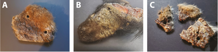
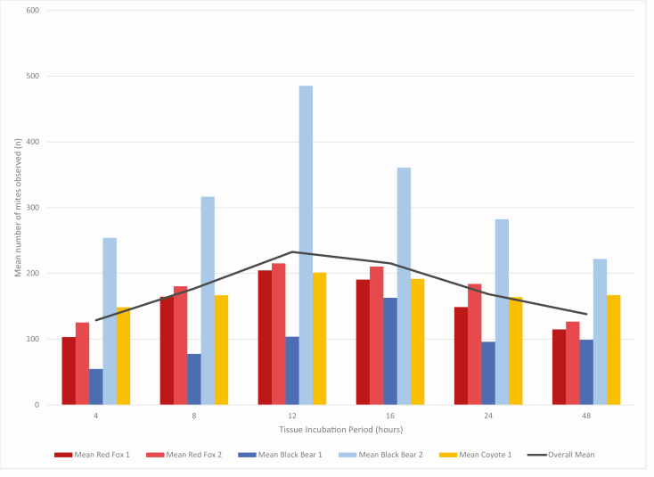
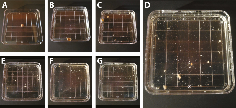
Sarcoptic mange is a parasitic skin disease caused by the burrowing mite Sarcoptes scabiei that affects a diversity of mammals, including humans, worldwide. In North America, the most commonly affected wildlife includes wild canids, such as coyotes and red foxes, and more recently American black bears in the Mid-Atlantic and Northeast United States. Currently, surveillance for sarcoptic mange in wildlife is syndromic, relying on detection of clinical signs and lesions, such as alopecia and crusting of skin. When possible, skin scrapes are used to identify the causative mite. While skin scrapes are a valuable diagnostic tool to identify mites, this approach has significant limitations when used for quantification of mite burden. To further investigate mite burden in cases of sarcoptic mange, 6-mm punch biopsies were collected from affected skin of red foxes (Vulpes vulpes Linnaeus [Carnivora: Canidae]), a species historically affected by sarcoptic mange, frequently with high mite burdens and severe skin disease, and validated on skin tissue from mange-affected American black bears (Ursus americanus Pallas [Carnivora: Ursidae]) and coyotes (Canis latrans Say [Carnivora: Canidae]). Biopsies were digested by incubating the tissue in potassium hydroxide (KOH) at 55°C. The greatest tissue clearance and lowest mite degradation resulted after 12 h of tissue digestion. The purpose of this manuscript is to describe a methodology for host tissue digestion and mite quantification in cases of sarcoptic mange. This method will provide a valuable surveillance and research tool to better understand sarcoptic mange in wild and domestic animals, with applications to a diversity of other ectoparasitic diseases.
Keywords: Sarcoptes scabiei; canid; mange; tissue digestion; wildlife.
© The Author(s) 2020. Published by Oxford University Press on behalf of Entomological Society of America.
Figures
Similar articles
-
Of microbes and mange: consistent changes in the skin microbiome of three canid species infected with Sarcoptes scabiei mites.Parasit Vectors. 2019 Oct 16;12(1):488. doi: 10.1186/s13071-019-3724-0. Parasit Vectors. 2019. PMID: 31619277 Free PMC article.
-
Genetic Characterization of Sarcoptes scabiei from Black Bears (Ursus americanus) and Other Hosts in the Eastern United States.J Parasitol. 2017 Oct;103(5):593-597. doi: 10.1645/17-26. Epub 2017 Jun 22. J Parasitol. 2017. PMID: 28639466
-
ASSAYS FOR DETECTION AND IDENTIFICATION OF THE CAUSATIVE AGENT OF MANGE IN FREE-RANGING BLACK BEARS ( URSUS AMERICANUS).J Wildl Dis. 2018 Jul;54(3):471-479. doi: 10.7589/2017-06-148. Epub 2018 Mar 2. J Wildl Dis. 2018. PMID: 29498895
-
A review of sarcoptic mange in North American wildlife.Int J Parasitol Parasites Wildl. 2019 Jun 13;9:285-297. doi: 10.1016/j.ijppaw.2019.06.003. eCollection 2019 Aug. Int J Parasitol Parasites Wildl. 2019. PMID: 31304085 Free PMC article. Review.
-
Spatiotemporal spread of sarcoptic mange in the red fox (Vulpes vulpes) in Switzerland over more than 60 years: lessons learnt from comparative analysis of multiple surveillance tools.Parasit Vectors. 2019 Nov 5;12(1):521. doi: 10.1186/s13071-019-3762-7. Parasit Vectors. 2019. PMID: 31690337 Free PMC article. Review.
Cited by
-
Special Collection: Protocols in Medical and Veterinary Entomology.J Insect Sci. 2020 Nov 1;20(6):1. doi: 10.1093/jisesa/ieaa122. J Insect Sci. 2020. PMID: 33135744 Free PMC article. No abstract available.
-
The use of haystacks by wolves may facilitate the transmission of sarcoptic mange.Sci Rep. 2024 Nov 16;14(1):28304. doi: 10.1038/s41598-024-78026-w. Sci Rep. 2024. PMID: 39550401 Free PMC article.
References
-
- Alasaad S, Granados J E, Cano-Manuel F J, Meana A, Zhu X Q, and Pérez J M. . 2008. Epidemiology of fasciolosis affecting Iberian ibex (Capra pyrenaica) in southern Spain. Parasitol. Res. 102: 751–755. - PubMed
-
- Alasaad S, Rossi L, Soriguer R C, Rambozzi L, Soglia D, Pérez J M, and Zhu X Q. . 2009. Sarcoptes mite from collection to DNA extraction: the lost realm of the neglected parasite. Parasitol. Res. 104: 723–732. - PubMed
-
- Bornstein S., T. Mörner, and W. M. Samuel. 2001. Sarcoptes scabiei and sarcoptic mange, pp. 107-119. In W. M. Samuel, M. J. Pybus, and A. A. Kocan (eds.), Parasitic diseases of wild mammals, 2nd ed. Iowa State University Press, Ames, IA.
Publication types
MeSH terms
LinkOut - more resources
Full Text Sources
Medical
