. 2021 Jan 8;128(1):42-58.
doi: 10.1161/CIRCRESAHA.120.317372. Epub 2020 Nov 2.

Live Imaging of Intracranial Lymphatics in the Zebrafish

Affiliations

Live Imaging of Intracranial Lymphatics in the Zebrafish

Daniel Castranova et al. Circ Res. .

Abstract

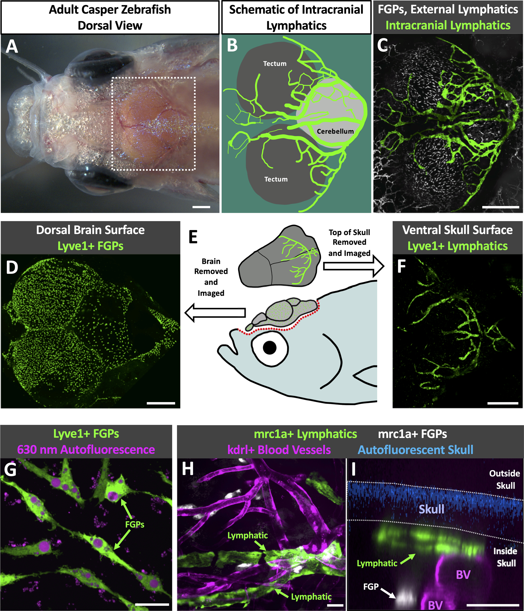
Rationale: The recent discovery of meningeal lymphatics in mammals is reshaping our understanding of fluid homeostasis and cellular waste management in the brain, but visualization and experimental analysis of these vessels is challenging in mammals. Although the optical clarity and experimental advantages of zebrafish have made this an essential model organism for studying lymphatic development, the existence of meningeal lymphatics has not yet been reported in this species.

Objective: Examine the intracranial space of larval, juvenile, and adult zebrafish to determine whether and where intracranial lymphatic vessels are present.

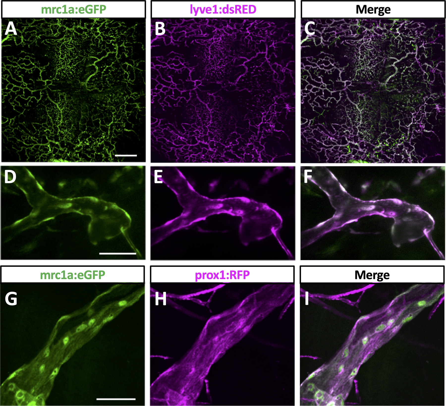
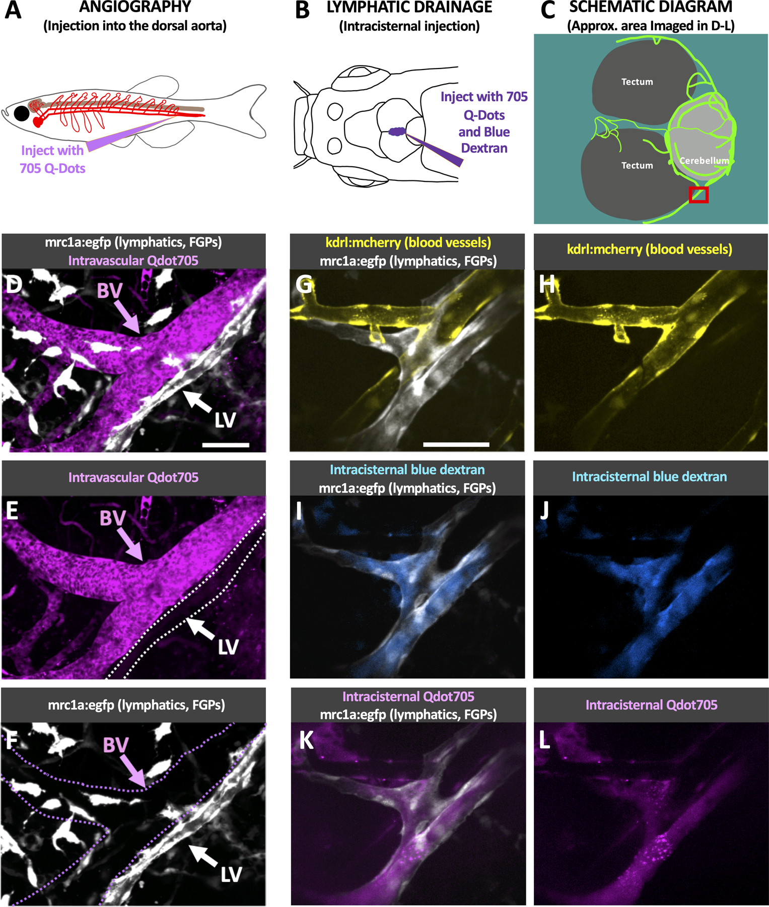
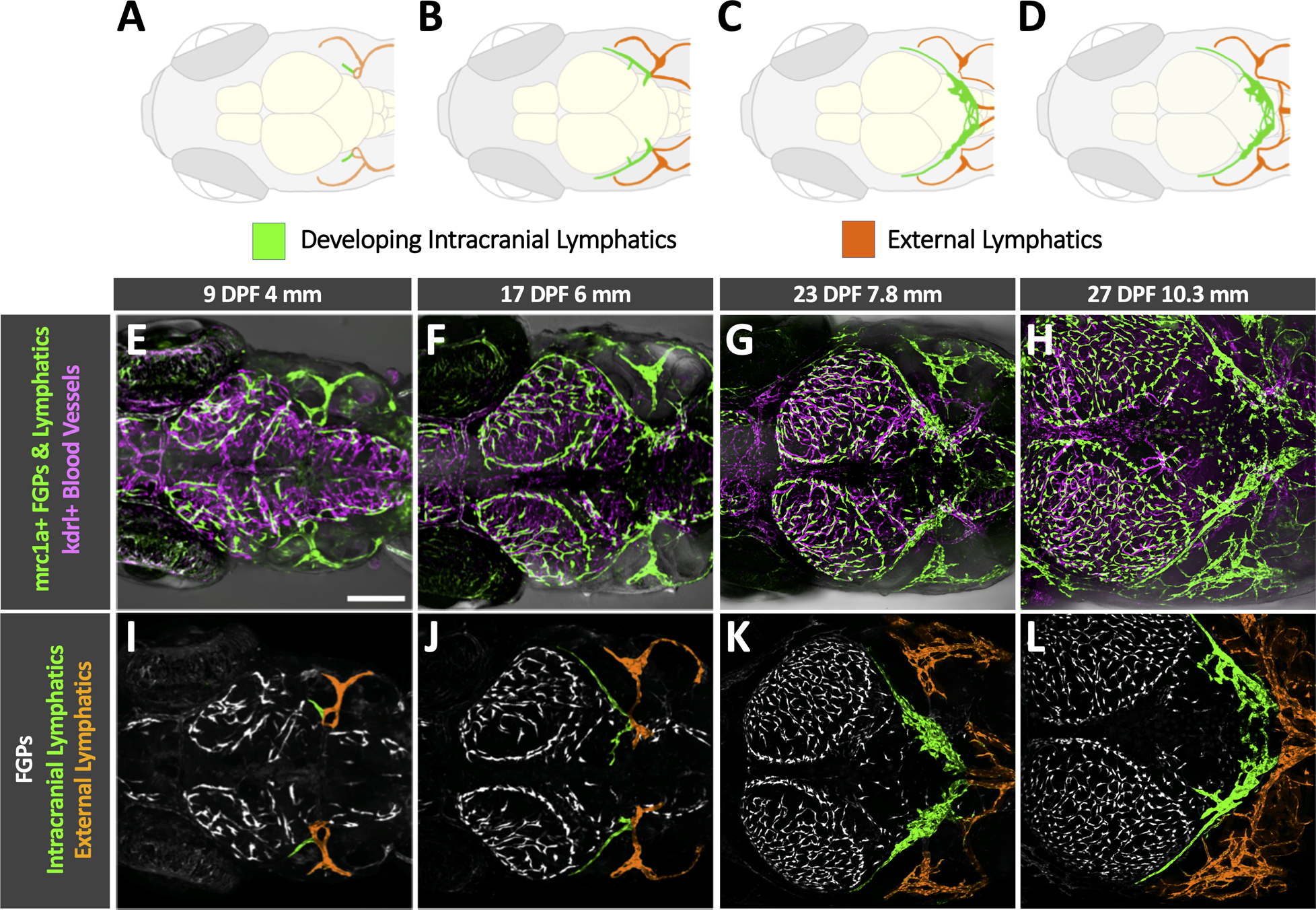
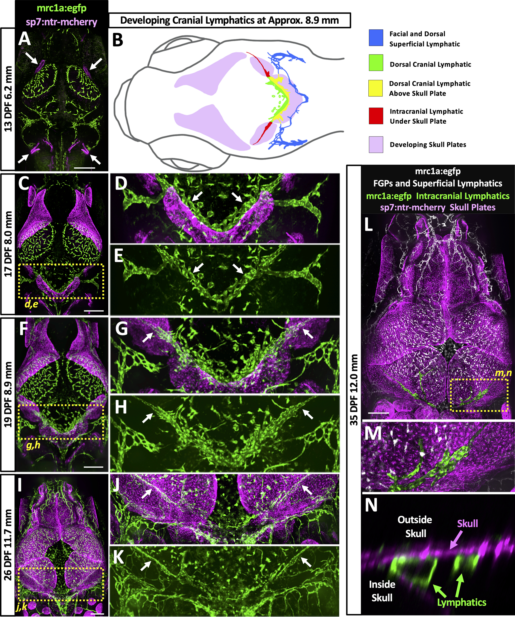
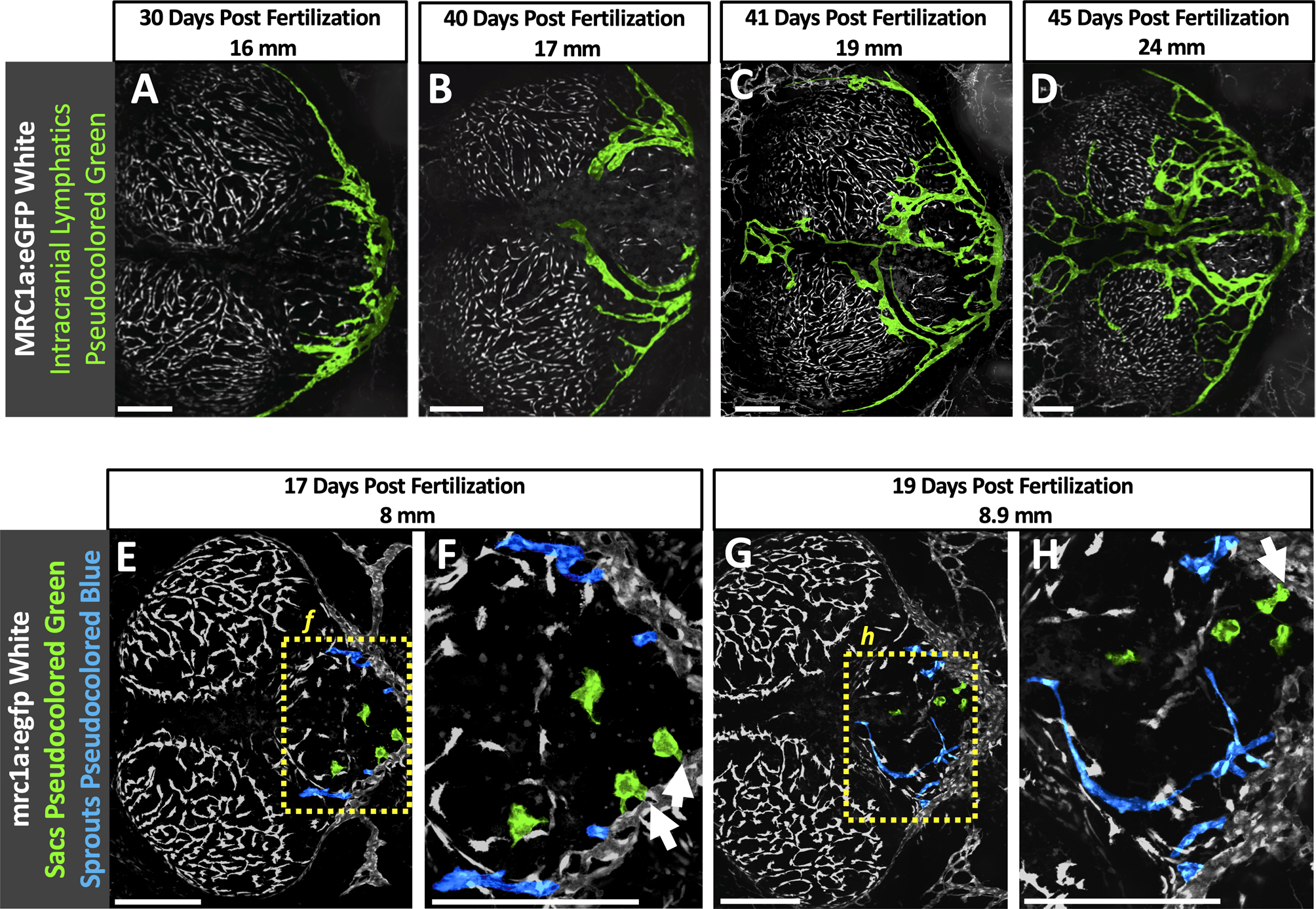
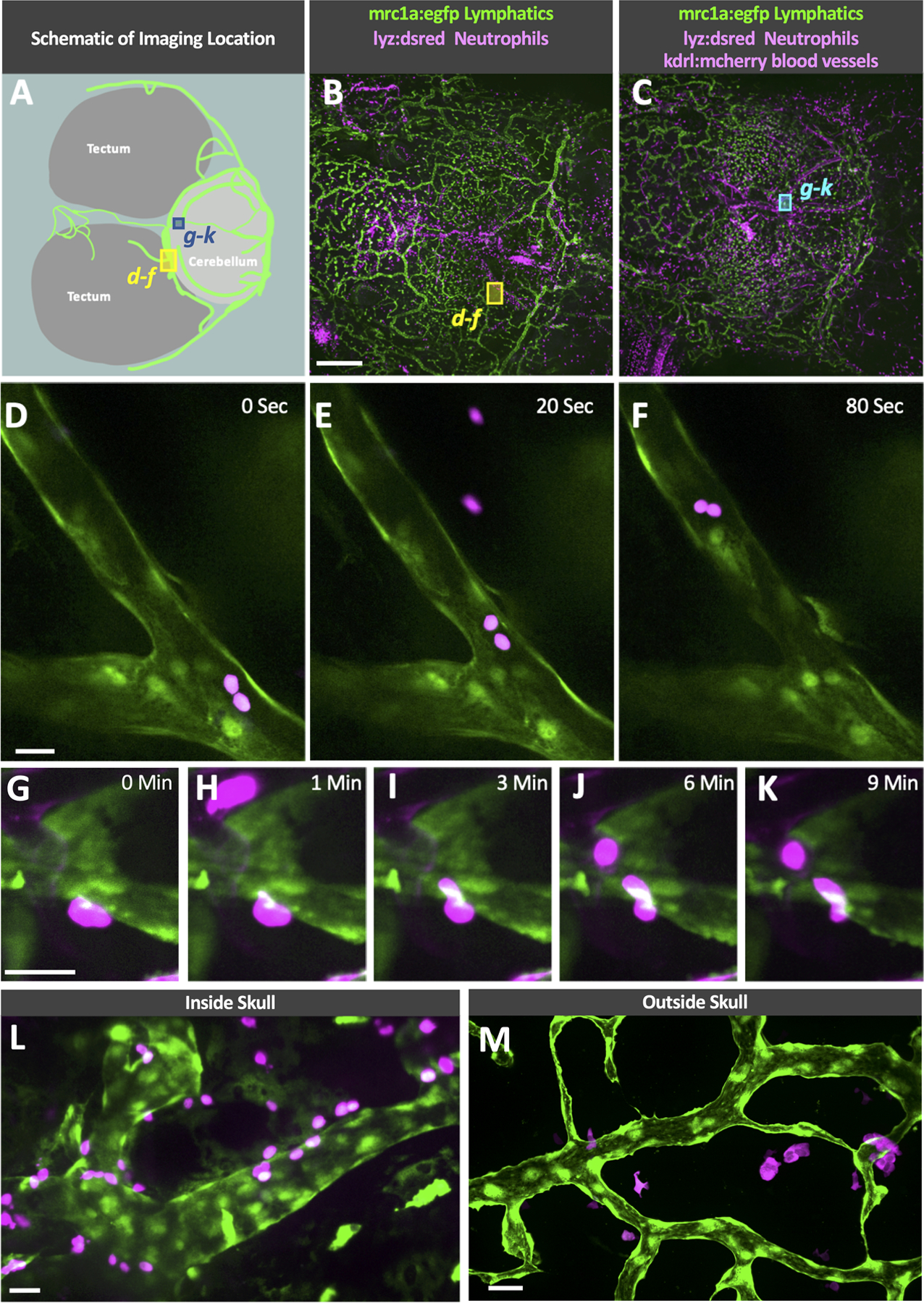
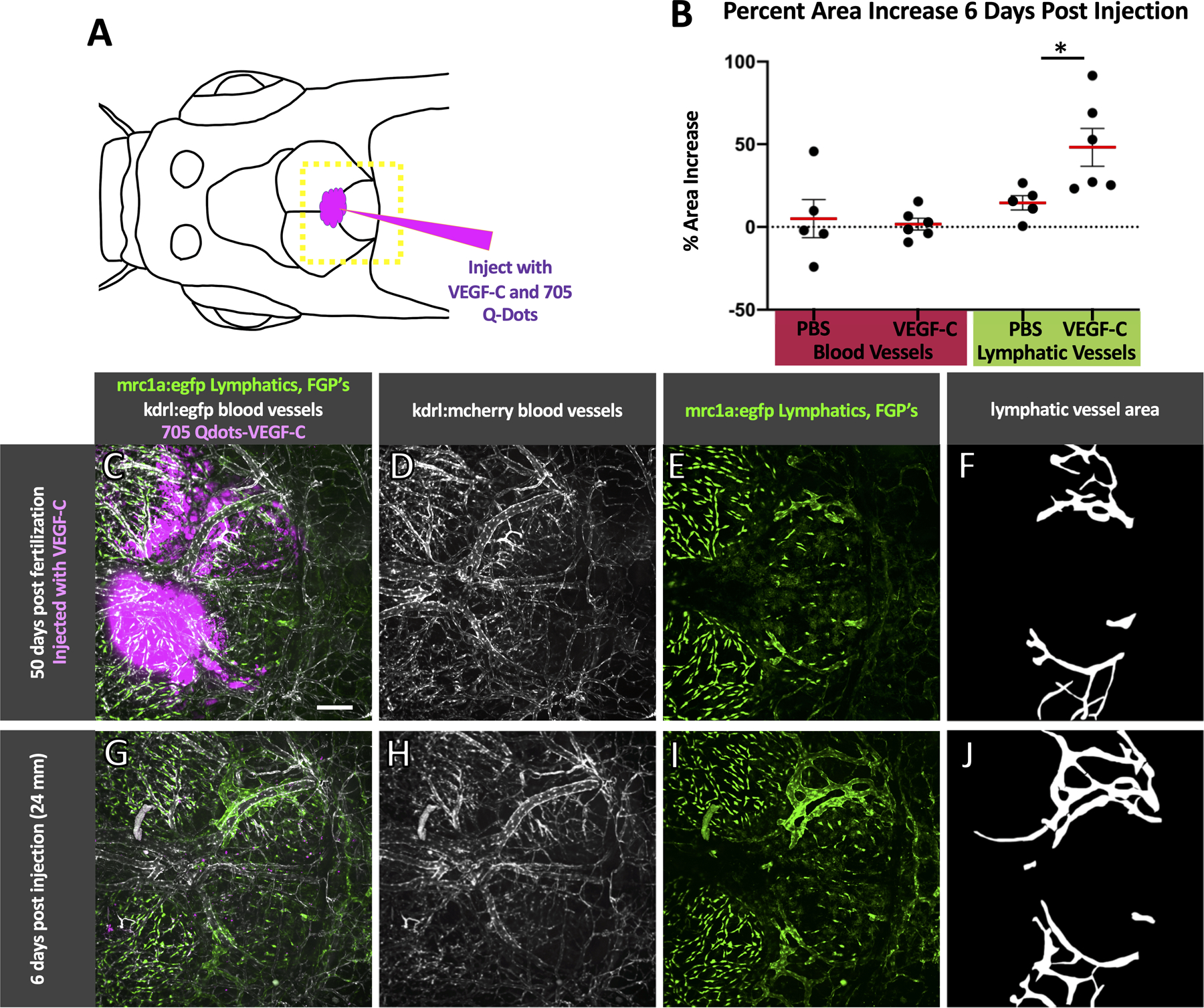
Methods and results: Using high-resolution optical imaging of the meninges in living animals, we show that zebrafish possess a meningeal lymphatic network comparable to that found in mammals. We confirm that this network is separate from the blood vascular network and that it drains interstitial fluid from the brain. We document the developmental origins and growth of these vessels into a distinct network separated from the external lymphatics. Finally, we show that these vessels contain immune cells and perform live imaging of immune cell trafficking and transmigration in meningeal lymphatics.

Conclusions: This discovery establishes the zebrafish as a important new model for experimental analysis of meningeal lymphatic development and opens up new avenues for probing meningeal lymphatic function in health and disease.

Keywords: brain; developmental biology; meninges; zebrafish.


Figures

Fig. 1
Fig. 1. Intracranial lymphatic vessels in the adult zebrafish
A. Dorsal view image of the head of an adult casper (roy, nacre double mutant) zebrafish. B. Schematic diagram of the boxed region in panel A, showing the optic tecta and cerebellum with a typical network of intracranial lymphatic vessels in a young adult zebrafish. C. Confocal image of the dorsal head of a Tg(mrc1a:egfp)y251, casper adult zebrafish, with mrc1a+FGPs and superficial lymphatics in grey and intracranial meningeal lymphatics pseudocolored green. D. Confocal image of the dorsal surface of a dissected brain from a Tg(−5.2lyve1b:DsRed)nz101, casper adult zebrafish, with lyve1+ FGPs but no lymphatic vessels. E. Schematic diagram of dissection of an adult zebrafish head for imaging the dorsal surface of the brain and the ventral surface of the skull. F. Confocal image of the ventral (inner) surface of a dissected brain from a Tg(−5.2lyve1b:DsRed)nz101, casper adult zebrafish, with lyve1+ lymphatic vessels but no FGPs. G. Higher magnification confocal image of the dorsal surface of a dissected brain removed from a Tg(−5.2lyve1b:DsRed)nz101, casper adult zebrafish, showing individual separated lyve1+ FGPs with characteristic large autofluorescent internal vacuoles. H. Higher magnification confocal image of the outer layer of the brain imaged through the skull of a living casper adult double transgenic zebrafish Tg(mrc1a:egfp)y251, Tg(kdrl:mcherry) y206, showing mrc1a+ lymphatic vessels and kdrl+ blood vessels. I. Higher magnification confocal image of the dorsal head of a Tg(mrc1a:egfp)y251, Tg(kdrl:mcherry) y206, casper adult zebrafish. This orthogonal view shows a cross section of an mrc1a+ lymphatic vessel immediately below the blue autofluorescent skull and an mrc1a+ FGP in a deeper layer immediately adjacent to a kdrl+ blood vessel. Unless otherwise noted all images are dorsal views, rostral to the left. Scale bars: 500 um (A,C,E,F) 25 um (G,H,I). (BV- blood vessels, roy- roy orbison, mrc1a-mannose receptor C, type 1a, eGFP- green fluorescent protein, Tg- transgenic, lyve1b- lymphatic vessel endothelial hyaluronic receptor 1b, FGP- fluorescent granular perithelial cells, kdrl-kinase insert domain receptor like)
Fig. 2
Fig. 2. Molecular validation of zebrafish intracranial lymphatics
A-C. Confocal imaging overview intracranial and extracranial lymphatic vessel in the dorsal head of an adult casper, Tg(mrc1a:egfp)y251, Tg(−5.2lyve1b:DsRed)nz101 double transgenic zebrafish, showing mrc1a:egfp (A), lyve1b:dsred (B), and combined mrc1a:egfp and lyve1b:dsred (C) images. D-F. Confocal imaging of an intracranial lymphatic vessel in the dorsal head of an adult casper, Tg(mrc1a:egfp)y251, Tg(−5.2lyve1b:DsRed)nz101 double transgenic zebrafish, showing mrc1a:egfp (D), lyve1b:dsred (E), and combined mrc1a:egfp and lyve1b:dsred (F) images. D-F. Confocal imaging of an intracranial lymphatic vessel in the dorsal head of an adult casper, Tg(mrc1a:egfp)y251, Tg(prox1aBAC:KalTA4–4xUAS-E1b:uncTagRFP)nim5 double transgenic zebrafish, showing mrc1a:egfp (D), prox1:rfp (E), and combined mrc1a:egfp and prox1:rfp (F) images. Scale bars: 500 um (A), 50 um (D,G). (Tg-transgenic, mrc1a- mannose receptor C, type 1a, egfp-green fluorescent protein, lyve1b- lymphatic vessel endothelial hyaluronic receptor 1b, prox1- prospero homeobox 1a)
Fig. 3
Fig. 3. Functional validation of zebrafish intracranial lymphatics
A. Schematic diagram of the intravascular injection procedure for filling blood vessels. B. Schematic diagram of the intracisternal injection procedure for filling intracranial lymphatics. C. Schematic diagram of the optic tecta, cerebellum, and intracranial lymphatics in the dorsal head of an adult casper mutant, Tg(mrc1a:egfp)y251 zebrafish. The red box notes the approximate area shown in the high-magnification images in panels D-L. D-F. Confocal images of intracranial blood vessels (BV) and lymphatic vessels (LV) in the dorsal head of a Tg(mrc1a:egfp)y251 adult fish injected intravascularly with Qdot705, showing mrc1a:egfp+ lymphatics and FGPs + Qdot705 (D), Qdot705 alone (E), or mrc1a:egfp+ alone (F). G-L. Confocal images of intracranial blood vessels (BV) and lymphatic vessels (LV) in the dorsal head of a Tg(mrc1a:egfp)y251, Tg(kdrl:mcherry) y206 adult fish injected intracranially with Qdot705 and blue dextran, showing mrc1a:egfp+ lymphatics and kdrl:mcherry+ blood vessels (G), kdrl:mcherry+ blood vessels (H), mrc1a:egfp+ lymphatics and blue dextran (I), blue dextran (J), mrc1a:egfp+ lymphatics and Qdot705 (K), Qdot705 (L). Scale bars: 50 um. See Online Video III for 3D renderings and real-time imaging of the vessels shown in panels G-L. (mrc1a- mannose receptor C, type 1a, FGPs- fluorescent granular perithelial cells, kdrl- kinase insert domain receptor like, BV- blood vessels, LV-lymphatic vessels)
Fig. 4
Fig. 4. Initial development of intracranial lymphatics in larval zebrafish
A-D. Schematic diagrams showing brain structures, developing intracranial lymphatics (green), and external lymphatics (orange) in the dorsal heads of 9 dpf/4 mm (A), 17 dpf/6 mm (B), 23 dpf/7.8 mm (C), and 27 dpf/10.3 mm (D) zebrafish. Each diagram illustrates features in the corresponding confocal micrograph below (panels E-H). OT, optic tecta; C, cerebellum. E-H. Confocal images of kdrl:mcherry positive blood vessels (magenta) and mrc1a:egfp positive lymphatic vessels and FGPs (green) in the dorsal heads of 9 dpf/4 mm (E), 17 dpf/6 mm (F), 23 dpf/7.8 mm (G), and 27 dpf/10.3 mm (H) Tg(mrc1a:egfp)y251, kdrl:mcherry) double-transgenic zebrafish. I-L. Colored versions of the same confocal image fields shown in panels E-H with only the mrc1a:egfp fluorescence channel visible, revealing FGPs (white, uncolored), external lymphatics (colored orange), and developing intracranial lymphatics (colored green). Scale bar: 250 um. a See Online Video IV for 3D renderings of the same image stacks shown in this figure. Intracranial and extracranial vessels were distinguished by careful examination of Z-stacks and by comparison to similar staged transgenic or stained animals marking developing bone (see Figure 5 for examples). Dpf- days post fertilization, mm- millimeters, mrc1a- mannose receptor C, type 1a, FGPS- fluorescent granular perithelial cells, kdrl-kinase insert domain receptor like)
Fig. 5
Fig. 5. Intracranial lymphatic development and skull formation
A,C,F,I,L. Overview confocal images of mrc1a:egfp+ lymphatic vessels and FGPs (green) and sp7:mcherry+ developing skull plates (magenta) in the dorsal heads of 13 dpf/6.2 mm (A), 17 dpf/8.0 mm (C), 19 dpf/8.9 mm (F), 26 dpf/11.7 mm (I), and 35 dpf/12.0 mm (L) Tg(mrc1a:egfp)y251, Tg(Ola.Sp7:mCherry-Eco.NfsB)pd46 double-transgenic zebrafish. Arrows in panel A note beginning of skull plate formation. Yellow dashed boxes in panels C, F, I, and L note the areas shown in the corresponding higher magnification images in the panels to the right (D,E,G,H,J,K) or below (M,N). B. Schematic diagram corresponding to the confocal image of a 19 dpf/8.9 mm Tg(mrc1a:egfp)y251, Tg(Ola.Sp7:mCherry-Eco.NfsB)pd46 double-transgenic zebrafish shown in panel F. Diagram shows developing skull plates (pink), superficial lymphatics (blue), still-uncovered intracranial lymphatic (green), portions of intracranial lymphatic network present above edge of growing skull plates (yellow), and portions of intracranial lymphatics already covered by the growing skull plates (red). D,G,J. Higher magnification confocal images of the yellow boxed regions in the panels to the left, showing mrc1a:egfp+ lymphatics (green) and sp7:mcherry+ developing skull plates (magenta). White arrows note lymphatics adjacent to (D) or under (G,J) skull plates. E,H,K. Higher magnification confocal images of the yellow boxed regions in the panels to the left, showing only mrc1a:egfp+ lymphatics (green). White arrows note lymphatics adjacent to (E) or under (H,K) skull plates. M. Higher magnification confocal image of the yellow boxed region in panel L, showing mrc1a:egfp+ meningeal lymphatics (green) below the nearly completed sp7:mcherry+ skull (magenta). N. Higher magnification orthogonal (side) view confocal image of the yellow boxed region in panel L, showing mrc1a:egfp+ meningeal lymphatic tubes (green) inside the skull below the sp7:mcherry+ skull (magenta). Scale bars: 250 um. (dpf- days post fertilization, mm- millimeters, mrc1a-mannose receptor C type 1a, GFP- green fluorescent protein, ntr-nitro-reductase, FGPs- fluorescent granular perithelial cells, sp7- sp7 transcription factor)
Fig. 6
Fig. 6. Later development of intracranial lymphatics in juvenile zebrafish
A-D. Colored confocal images of mrc1a:egfp+ FGPs (white, uncolored) and intracranial lymphatic vessels (colored green) in the dorsal heads of 30 dpf/16 mm (A), 40 dpf/17 mm (B), 41 dpf/19 mm (C), and 45 dpf/24 mm (D) Tg(mrc1a:egfp)y251 transgenic zebrafish. E-H. Colored confocal images of mrc1a:egfp+ FGPs and the primary intracranial lymphatic plexus (white, uncolored), sprouting intracranial lymphatic vessels (colored blue) and lymphatic sacs (colored green) in the dorsal head of a 15 dpf/6.7 mm Tg(mrc1a:egfp)y251 transgenic zebrafish (E), Higher magnification view of the boxed region in panel E, showing sprouting intracranial lymphatics (arrows) (F), and a 17 dpf/8 mm Tg(mrc1a:egfp)y251 transgenic zebrafish (G) with higher magnification view of the boxed region in panel G, showing presumptive lymphatic sacs fusing with the growing lymphatic vessels (arrows) (H). Scale Bars: 250 um. (dpf- days post fertilization, mm-millimeters, mrc1a- mannose receptor C type 1a, GFP- green fluorescent protein)
Fig. 7
Fig. 7. Immune cell trafficking in zebrafish intracranial lymphatics
A. Schematic diagram of the optic tecta, cerebellum, and intracranial lymphatics in the dorsal head of an adult casper, Tg(mrc1a:egfp)y251 zebrafish. The blue and yellow boxes note the approximate area shown in the high-magnification images in panels D-F and G-K. B-C. Confocal overview images series through the skull of a living adult casper, Tg(mrc1a:egfp)y251, Tg(lyz:DsRed2)nz50 double transgenic zebrafish (B) and Tg(mrc1a:egfp)y251, Tg(lyz:DsRed2)nz50, Tg(kdrl:mcherry) y206 tripple transgenic zebrafish showing the imaging locations of D-F (yellow box) and G-K (blue box). D-F. Selected frames from a time-lapse confocal image series taken through the skull of a living adult casper, Tg(mrc1a:egfp)y251, Tg(lyz:DsRed2)nz50 double transgenic zebrafish showing neutrophils trafficking through an intracranial lymphatic vessel (see Online Video VII). G-K. Selected frames from a time-lapse confocal image series taken through the skull of a living adult casper, Tg(mrc1a:egfp)y251, Tg(lyz:DsRed2)nz50, Tg(kdrl:mcherry) y206 triple transgenic zebrafish showing a neutrophil transmigrating into an intracranial lymphatic vessel (see Online Video VIII). L. Image taken through the skull of a living adult casper, Tg(mrc1a:egfp)y251, Tg(lyz:DsRed2)nz50 double transgenic zebrafish showing neutrophils on the inside and outside of an intracranial lymphatic vessel. M. Image taken of superficial lymphatics on the dorsal head of a living adult casper, Tg(mrc1a:egfp)y251, Tg(lyz:DsRed2)nz50 double transgenic zebrafish showing neutrophils interacting with a lymphatic vessel outside of the skull. Scale bars: 500 um (B), 25 um (D, G, L, M). (mrc1a- mannose receptor C type 1a, eGFP- green fluorescent protein, lyz-lysozyme)
Fig. 8
Fig. 8. Injection of VEGF-C stimulates intracranial lymphatic growth in zebrafish
A. Schematic diagram of the intracranial injection of VEGF-C (n = 6) or PBS (n = 5) into 50 dpf casper, Tg(mrc1a:egfp)y251; Tg(kdrl:mcherry) y206 zebrafish (19–25 mm) with the approximate area imaged shown by the dashed yellow box. B. Strip plots with a red line showing the average percent increase in area of blood and lymphatic vessels from the time of injection to six days post injection with error bars showing the standard error of the mean. VEGF-C injected fish showed a significant increase in lymphatic area (Bonferroni-adjusted p = 0.034 Mann-Whitney U test, p < 0.05). C-E. Maximum projection confocal image of the dorsal head of a casper mutant, Tg(mrc1a:egfp)y251; Tg(kdrl:mcherry) y206 double transgenic animal at the time of VEGF-C/705Qdot injection with 705Qdots marking the VEGF-C injection site shown in magenta (C), blood vessels in white (C,D), and FGPs and lymphatics in green (C,E). G-I. The same fish as shown in C-E six days post-injection, showing an increase in lymphatic vessel growth,. 705Qdots shown in magenta (G), blood vessels in white (G,H), and FGPs and lymphatics in green (G,I). F,J. Area selections of lymphatic vessels based on E and I, used for percent area calculations in B. Scale bar: 200 um. (VEGF-C – vascular endothelial growth factor C, dpf- days post fertilization, mm- millimeters, mrc1a- mannose receptor C, type 1a, kdrl-kinase insert domain receptor like, FGPs- fluorescent granular perithelial cells)

Comment in

Similar articles

Cited by

References

    1. Louveau A, Smirnov I, Keyes TJ, Eccles JD, Rouhani SJ, Peske JD, Derecki NC, Castle D, Mandell JW, Lee KS, et al. Structural and functional features of central nervous system lymphatic vessels. Nature. 2015;523:337–341 - PMC - PubMed
    1. Aspelund A, Antila S, Proulx ST, Karlsen TV, Karaman S, Detmar M, Wiig H, Alitalo K. A dural lymphatic vascular system that drains brain interstitial fluid and macromolecules. J Exp Med. 2015;212:991–999 - PMC - PubMed
    1. Ahn JH, Cho H, Kim JH, Kim SH, Ham JS, Park I, Suh SH, Hong SP, Song JH, Hong YK, et al. Meningeal lymphatic vessels at the skull base drain cerebrospinal fluid. Nature. 2019;572:62–66 - PubMed
    1. Antila S, Karaman S, Nurmi H, Airavaara M, Voutilainen MH, Mathivet T, Chilov D, Li Z, Koppinen T, Park JH, et al. Development and plasticity of meningeal lymphatic vessels. J Exp Med. 2017;214:3645–3667 - PMC - PubMed
    1. Louveau A, Herz J, Alme MN, Salvador AF, Dong MQ, Viar KE, Herod SG, Knopp J, Setliff JC, Lupi AL, et al. Cns lymphatic drainage and neuroinflammation are regulated by meningeal lymphatic vasculature. Nat Neurosci. 2018;21:1380–1391 - PMC - PubMed

Publication types

Substances

LinkOut - more resources