. 2020 Nov 2;16(11):e1009185.
doi: 10.1371/journal.pgen.1009185. eCollection 2020 Nov.

Opposing functions of Fng1 and the Rpd3 HDAC complex in H4 acetylation in Fusarium graminearum

Affiliations

Opposing functions of Fng1 and the Rpd3 HDAC complex in H4 acetylation in Fusarium graminearum

Hang Jiang et al. PLoS Genet. .

Abstract

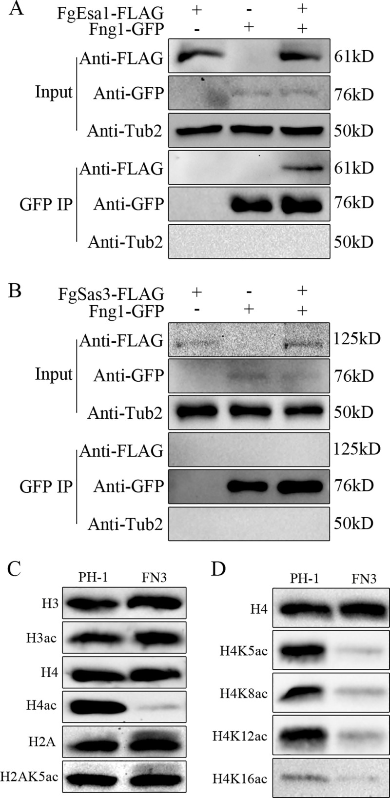
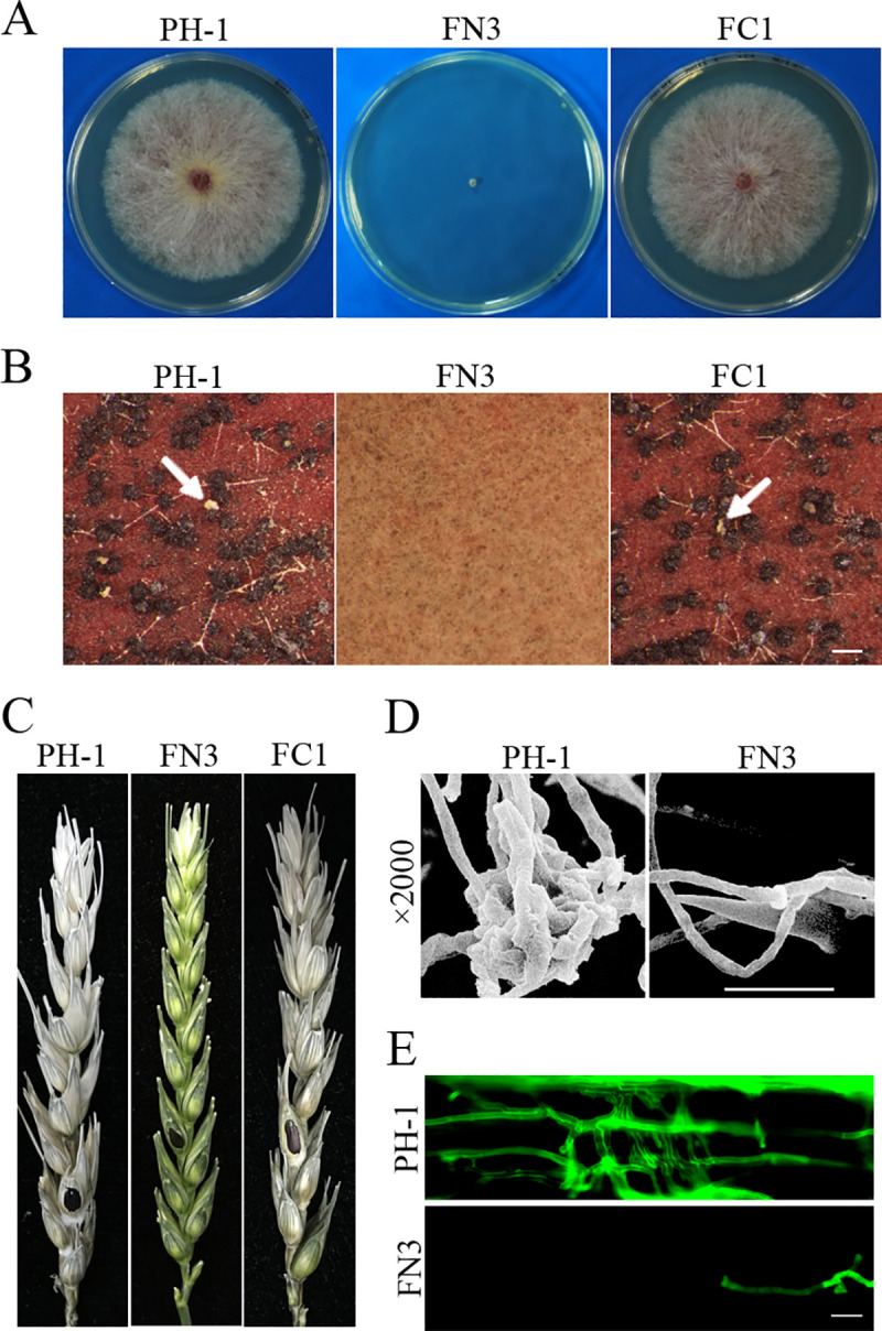
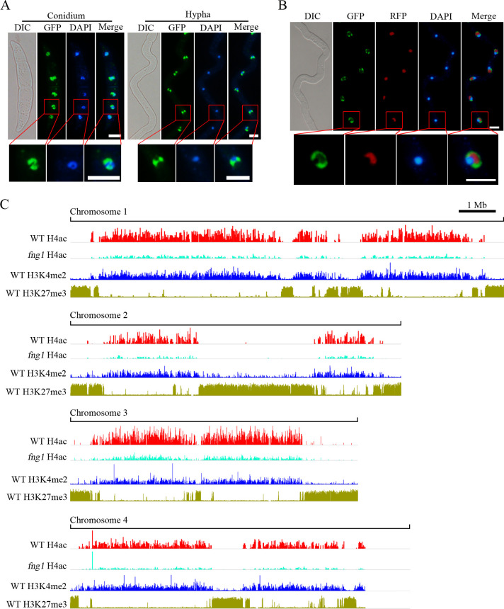
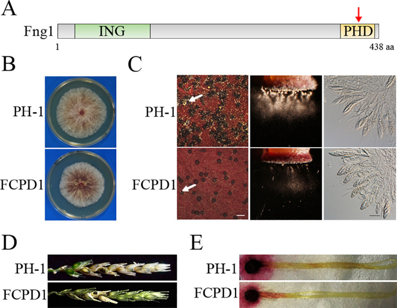
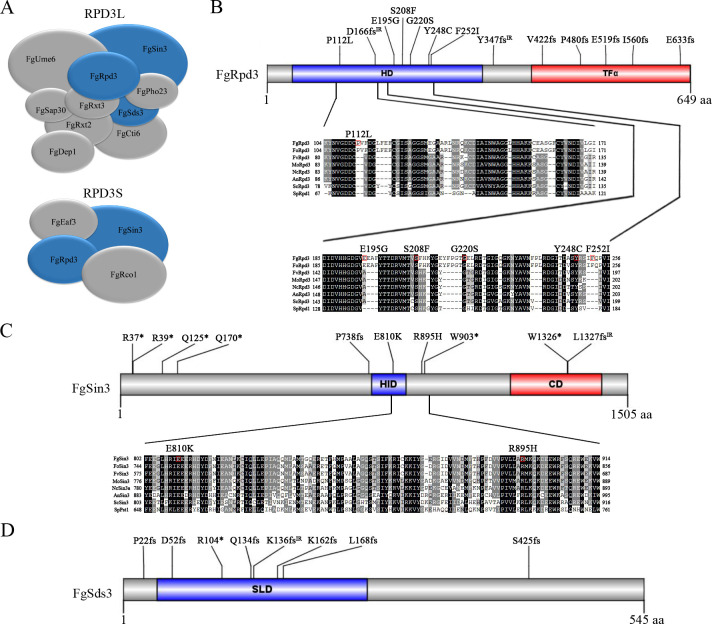
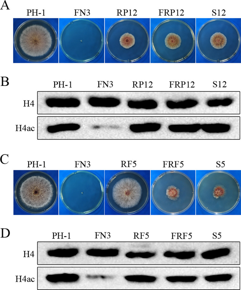
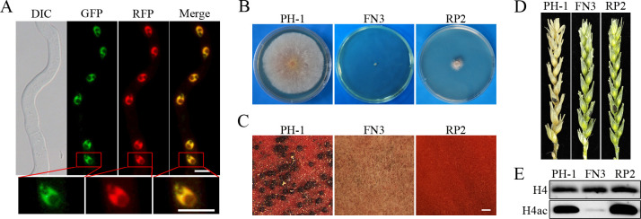
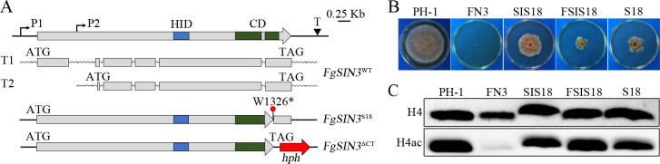
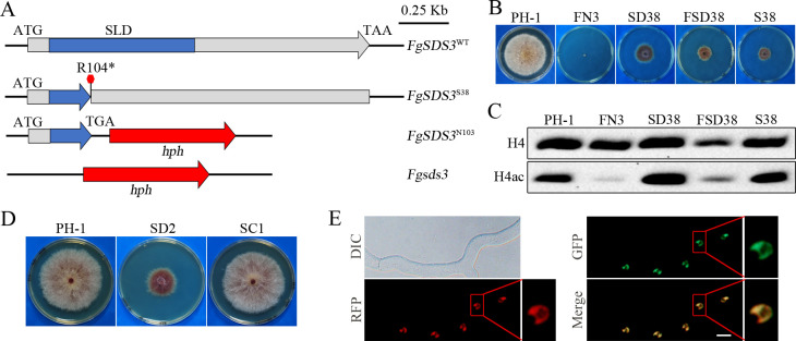
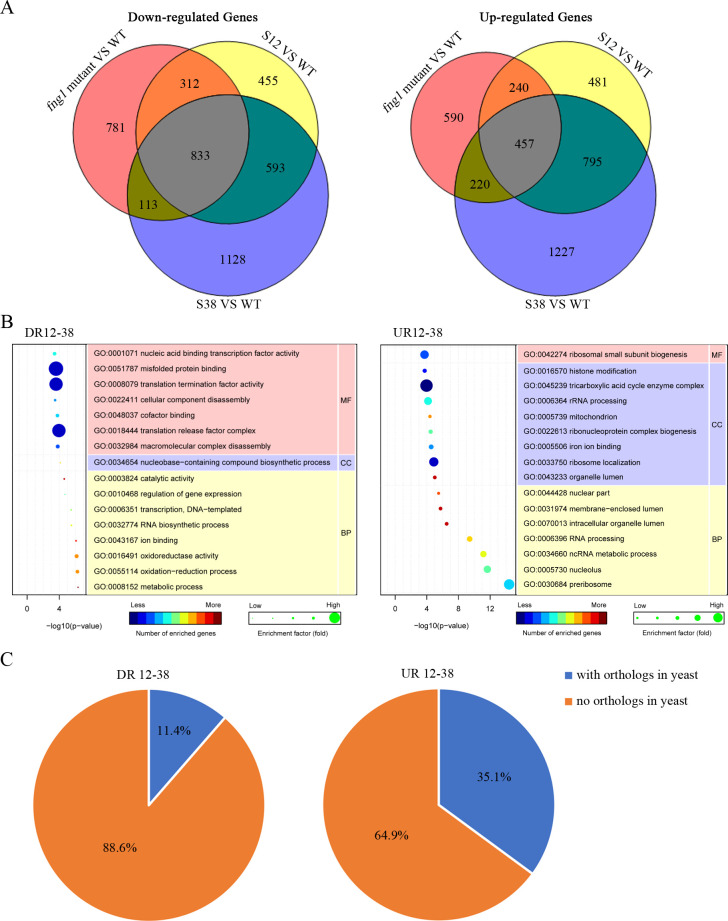
Histone acetylation, balanced by histone acetyltransferase (HAT) and histone deacetylase (HDAC) complexes, affects dynamic transitions of chromatin structure to regulate transcriptional accessibility. However, little is known about the interplay between HAT and HDAC complexes in Fusarium graminearum, a causal agent of Fusarium Head Blight (FHB) that uniquely contains chromosomal regions enriched for house-keeping or infection-related genes. In this study, we identified the ortholog of the human inhibitor of growth (ING1) gene in F. graminearum (FNG1) and found that it specifically interacts with the FgEsa1 HAT of the NuA4 complex. Deletion of FNG1 led to severe growth defects and blocked conidiation, sexual reproduction, DON production, and plant infection. The fng1 mutant was normal in H3 acetylation but significantly reduced in H4 acetylation. A total of 34 spontaneous suppressors of fng1 with faster growth rate were isolated. Most of them were still defective in sexual reproduction and plant infection. Thirty two of them had mutations in orthologs of yeast RPD3, SIN3, and SDS3, three key components of the yeast Rpd3L HDAC complex. Four mutations in these three genes were verified to suppress the defects of fng1 mutant in growth and H4 acetylation. The rest two suppressor strains had a frameshift or nonsense mutation in a glutamine-rich hypothetical protein that may be a novel component of the FgRpd3 HDAC complex in filamentous fungi. FgRpd3, like Fng1, localized in euchromatin. Deletion of FgRPD3 resulted in severe growth defects and elevated H4 acetylation. In contract, the Fgsds3 deletion mutant had only a minor reduction in growth rate but FgSIN3 appeared to be an essential gene. RNA-seq analysis revealed that 48.1% and 54.2% of the genes with altered expression levels in the fng1 mutant were recovered to normal expression levels in two suppressor strains with mutations in FgRPD3 and FgSDS3, respectively. Taken together, our data showed that Fng1 is important for H4 acetylation as a component of the NuA4 complex and functionally related to the FgRpd3 HDAC complex for transcriptional regulation of genes important for growth, conidiation, sexual reproduction, and plant infection in F. graminearum.


Conflict of interest statement

The authors have declared that no competing interests exist.

Figures

Fig 1
Fig 1. Fng1 is associated with FgEsa1 and required for H4 acetylation.
(A). Co-immunoprecipitation (co-IP) assays for the interaction between Fng1 and FgEsa1. Western blots of total protein isolated from transformants expressing the FNG1-GFP and/or FgESA1-3×FLAG (input) and proteins eluted from anti-GFP affinity beads (GFP IP) were detected with anti-FLAG and anti-GFP antibodies. (B). Co-IP assays for the interaction between Fng1 and FgSas3. Western blots of total protein isolated from transformants expressing the FNG1-GFP and/or FgSAS3-3×FLAG (input) and proteins eluted from anti-GFP affinity beads (GFP IP) were detected with anti-FLAG and anti-GFP antibodies. (C). Western blots of total proteins isolated from the wild type (PH-1) and fng1 mutant (FN3) were detected with the anti-H4ac, anti-H3ac, and H2AK5ac antibodies. Detection with the anti-H4, anti-H3, or anti-H2A antibodies was used as loading control. (D). Western blots of total proteins isolated from PH-1 and the fng1 mutant were detected with the antibodies specific for H4K5ac, H4K8ac, H4K12ac, and H4K16ac.
Fig 2
Fig 2. Assays for the function of Fng1 in growth, reproduction, and pathogenesis.
(A). Three-day-old PDA cultures of the wild type (PH-1), fng1 mutant (FN3), and fng1/FNG1 transformant (FC1). (B). Perithecia from mating cultures of the same set of strains were examined at 8 dpf. Ascospore cirrhi are marked with arrows. Bar, 1 mm. (C). Wheat heads inoculated with the indicated strains were examined for head blight symptoms at 14 days post-inoculation (dpi). Black dots mark the inoculated spikelets. (D). Infection cushions formed by PH-1 and fng1 mutant FN3 on wheat lemma were examined by SEM under ×2,000 amplification at 2 dpi. Bar, 20 μm. (E). Wheat coleoptiles infected with PH-1 and FN3 were examined for invasive hyphae at 3 dpi after staining with Alexa Fluor 488. Bar, 20 μm.
Fig 3
Fig 3. Subcellular localization of Fng1-GFP and chromosomal regions enriched for Fng1-dependent H4ac.
(A). Conidia and germlings of the FNG1-GFP transformant were stained with DAPI and examined by DIC and epifluorescence microscopy. Bar, 5 μm. (B). Germlings of the FNG1-GFP FgNOP1-RFP transformant (FCNR1) were stained with DAPI and examined by DIC and epifluorescence microscopy. Bar, 5 μm. The lower panels are close-up view of the indicated nuclei. (C). The distribution of DNA sequences co-immunoprecipitated with the anti-H4ac antibody in the wild-type strain PH-1 and fng1 mutant (chromosomes 1–4) was compared with chromosomal regions enriched for H3K27me3 and H3K4me2 in the genome of F. graminearum.
Fig 4
Fig 4. The effects of PHD domain deletion on FNG1 functions.
(A). Schematic drawing of the Fng1 protein with the ING and PHD domains indicated by red arrow. (B). Three-day-old PDA cultures of PH-1 and fng1/FNG1ΔPHD transformant (FCPD1). (C). Mating cultures were examined for perithecium formation (left), ascospore discharge (middle), and asci with ascospores (right) at 8 dpf. Ascospore cirrhi are indicated by arrows. White bar, 1 mm; Black bar, 20 μm. (D). Wheat heads inoculated with the indicated strains were examined for head blight symptoms at 14 dpi. Black dots mark the inoculated spikelets. (E). Corn silks inoculated with culture blocks were photographed at 5 dpi.
Fig 5
Fig 5. Spontaneous suppressors of the fng1 mutant.
(A). PDA cultures of the fng1 mutant strains after incubation for more than two weeks. Suppressor strains are indicated by the red arrows. (B). Four-day-old PDA cultures of the wild type (PH-1) and representative suppressor mutants which were divided into three types based on growth rate. (C). Mating cultures of PH-1 and indicated suppressor strains were examined at 8 dpf. Bar, 1 mm. Perithecia are indicated by arrows. (D). Flowering wheat heads were inoculated with PH-1 and indicated suppressor strains, and photographed at 14 dpi. Black dots mark the inoculated spikelets.
Fig 6
Fig 6. Suppressors identified in the RPD3 HDAC complex.
(A). Schematic diagrams showing components of the yeast Rpd3L and Rpd3S complexes with orthologs in F. graminearum. (B). Spontaneous suppressor mutations identified in FgRpd3. The histone deacetylase (HD) domain and transcription initiation factor IIF-alpha (TFα) domain are shaded in blue and red. Sequence alignments of the marked region of FgRpd3 with its orthologs from F. oxysporum (Fo), F. verticillioides (Fv), M. oryzae (Mo), N. crassa (Nc), A. nidulans (An), S. cerevisiae (Sc), and S. pombe (Sp). (C). Schematic drawing of FgSin3 and spontaneous suppressor mutations. Sequence alignments of the marked region of FgSin3 with its orthologs from Fo, Fv, Mo, Nc, An, Sc, and Sp. HID, histone deacetylase interacting domain; CD, C-terminal domain. (D). Schematic drawing of FgSds3 and spontaneous suppressor mutations. SLD, Sds3-like domain.
Fig 7
Fig 7. Mutations in FgRPD3 suppressive to the growth defects of the fng1 mutant.
(A). Three-day-old PDA cultures of wild type (PH-1), fng1 mutant (FN3), FgRPD3Y248C (RP12), fng1 FgRPD3Y248C (FRP12), and S12. (B). Western blots of total proteins isolated from the indicated strains were detected with the anti-H4ac antibody. Detection with the anti-H4 antibody was used as a loading control. (C). Three-day-old PDA cultures of PH-1, FN3, FgRPD3Δ480–649 (RF5), fng1 FgRPD3Δ480–649 (FRF5), and S5. (D). Western blots of total proteins isolated from the indicated strains were detected with the anti-H4ac antibody. Detection with the anti-H4 antibody was used as a loading control.
Fig 8
Fig 8. Localization of FgRpd3 and phenotypes of the Fgrpd3 deletion mutant.
(A). Germlings of the FNG1-GFP FgRPD3-RFP transformant (FCRR1) were examined by DIC and epifluorescence microscopy. The lower panels are close-up view of the indicated nuclei. Bar, 5 μm. (B). Three-day-old PDA cultures of PH-1, fng1 mutant (FN3), and Fgrpd3 mutant (RP2). (C). 8 days post-fertilization (dpf) mating cultures of the indicated strains. Bar, 1 mm. (D). Flowering wheat heads inoculated with PH-1, FN3, and RP2 were photographed at 14 dpi. Black dots mark the inoculated spikelets. (E). Western blots of total proteins isolated from PH-1, FN3, and RP2 were detected with an anti-H4ac antibody. Detection with the anti-H4 antibody was used as a loading control.
Fig 9
Fig 9. Mutations in FgSIN3 suppress the growth defect of the fng1 mutant.
(A). Schematic drawing of different alleles of FgSIN3 and its transcripts. The longer (T1) and shorter (T2) transcripts were generated with alternative transcript initiation sites (P1 and P2) but the same termination site (T). While suppressor strain S18 had the non-sense mutation at W1326, the FgSIN3ΔCT mutant had the C-terminal 180 amino acid residues replaced with the hygromycin phosphotransferase (hph) cassette. HID, histone deacetylase-interacting domain; CD, C-terminal domain. (B). Three-day-old PDA cultures of wild type (PH-1), fng1 mutant (FN3), FgSIN3ΔCT (SIS18), fng1 FgSIN3ΔCT (FSIS18), and suppressor strain S18. (C). Western blots of total proteins isolated from the indicated strains were detected with an anti-H4ac antibody. Detection with the anti-H4 antibody was used as a loading control.
Fig 10
Fig 10. Null mutations in FgSDS3 suppress the growth defect of the fng1 mutant.
(A). Schematic drawing of different alleles of FgSDS3. In suppressor strain S38, the nonsense mutation at R104 resulted in the truncation of part of the SLD (sds3-like domain) and the rest of FgSds3 protein. While the entire FgSDS3 gene was deleted in the Fgsds3 deletion, only the region after R104 was replaced with the hph cassette in the FgSDS3N103 mutant. (B). Three-day-old PDA cultures of PH-1, FN3, FgSDS3N103 (SD38), fng1 FgSDS3N103 (FSD38), and suppressor strain S38. (C). Western blots of total proteins isolated from the indicated strains were detected with an anti-H4ac antibody. Detection with the anti-H4 antibody was used as a loading control. (D). Three-day-old PDA cultures of the wild type (PH-1), Fgsds3 mutant (SD2), and Fgsds3/FgSDS3 transformant (SC1). (E). Germlings of the FgSDS3-GFP FgRPD3-RFP transformant (SCRR1) were examined by DIC and epifluorescence microscopy. The lower panels are close-up view of the indicated nuclei. Bar, 5 μm.
Fig 11
Fig 11. RNA-seq analysis of fng1 mutant and suppressor strains S12 and S38.
(A). Venn diagram showing the numbers of genes down-regulated (left panel) and up-regulated (right panel) in the fng1 mutant, suppressor strain S12 and suppressor strain S38. (B). GO enrichment analysis of the down-regulated (DR12-38, left panel) and up-regulated (UR12-38, right panel) genes in fng1 mutant and recovered in both suppressor strains S12 and S38. BP, MF, and CC stand for biological process, molecular function, and cellular component, respectively. (C). Pie chart showing the proportion of genes (DR12-38 and UR12-38) with and without orthologs in budding yeast.

Similar articles

Cited by

References

    1. Eberharter A, Becker PB. Histone acetylation: a switch between repressive and permissive chromatin. Second in review series on chromatin dynamics. EMBO Rep. 2002;3(3):224–9. 10.1093/embo-reports/kvf053 . - DOI - PMC - PubMed
    1. Freitag M. Histone methylation by SET domain proteins in fungi. Annu Rev Microbiol. 2017;71:413–39. 10.1146/annurev-micro-102215-095757 - DOI - PubMed
    1. Shahbazian MD, Grunstein M. Functions of site-specific histone acetylation and deacetylation. Annu Rev Biochem. 2007;76:75–100. 10.1146/annurev.biochem.76.052705.162114 - DOI - PubMed
    1. Kurdistani SK, Grunstein M. Histone acetylation and deacetylation in yeast. Nat Rev Mol Cell Biol. 2003;4(4):276–84. 10.1038/nrm1075 - DOI - PubMed
    1. Allard S, Utley RT, Savard J, Clarke A, Grant P, Brandl CJ, et al. NuA4, an essential transcription adaptor/histone H4 acetyltransferase complex containing Esa1p and the ATM-related cofactor Tra1p. EMBO J. 1999;18(18):5108–19. 10.1093/emboj/18.18.5108 - DOI - PMC - PubMed

Publication types

Supplementary concepts