. 2020 Nov 2;15(11):e0241186.
doi: 10.1371/journal.pone.0241186. eCollection 2020.

Docosahexaenoic acid inhibits the proliferation of Kras/TP53 double mutant pancreatic ductal adenocarcinoma cells through modulation of glutathione level and suppression of nucleotide synthesis

Affiliations

Docosahexaenoic acid inhibits the proliferation of Kras/TP53 double mutant pancreatic ductal adenocarcinoma cells through modulation of glutathione level and suppression of nucleotide synthesis

Wei-Chia Hung et al. PLoS One. .

Abstract

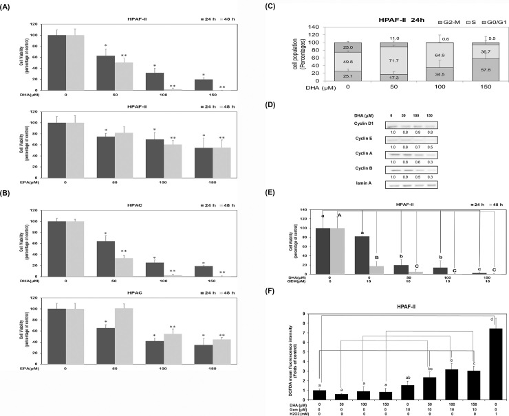
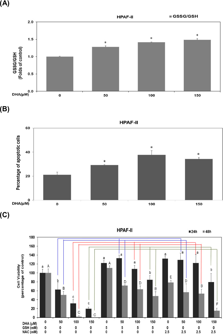
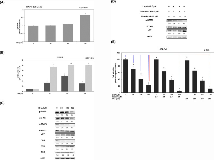
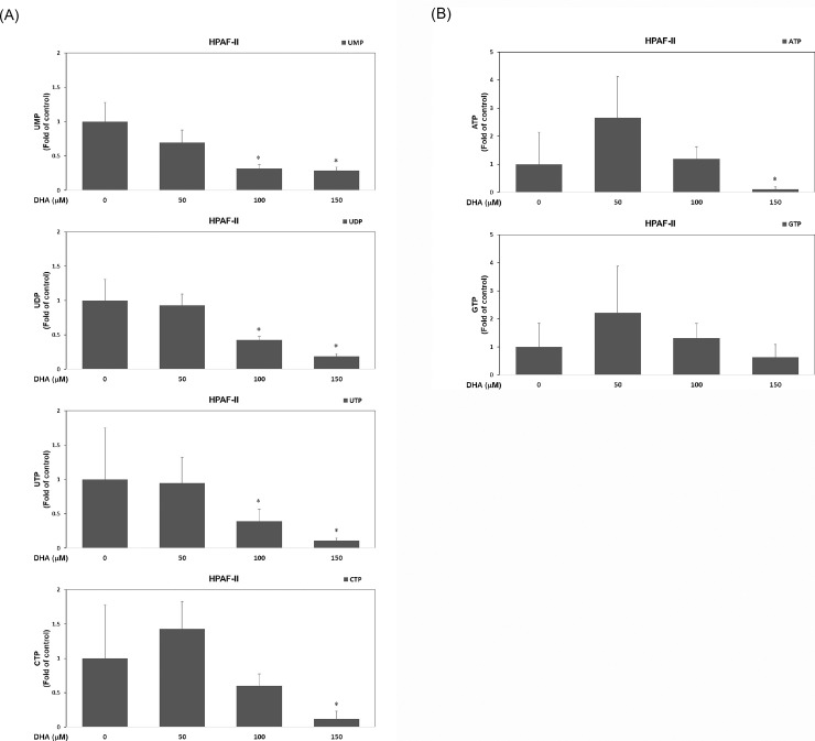
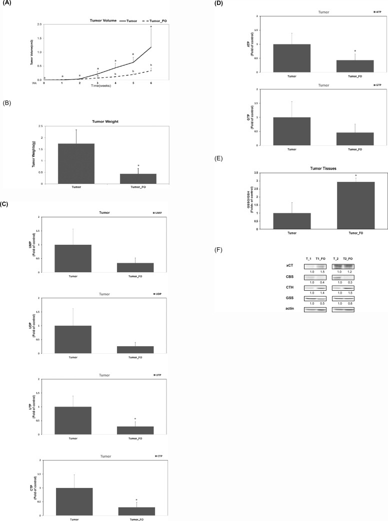
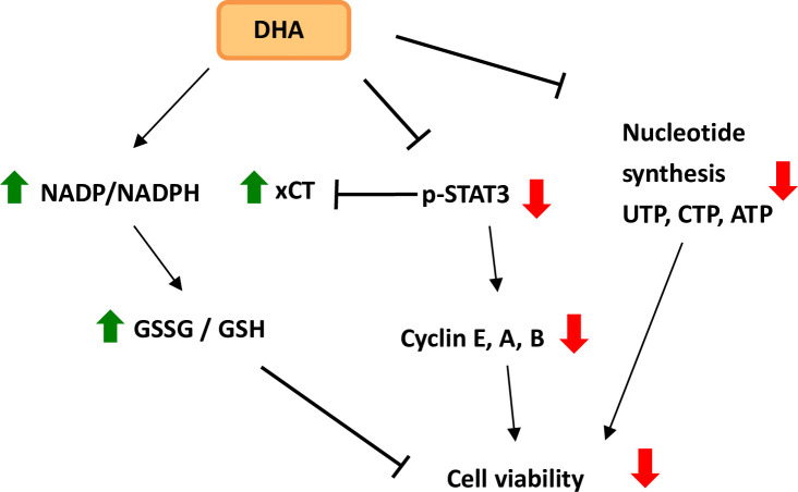
The treatment of cancer cells obtained by blocking cellular metabolism has received a lot of attention recently. Previous studies have demonstrated that Kras mutation-mediated abnormal glucose metabolism would lead to an aberrant cell proliferation in human pancreatic ductal adenocarcinoma (PDAC) cells. Previous literature has suggested that consumption of fish oil is associated with lower risk of pancreatic cancer. In this study, we investigated the anti-cancer effects of docosahexaenoic acid (DHA) in human PDAC cells in vitro and in vivo. Omega-3 polyunsaturated fatty acids (PUFAs) such as DHA and eicosapentaenoic acid (EPA) significantly inhibited the proliferation of human PDAC cells. The actions of DHA were evaluated through an induction of cell cycle arrest at G1 phase and noticed a decreased expression of cyclin A, cyclin E and cyclin B proteins in HPAF-II cells. Moreover, it was found that co-treatment of DHA and gemcitabine (GEM) effectively induced oxidative stress and cell death in HPAF-II cells. Interestingly, DHA leads to an increased oxidative glutathione /reduced glutathione (GSSG/GSH) ratio and induced cell apoptosis in HPAF-II cells. The findings in the study showed that supplementation of GSH or N-Acetyl Cysteine (NAC) could reverse DHA-mediated cell death in HPAF-II cells. Additionally, DHA significantly increased cellular level of cysteine, cellular NADP/NADPH ratio and the expression of cystathionase (CTH) and SLCA11/xCT antiporter proteins in HPAF-II cells. The action of DHA was, in part, associated with the inactivation of STAT3 cascade in HPAF-II cells. Treatment with xCT inhibitors, such as erastin or sulfasalazine (SSZ), inhibited the cell survival ability in DHA-treated HPAF-II cells. DHA also inhibited nucleotide synthesis in HPAF-II cells. It was demonstrated in a mouse-xenograft model that consumption of fish oil significantly inhibited the growth of pancreatic adenocarcinoma and decreased cellular nucleotide level in tumor tissues. Furthermore, fish oil consumption induced an increment of GSSG/GSH ratio, an upregulation of xCT and CTH proteins in tumor tissues. In conclusion, DHA significantly inhibited survival of PDAC cells both in vitro and in vivo through its recently identified novel mode of action, including an increment in the ratio of GSSG/GSH and NADP/NADPH respectively, and promoting reduction in the levels of nucleotide synthesis.


Conflict of interest statement

The authors have declared that no competing interests exist.

Figures

Fig 1
Fig 1. DHA inhibits cell survival through an induction of cell cycle arrest in human PDAC cells in vitro.
Human PDAC cells such as HPAF-II (A) and HPAC (B) cells were cultured in MEM medium with DHA or EPA (at concentrations of 0, 50, 100 and 150 μM) for 24 hours (24 h) and 48 hours (48 h). Trypan blue exclusion assay was performed to measure cell viability as described in Materials and Methods. A single asterisk (*) represents a statistically significant difference in comparison with the untreated subgroup at 24 h, at P <0.05. A double asterisks (**) represents a statistically significant difference in comparison with the untreated subgroup at 48 h, at P <0.05. (C)To measure cell cycle distribution, human PDAC HPAF-II cells were cultured in the presence of DHA (0, 50, 100 and 150 μM) and cultured in 10% FBS MEM medium for 24 h. The measurement of the cell population at different cell cycle phases was performed using flow cytometry analysis, as described in Materials and Methods. (D) Human PDAC HPAF-II cells were treated with DHA (at concentrations of 0, 50, 100 and 150 μM) in 10% FBS MEM medium for 24 h. Western blot analysis of nuclear proteins was performed using monoclonal antibodies against cyclin D1, cyclin E, cyclin A, cyclin B and lamin A antibodies, as described under Materials and Methods. Band intensities represent the amounts of cyclin D1, cyclin E, cyclin A and cyclin B in the nuclei of human PDAC HPAF-II cells. (E) Human PDAC HPAF-II cells were cultured in MEM medium with DHA (at concentrations of 0, 50, 100 and 150 μM) in the presence or absence of 10 μM gemcitabine (GEM) for 24 h and 48 h. Trypan blue exclusion assay was performed to measure cell survival as described in Materials and Methods. Different lower-case letter represents a statistically significant difference within different subgroups at 24 h, at P <0.05 (indicated with blue line). Different upper-case letter represents a statistically significant difference within different subgroups at 48 h, at P <0.05 (indicated with red line). (F) Human PDAC HPAF-II cells were cultured in MEM medium with DHA (at concentrations of 0, 50, 100 and 150 μM) in the presence or absence of 10 μM gemcitabine (GEM) for 24 h. H2O2 (at a dosage of 1 mM) was used as a positive control. The intracellular ROS level was determined by using the DCFDA assay as described in Materials and Methods. Different letters represent a statistically significant difference among different subgroups at 24 h, at P <0.05. A blue line represents a significant difference of DHA_GEM groups in comparison with the untreated control group. Red lines represent a significant difference between DHA subgroups (at dosages of 50, 100 and 150 μM) in the absence or presence of GEM (10 μM). A green line represents a significant difference between the positive control group (H2O2 group) and untreated control group.
Fig 2
Fig 2. DHA inhibits cell survival through modulation of GSSG/GSH level in PDAC cells.
(A) Human PDAC HPAF-II cells were cultured in MEM medium with DHA (at concentrations of 0, 50, 100 and 150 μM) for 24 h. Analysis of thiol compounds (GSSG and GSH) was performed as described in Materials and Methods. A single asterisk (*) represents a statistically significant difference in comparison with untreated subgroup of HPAF-II cells, at P <0.05. (B) The plots indicate the levels of apoptotic cell populations after treatment of DHA for 24 h in human PDAC HPAF-II cells. The quantitative result of the apoptotic cell populations was presented as apoptotic levels. Statistical significance is expressed as the mean ± SD (standard deviation) of three independent experiments. A single asterisk (*) represents a statistically significant difference in comparison with untreated subgroup of HPAF-II cells, at P <0.05. (C) Human PDAC HPAF-II cells were cultured in MEM medium with DHA (at concentrations of 0, 50, 100 and 150 μM) for 24 h and 48 h. The effect of GSH or NAC on DHA-treated HPAF-II cells were evaluated by using trypan blue exclusion assay described above. Different lower-case letter represents a statistically significant difference among different subgroups at 24 h, at P <0.05. Different upper-case letter represents a statistically significant difference among different subgroups at 48 h, at P <0.05. Blue lines represent a significant difference between DHA subgroups (at dosage of 50 μM) in the presence of GSH or NAC. Red lines represent a significant difference between DHA subgroups (at dosage of 100 μM) in the presence of GSH or NAC. Green lines represent a significant difference between DHA subgroups (at dosage of 150 μM) in the presence of GSH or NAC.
Fig 3
Fig 3. DHA modulates the cellular NADPH level and the expression of xCT antiporter, phosphorylated STAT3, CBS and CTH proteins in PDAC cells.
Human HPAF-II cells treated with DHA (at concentrations of 0, 50, 100 and 150 μM) in 10% FBS MEM medium for 24 h. (A) Analysis of cellular cysteine level was performed as described in Materials and Methods. A single asterisk (*) represents a statistically significant in comparison with untreated subgroup of HPAF-II cells, at P <0.05. (B) Analysis of cellular NADP/NADPH level was performed as described in Materials and Methods. A single asterisk (*) represents a statistically significant difference in comparison with untreated subgroup of HPAF-II cells at 24 h, at P <0.05. A double asterisk (**) represents a statistically significant difference in comparison with untreated subgroup of HPAF-II cells at 48 h, at P <0.05. (C) Western blot analysis of cytoplasmic proteins (at 24 h time point) using monoclonal antibodies against p-EGFR (Tyr 1068), p-c-Met (Tyr1234/1235), p-STAT3 (Tyr705), t-STAT3, xCT, CBS, CTH, GSS and internal controls (actin). Band intensities represent the amounts of p-EGFR (Tyr 1068), p-c-Met (Tyr1234/1235), p-STAT3 (Tyr705), xCT, CBS, CTH and GSS in the cytoplasm of HPAF-II cells. (D) Human HPAF-II cells treated with Lapatinib (EGFR inhibitor, 5 μM), PHA-665752 (c-Met inhibitor, 0.5 μM) or Ruxolitinib (STAT3 inhibitor, 15 μM) for 24 h. Western blot analysis of cytoplasmic proteins (at 24 h time point) using monoclonal antibodies against p- p-STAT3 (Tyr705), t-STAT3, xCT and actin. Band intensities represent the amounts of p-STAT3 (Tyr705) and xCT in the cytoplasm of HPAF-II cells. (E) HPAF-II cells were pretreated with 2.5 μM erastin or 250 μM sulfasalazine (SSZ) for 24h and followed by treatment of DHA (0, 50, 100 and 150 μM) for 24 h. The effects of erastin or SSZ was measured by using trypan blue exclusion assay described above. Statistical significance is expressed as the mean ± SD (standard deviation) of two independent experiments. Different letters represent a statistically significant difference among different subgroups (at P<0.05). A blue line represents a significant difference between DHA subgroups (at dosages of 50, 100 and 150 μM) and the untreated control subgroup. A red line represents a significant difference between DHA subgroups (at dosage of 150 μM) in the presence of erastin or SSZ.
Fig 4
Fig 4. DHA suppresses the nucleotide synthesis in PDAC cells.
(A) Human PDAC HPAF-II cells were treated with DHA (at concentrations of 0, 50, 100 and 150 μM) in 10% FBS MEM medium for 24 h. Analysis of cellular UMP, UDP, UTP and CTP levels was performed as described in Materials and Methods. A single asterisk (*) represents a statistically significant difference in comparison with untreated subgroup of HPAF-II cells, at P <0.05. (B) Analysis of cellular ATP and GTP levels was performed as described in Materials and Methods. A single asterisk (*) represents a statistically significant difference in comparison with untreated subgroup of HPAF-II cells, at P <0.05.
Fig 5
Fig 5. Consumption of fish oil inhibits the growth of pancreatic adenocarcinoma by suppressing nucleotide synthesis in a mouse xenograft model.
Xenograft NOD SCID mice (n = 6 for each subgroup) were divided into two subgroups (the tumor subgroup and tumor_FO subgroup). The tumor_FO subgroup was given FO (at a dosage of 4% w/w diet per day) for 6 weeks. Data represent the change in the tumor volume (A) and tumor weight (B) between the tumor subgroup (Tumor; the control subgroup) and tumor with FO at dosage of 4% w/w diet per day (Tumor_FO; the experimental subgroup). (A) For the analysis of tumor volume, different letters represent a statistically significant difference in comparison to the control subgroup at the same time points (P<0.05). (B) For the analysis of tumor weight, a single asterisk (*) indicates a significant difference in comparison to the control subgroup (P<0.05). (C) Analysis of UMP, UDP, UTP and CTP levels in tumor tissues was performed as described in Materials and Methods. A single asterisk represents a statistically significant difference in comparison to the control subgroup, at P <0.05. (D) Analysis of ATP and GTP levels in tumor tissues was performed as described in Materials and Methods. A single asterisk represents a statistically significant difference in comparison to the control subgroup, at P <0.05. (E) Analysis of thiol compounds (GSSG and GSH) was performed as described in Materials and Methods. A single asterisk represents a statistically significant difference of GSSG/GSH ratio in tumor tissues in tumor_FO subgroup in comparison to the control subgroup, at P <0.05. (F) Western blot analysis of proteins in different sets of tumor tissues using monoclonal antibodies against xCT, CBS, CTH, GSS and internal controls (actin). Band intensities represent the amounts of xCT, CBS, CTH and GSS proteins.
Fig 6
Fig 6. Proposed mechanisms of DHA-mediated modulation of GSSG/GSH and NADP/NADPH ratios and suppression of nucleotide synthesis in human PDAC cells.
Red arrows indicate decreased level. Green arrows indicate increased level.→: induction; −│: suppression.

Similar articles

Cited by

References

    1. Siegel RL, Miller KD, Jemal A. Cancer statistics, 2018. CA Cancer J Clin. 2018;68(1):7–30. 10.3322/caac.21442 . - DOI - PubMed
    1. Modolell I, Guarner L, Malagelada JR. Vagaries of clinical presentation of pancreatic and biliary tract cancer. Annals of oncology: official journal of the European Society for Medical Oncology. 1999;10 Suppl 4:82–4. . - PubMed
    1. Ryan DP, Hong TS, Bardeesy N. Pancreatic adenocarcinoma. N Engl J Med. 2014;371(11):1039–49. 10.1056/NEJMra1404198 . - DOI - PubMed
    1. Witkiewicz AK, McMillan EA, Balaji U, Baek G, Lin WC, Mansour J, et al. Whole-exome sequencing of pancreatic cancer defines genetic diversity and therapeutic targets. Nat Commun. 2015;6:6744 Epub 2015/04/10. 10.1038/ncomms7744 - DOI - PMC - PubMed
    1. Ying H, Kimmelman AC, Lyssiotis CA, Hua S, Chu GC, Fletcher-Sananikone E, et al. Oncogenic Kras maintains pancreatic tumors through regulation of anabolic glucose metabolism. Cell. 2012;149(3):656–70. Epub 2012/05/01. 10.1016/j.cell.2012.01.058 - DOI - PMC - PubMed

Publication types

MeSH terms