. 2020 Nov 2;18(11):e3000936.
doi: 10.1371/journal.pbio.3000936. eCollection 2020 Nov.

Aequorea's secrets revealed: New fluorescent proteins with unique properties for bioimaging and biosensing

Affiliations

Aequorea's secrets revealed: New fluorescent proteins with unique properties for bioimaging and biosensing

Gerard G Lambert et al. PLoS Biol. .

Abstract

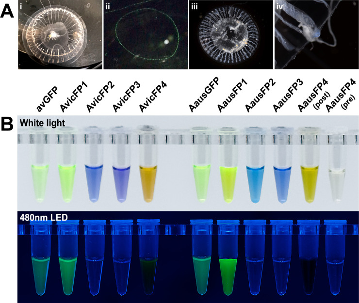
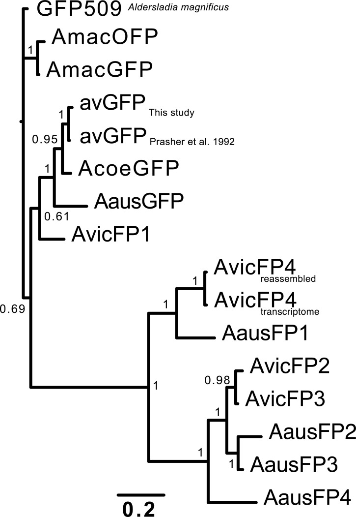
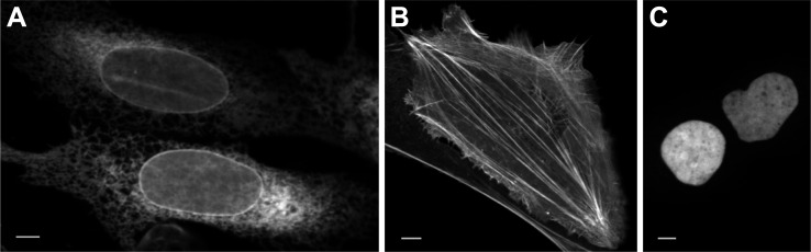
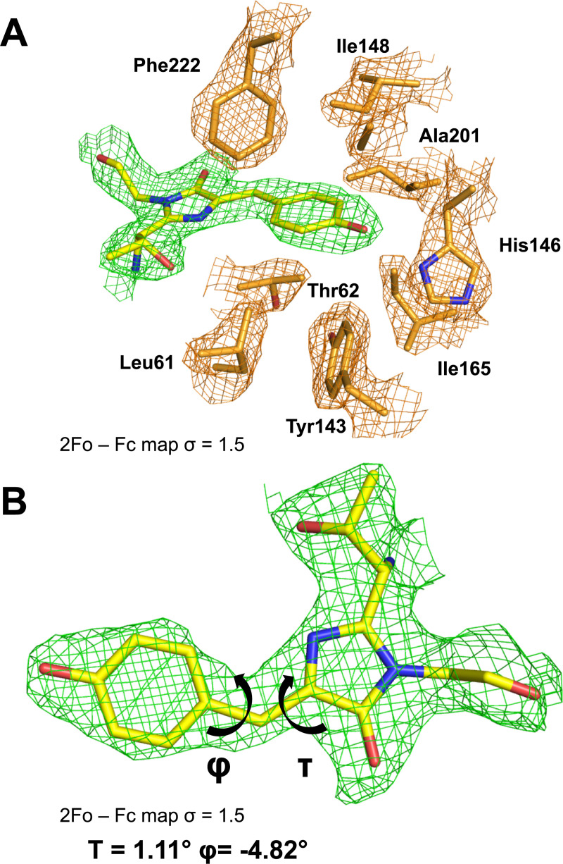
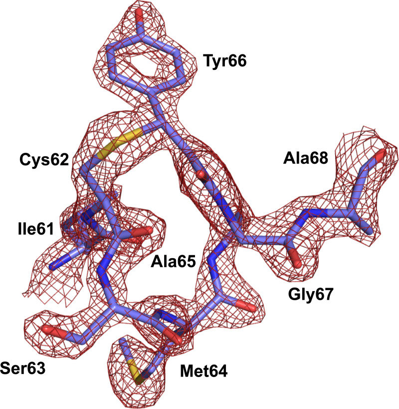
Using mRNA sequencing and de novo transcriptome assembly, we identified, cloned, and characterized 9 previously undiscovered fluorescent protein (FP) homologs from Aequorea victoria and a related Aequorea species, with most sequences highly divergent from A. victoria green fluorescent protein (avGFP). Among these FPs are the brightest green fluorescent protein (GFP) homolog yet characterized and a reversibly photochromic FP that responds to UV and blue light. Beyond green emitters, Aequorea species express purple- and blue-pigmented chromoproteins (CPs) with absorbances ranging from green to far-red, including 2 that are photoconvertible. X-ray crystallography revealed that Aequorea CPs contain a chemically novel chromophore with an unexpected crosslink to the main polypeptide chain. Because of the unique attributes of several of these newly discovered FPs, we expect that Aequorea will, once again, give rise to an entirely new generation of useful probes for bioimaging and biosensing.


Conflict of interest statement

The authors have declared that no competing interests exist.

Figures

Fig 1
Fig 1. Photographs of Aequorea individuals from this study and purified fluorescent proteins cloned from these samples.
(A) White-light (i) and fluorescence (400-nm LED illumination) (ii) photographs of A. victoria and white-light photographs of A. cf. australis (iii, iv). The blue coloration of A. cf. australis is shown in the higher magnification image of one of its tentacle bulbs (iv). (B) Purified recombinant proteins from Aequorea species, shown under white light and 480-nm LED without emission filters. Protein concentrations were adjusted to display similar optical density as judged by eye and were between 0.5 and 2 mg/ml for all samples.
Fig 2
Fig 2. Absorbance and emission spectra (where measurable) for FP homologs in this study.
Proteins from each species were designated AvicFP or AausFP and numbered in order of discovery, with chromoproteins retaining the “FP” nomenclature for consistency. For photoswitchable and photoconvertible proteins, pre-illumination absorbance spectra are shown as dotted lines, and post-illumination absorbance spectra as solid lines. Emission spectra are shown as green solid lines. The emission spectra for AvicFP2 and AvicFP3 were measured using 460-nm excitation prior to photoconversion. The emission spectrum of AausFP4 was measured using 440-nm excitation after photoswitching to the blue-absorbing state. Red arrows indicate peaks that increase or decrease upon photoconversion or switching. For ease of display, spectra are normalized to the maximum visible absorbance for non-photoactive proteins, and to the pre- (for AvicFP2) or post-illumination (for AvicFP3 and AausFP4) maximum for photoactive proteins. All plots share the same x-axis scale as shown for AausGFP. The data underlying this figure may be found at FPbase (https://www.fpbase.org). FP, fluorescent protein.
Fig 3
Fig 3. Phylogenetic tree for FPs cloned in this study, with Aequorea macrodactyla and Aldersladia magnificus green FPs included as outgroups.
Green-emitting FPs with avGFP-like properties, including AvicFP1, fall into 1 cluster of fairly closely related sequences, while the novel fluorescent (AausFP1 and AvicFP4) and non-fluorescent homologs form 2 additional families. The data underlying this figure (nucleotide sequences of the FPs from this study) may be found in GenBank, accession numbers MN114103 through MN114112. avGFP, Aequorea victoria green fluorescent protein; FP, fluorescent protein.
Fig 4
Fig 4. Expression of mAvicFP1-tagged proteins in mammalian cells.
mAvicFP1 fusions to (A) CytERM, (B) LifeAct, and (C) H2B. U2-OS cells display expected localization. Scale bar is 10 mm. The data underlying this figure (raw image data) may be found at https://doi.org/10.26300/4x48-y393.
Fig 5
Fig 5. The AausFP1 chromophore environment.
(A) 2FobsFcalc electron-density map contoured at a 1.5 σ level superimposed over the model of the chromophore and the neighboring residues in the structure of AausFP1. (B) Dihedral angle definition around the chromophore methylene bridge. The data underlying this figure may be found in PDB 6S67.
Fig 6
Fig 6. 2FobsFcalc electron-density map contoured at a 2.0 σ level superimposed over the model of the chromophore and the 3 covalently bonded residues in the structure of AausFP2.
The data underlying this figure may be found in PDB 6S68.

References

    1. Prasher DC, Eckenrode VK, Ward WW, Prendergast FG, Cormier MJ. Primary structure of the Aequorea victoria green-fluorescent protein. Gene. 1992;111:229–33. 10.1016/0378-1119(92)90691-h - DOI - PubMed
    1. Tsien RY. The green fluorescent protein. Annu Rev Biochem. 1998;67:509–44. 10.1146/annurev.biochem.67.1.509 - DOI - PubMed
    1. Shaner NC, Patterson GH, Davidson MW. Advances in fluorescent protein technology. J Cell Sci. 2007;120:4247–60. 10.1242/jcs.005801 - DOI - PubMed
    1. Rodriguez EA, Campbell RE, Lin JY, Lin MZ, Miyawaki A, Palmer AE, et al. The growing and glowing toolbox of fluorescent and photoactive proteins. Trends Biochem Sci. 2017;42:111–29. 10.1016/j.tibs.2016.09.010 - DOI - PMC - PubMed
    1. Mishin AS, Subach FV, Yampolsky IV, King W, Lukyanov KA, Verkhusha VV. The first mutant of the Aequorea victoria green fluorescent protein that forms a red chromophore. Biochemistry. 2008;47:4666–73. 10.1021/bi702130s - DOI - PMC - PubMed

Publication types

Substances