. 2020 Nov 2;18(11):e3000901.
doi: 10.1371/journal.pbio.3000901. eCollection 2020 Nov.

Membrane progesterone receptor induces meiosis in Xenopus oocytes through endocytosis into signaling endosomes and interaction with APPL1 and Akt2

Affiliations

Membrane progesterone receptor induces meiosis in Xenopus oocytes through endocytosis into signaling endosomes and interaction with APPL1 and Akt2

Nancy Nader et al. PLoS Biol. .

Erratum in

Abstract

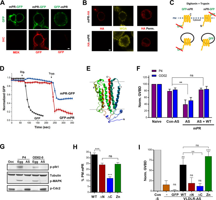
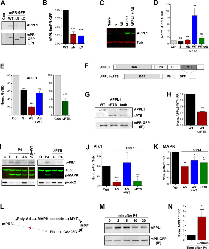
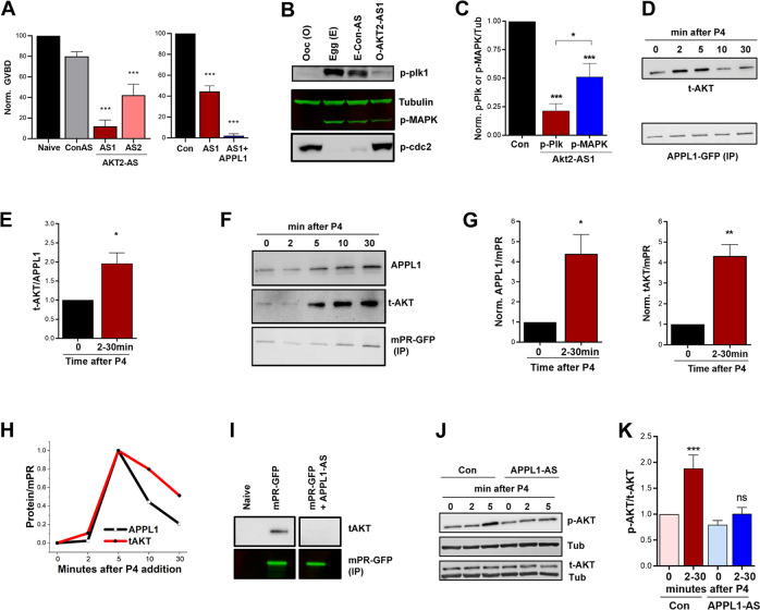
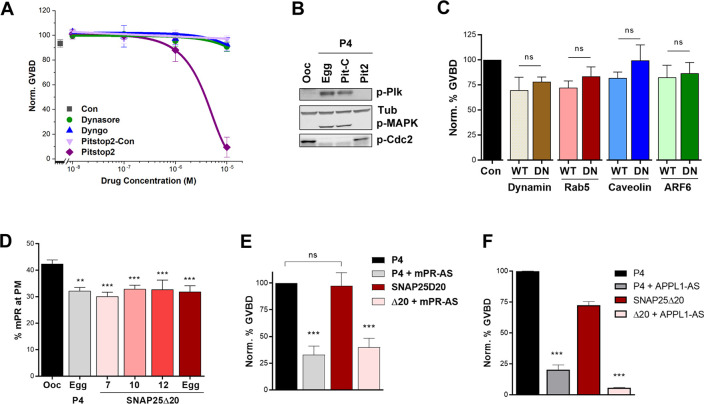
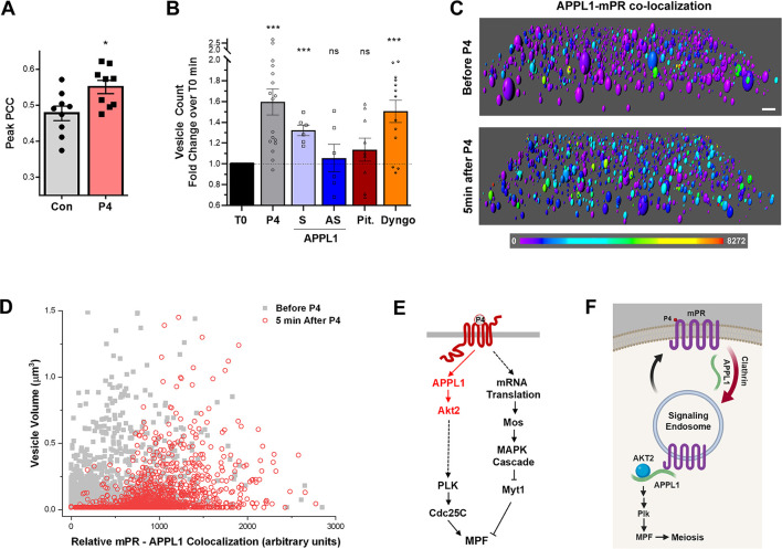
The steroid hormone progesterone (P4) mediates many physiological processes through either nuclear receptors that modulate gene expression or membrane P4 receptors (mPRs) that mediate nongenomic signaling. mPR signaling remains poorly understood. Here we show that the topology of mPRβ is similar to adiponectin receptors and opposite to that of G-protein-coupled receptors (GPCRs). Using Xenopus oocyte meiosis as a well-established physiological readout of nongenomic P4 signaling, we demonstrate that mPRβ signaling requires the adaptor protein APPL1 and the kinase Akt2. We further show that P4 induces clathrin-dependent endocytosis of mPRβ into signaling endosome, where mPR interacts transiently with APPL1 and Akt2 to induce meiosis. Our findings outline the early steps involved in mPR signaling and expand the spectrum of mPR signaling through the multitude of pathways involving APPL1.


Conflict of interest statement

I have read the journal's policy and the authors of this manuscript have the following competing interests: KM is a co-founder of Valdia Health.

Figures

Fig 1
Fig 1. mPRβ has an opposite topology to GPCRs and is required for oocyte meiosis.
(A) Nonpermeabilized CHO cells overexpressing mPR-GFP or GFP-mPR stained with anti-GFP or anti-MEK antibodies. Scale bar 2 μm. (B) Immunostaining of CHO cells overexpressing mPR-HA or HA-mPR in nonpermeabilized or permeabilized (HA Perm) conditions. PM stained with WGA. Scale bar 2 μm. (C) Schematic representation of the FPP assay. (D) Time course of GFP fluorescence in cells transfected as indicated and treated with Dig then Tryp at the indicated time points (mean ± SEM; n = 8–11 cells). (E) 3D model of Xenopus mPRβ. (F) Oocyte maturation in response to P4 or OD 0–02 in naive oocytes, oocytes injected with Con-AS, oocytes injected with specific mPR-AS, and oocytes injected with antisense oligos and expressing WT mPR-GFP (50 ng/oocyte) (AS + WT) (mean ± SEM; n = 3–4 donor females). (G) Activation state of MAPK and Plk for the treatments in panel (F). For the AS treatments, oocytes that did not mature (no white spot) in response to P4 were collected when the control group has reached maximal maturation levels. (H) Percent of the total mPR population at the PM for full-length mPR (WT) and the different mutants as indicated (mean ± SEM; 15–23 oocytes per condition, from 3 donor females). (I) GVBD rescue using the mPR mutants (20 ng/oocyte) following VLDLR-AS or sense oligos injection (Con-S). GFP: GFP RNA injection as a control (mean ± SEM; n = 3 donor females). ** p < 0.01, *** p < 0.001. Refer to S1 Data file. AS, injected with mPR antisense oligos; Con-AS, control antisense oligos; Dig, digitonin; Egg, eggs matured with P4; FPP, fluorescence protease protection; GFP, green fluorescent protein; GVBD, germinal vesicle breakdown; HA, hemagglutinin tag; MAPK, mitogen-activated protein kinase; mPR, membrane progesterone receptor; MEK, mitogen-activated protein kinase kinase; ns, not significant; Ooc, untreated oocyte; P4, progesterone; Plk1, Polo-like kinase 1; PM, plasma membrane; VLDLR, very-low-density lipoprotein receptor; VLDLR-AS, VLDLR antisense knockdown; Tryp, trypsin; WGA, wheat germ agglutinin; WT, wild type.
Fig 2
Fig 2. APPL1 is essential for P4-mPR-induced oocyte meiosis.
(A) IP of mPR-GFP from untreated oocytes (Con) and oocytes expressing mPR-GFP (WT) (20 ng/oocyte), the ΔN or ΔC mutants (20 ng/oocyte) probed for APPL1 and GFP. (B) Quantification of the IP experiments as the ratio of APPL1/mPR-GFP normalized to mPR-GFP WT expressing cells (mean ± SEM; n = 3). (C) WB analysis of APPL1 in untreated (naive), sense (S), antisense (AS) APPL1 oligos injected oocytes, in oocytes expressing APPL1.L (APPL1) (50 ng/oocyte), and in APPL1.L expressing oocytes co-injected with APPL1 antisense oligos (APPL1+AS). Tubulin is shown as a loading control. (D) Quantification of APPL1 expression normalized to tubulin for the conditions in panel (C) (mean ± SEM; n = 3). (E) Oocyte maturation measured as the levels of GVBD normalized to untreated oocytes (Con) in oocytes injected with APPL1 sense (S) or antisense oligos (AS), injected with APPL1 antisense oligos and APPL1 mRNA to overexpress APPL1 (AS + WT) (50 ng/oocyte) (left panel, mean ± SEM, n = 4 donor females), or injected APPL1-ΔPTB (ΔPTB) RNA (50 ng/oocyte) (right panel, mean ± SEM; n = 3 donor females). (F) Schematic of APPL1 full-length and APPL1-ΔPTB domains. (G) IP of mPR-GFP from oocytes expressing APPL1 full-length (WT) (20 ng/oocyte), APPL1-ΔPTB (ΔPTB) (20 ng/oocyte), or both proteins probed for APPL1 and GFP. (H) Quantification of the IP experiments in (G) as the ratio of APPL1/mPR-GFP normalized to mPR-GFP injected oocytes (mean ± SEM; n = 2). (I) Left Panel: WB of MAPK, Plk1, and Cdc2 phosphorylation from untreated oocytes (O), eggs matured with P4 (E), oocytes injected with APPL1 sense (S), antisense (AS) oligos, or antisense oligos and APPL1 RNA (AS+WT). Right Panel: WB of untreated oocytes (O) and P4 matured eggs (P4), and APPL1-ΔPTB injected oocytes untreated or P4 treated as indicated. Tub is shown as a loading control. For the AS and ΔPTB treatments with P4 immature oocytes with no white spot where collected at the end of the experiment when the control group has reached maximal GVBD levels. (J-K) Quantification of p-Plk1 as the ratio of p-Plk1/Tub (J) or p-MAPK as the ratio of p-MAPK/Tub (K) normalized to the ratios in untreated eggs (mean ± SEM; n = 3–5 donor females). (L) Current model for the signaling cascade downstream of mPR. (M) IP from oocytes overexpressing mPR-GFP (20 ng/oocyte) and treated with P4 (10−5 M) for the indicated times and probed for APPL1. (N) Quantification of the IP experiments in (M) as the ratio of APPL1/mPR-GFP normalized to untreated oocytes (time 0). The max response between 2 to 30 min from each donor frog was used for the analysis (2–30 minutes) (mean ± SEM; n = 5). *p < 0.05, **p < 0.01, *** p < 0.001. Refer to S1_Data file. APPL, adapter protein containing Pleckstrin homology domain, Phosphotyrosine binding domain and Leucine zipper motif 1; AS, antisense; BAR, bin, amphiphysin and Rvs domain; GFP, green fluorescent protein; GVBD, germinal vesicle breakdown; HA,; IP, immmunoprecipitation; MAPK, mitogen-activated protein kinase; mPR, membrane progesterone receptor; MEK, mitogen-activated protein kinase kinase; ns, not significant; P4, progesterone; Plk1, Polo-like kinase 1; PTB, phosphotyrosine-binding domain; Tub, tubulin; WB, western blot; WT, wild type.
Fig 3
Fig 3. Akt2 is required for mPR signaling.
(A) Oocyte maturation in response to P4 in untreated oocytes (Naive), oocytes injected with Con-AS oligos, oocyte injected with 2 different Akt2 antisense oligos (AS1 or AS2) (left panel), and in oocytes injected with Akt2-AS1 oligos with overexpression of APPL1 (1+APPL1) (50 ng/oocyte) (right panel). GVBD percentage is normalized to naive (mean ± SEM; n = 3–5 donor females). (B) Activation of the MAPK and Plk1 cascades in untreated oocytes (O), untreated eggs matured with P4 (E), eggs injected with Con-AS oligos and matured with P4 (E-Con-AS), or oocytes injected with Akt2 antisense oligos and treated with P4 (O-AKT-AS1). For the O-AKT-AS1 group immature oocyte with no white spot where collected when the control group has reached maximal GVBD levels. (C) Quantification of p-Plk1 and p-MAPK for the conditions in panel (B) as the ratio of p-Plk1 or p-MAPK to Tub, normalized to untreated eggs (Con) (mean ± SEM; n = 7). (D) IP over a P4 time course using overexpressed APPL1-GFP (20 ng/oocyte) as a bait and probed for t-AKT. (E) Quantification of Akt pulldown for the IP experiments in (D) as the ratio of t-AKT/APPL1-GFP. The maximal response between 2 to 30 minutes from each experiment is plotted (mean ± SEM; n = 5). (F) IP over a P4 time course using overexpressed mPR-GFP (20 ng/oocyte) as a bait and probed for APPL1 and total Akt. (G) Quantification of the IP experiments in (F) as the ratio of t-Akt/mPR-GFP. The max response between 2 to 30 minutes from each experiment is plotted (mean ± SEM; n = 5). (H) Normalized amounts of APPL1 and t-AKT that immunoprecipitate with mPR-GFP over time from a single experiment. (I) IP of mPR-GFP probed for t-AKT from oocytes expressing mPR-GFP without or with injection of APPL1 antisense oligos (mPR-GFP + APPL1-AS). (J) Western blot analysis of t-AKT and phospho-Akt S473 (p-AKT) over a P4 time course without (Con), or with APPL1 antisense oligos injection (APPL1-AS). Tub is used as a loading control. (K) Quantification of p-AKT for the conditions in panel (J). Maximal phospho/total Akt ratio between 2 to 30 minutes from each experiment is plotted. p-AKT and t-AKT levels were first normalized to Tub. Data are normalized to untreated oocytes (time 0) (mean ± SEM; n = 6 donor females). *p < 0.05, ** p < 0.01, ***p < 0.001. Refer to S1 Data file. APPL, adapter protein containing Pleckstrin homology domain, Phosphotyrosine binding domain and Leucine zipper motif 1; AS, antisense; Con-AS, control antisense oligos; GFP, green fluorescent protein; GVBD, germinal vesicle breakdown; IP, immmunoprecipitation; MAPK, mitogen-activated protein kinase; mPR, membrane progesterone receptor; ns, not significant; P4, progesterone; Plk1, Polo-like kinase 1; PTB, phosphotyrosine-binding domain; t-AKT, total Akt; Tub, tubulin; WT, wild type.
Fig 4
Fig 4. mPR is internalized through clathrin-dependent endocytosis in response to P4.
(A) Oocyte maturation in response to P4 after 4 hours pre-incubation with incremental concentrations of different compounds as indicated. Con: untreated oocytes (mean ± SEM; n = 3–5 donor females). (B) Activation of the MAPK and Plk cascades in the different indicated conditions. Tub is shown as a loading control. (C) GVBD normalized to untreated oocytes (Con) in oocytes overexpressing WT or DN forms of dynamin, Rab5, caveolin, or ARF6 following overnight treatment with P4. For the overexpression, oocytes were injected with 20 ng RNA/oocytes for all the clones. (mean ± SEM; n = 3–9 donor females per condition). (D) Percent of the total mPR population at the plasma membrane (PM) in oocytes and eggs at the indicated conditions. Oocytes were injected with 40 ng SNAP25Δ20 (mean ± SEM; n = 7–27 oocytes per condition, from 3 donor females). (E-F) Oocyte maturation in response to P4 or SNAP25Δ20 (Δ20) (40 ng/oocyte) following knockdown of mPRβ (mPR-AS) (mean ± SEM; n = 4 donor females) (E) or APPL1 (APPL1-AS) (F) (mean ± SEM; n = 3 donor females). **p < 0.01, ***p < 0.001. Refer to S1 Data file. ARF6, ADP-ribosylation factor 6; DN, dominant-negative; Egg, P4-matured eggs; GVBD, germinal vesicle breakdown; MAPK, mitogen-activated protein kinase; mPR, membrane progesterone receptor; ns, not significant; Ooc, untreated oocytes; Pit-C, Pitstop2 control; Pit2, Pitstop2; P4, progesterone; Rab5, Ras-related protein Rab-5A; SNAP25Δ20, dominant-negative synaptosome associate protein 25; Tub, tubulin; WT, wild-type.
Fig 5
Fig 5. APPL1-mPR colocalization is essential to mPR internalization in clathrin-dependent–dynamin-independent way.
(A) Peak PCC values measured in oocytes overexpressing mPR-GFP (20 ng/oocyte) with APPL1-mCherry (20 ng/oocyte), before (Con) and after P4 treatment (mean ± SEM; n = 9). (B) Fold increase in mPR-GFP-positive vesicles (oocytes were injected 20 ng of mPR-GFP RNA/oocyte) in response to P4. Each oocyte was imaged before P4 (T0) and 40 minutes after P4 and vesicles were counted across the z-stack 3D volume. Each oocyte served as its own control before P4 addition for normalization (mean ± SEM; n = 6–17 oocytes per condition). (C) Rendition of a confocal z-stack of images from an oocyte expressing APPL1-Ch and mPR-GFP before and 5 minutes after P4 treatment. Vesicles were rendered using Imaris software, and the heat map represents the colocalization intensity of the Cherry and GFP probes. Scale bar 2 μm. (D) Distribution of vesicle volume as a function of APPL1-mPR colocalization intensity calculated as in panel (C). (E) Signaling cascade downstream of mPR. (F) Cartoon model of P4-mPR signaling upstream of triggering the Plk1 cascade. * p < 0.05, *** p < 0.001. Refer to S1 Data file. APPL, adapter protein containing Pleckstrin homology domain, Phosphotyrosine binding domain and Leucine zipper motif 1; APPL1-S: oocytes injected with APPL1 sense oligos after P4; APPL1-AS: oocytes injected with APPL1 antisense oligos after P4; AS, antisense; control antisense oligos; Dyngo, oocytes treated with Dyngo (10−5 M) 4 hours prior to adding P4; GFP, green fluorescent protein; IP, immmunoprecipitation; MAPK, mitogen-activated protein; Mos, proto-oncogene serine/threonine kinase; MPF, maturation promoting factor; mPR, membrane progesterone receptor; Myt1, membrane-associated tyrosine- and threonine-specific cdc2-inhibitory kinase; ns, not significant; P4, progesterone; PCC, Pearson colocalization coefficient; Pit, oocytes treated with Pitstop (10−5 M) 4 hours prior to adding P4; Plk1, Polo-like kinase 1; PTB, phosphotyrosine-binding domain; t-AKT, total Akt; T0, normalized baseline; WT, wild type.

Similar articles

Cited by

References

    1. Singh M, Su C, Ng S. Non-genomic mechanisms of progesterone action in the brain. Front Neurosci. 2013;7:159 10.3389/fnins.2013.00159 - DOI - PMC - PubMed
    1. Dressing GE, Goldberg JE, Charles NJ, Schwertfeger KL, Lange CA. Membrane progesterone receptor expression in mammalian tissues: a review of regulation and physiological implications. Steroids. 2011;76(1–2):11–7. 10.1016/j.steroids.2010.09.006 - DOI - PMC - PubMed
    1. Valadez-Cosmes P, Vazquez-Martinez ER, Cerbon M, Camacho-Arroyo I. Membrane progesterone receptors in reproduction and cancer. Mol Cell Endocrinol. 2016;434:166–75. Epub 2016/07/03. 10.1016/j.mce.2016.06.027 . - DOI - PubMed
    1. Moussatche P, Lyons TJ. Non-genomic progesterone signalling and its non-canonical receptor. Biochem Soc Trans. 2012;40(1):200–4. 10.1042/BST20110638 . - DOI - PubMed
    1. Zhu Y, Rice CD, Pang Y, Pace M, Thomas P. Cloning, expression, and characterization of a membrane progestin receptor and evidence it is an intermediary in meiotic maturation of fish oocytes. Proc Natl Acad Sci USA. 2003;100(5):2231–6. 10.1073/pnas.0336132100 - DOI - PMC - PubMed

Publication types

MeSH terms