Review
. 2021 Mar 1;27(13):4230-4248.
doi: 10.1002/chem.202003697. Epub 2020 Dec 29.

Cyanine Dyes Containing Quinoline Moieties: History, Synthesis, Optical Properties, and Applications

Affiliations
Review

Cyanine Dyes Containing Quinoline Moieties: History, Synthesis, Optical Properties, and Applications

Kristina Ilina et al. Chemistry. .

Erratum in

Abstract

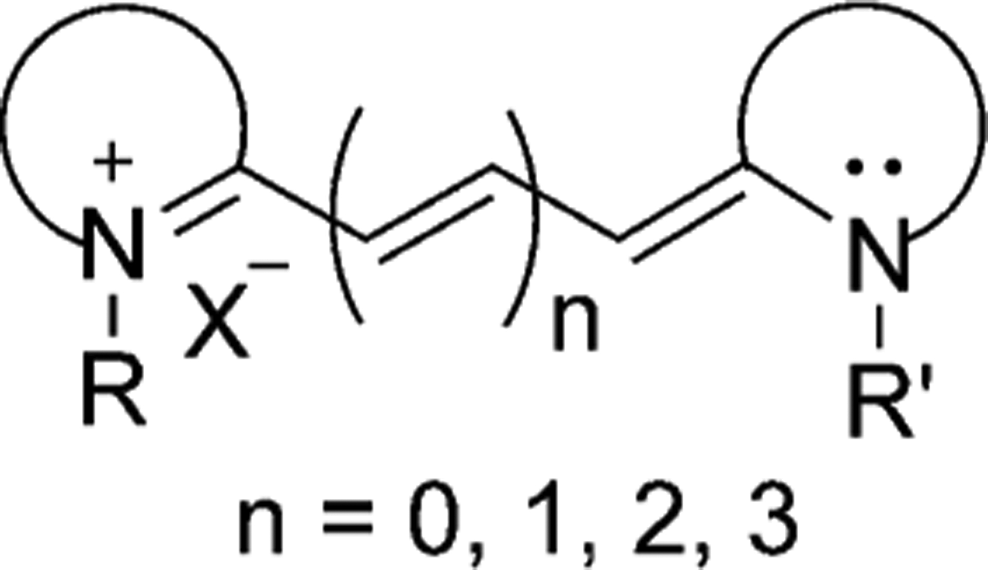
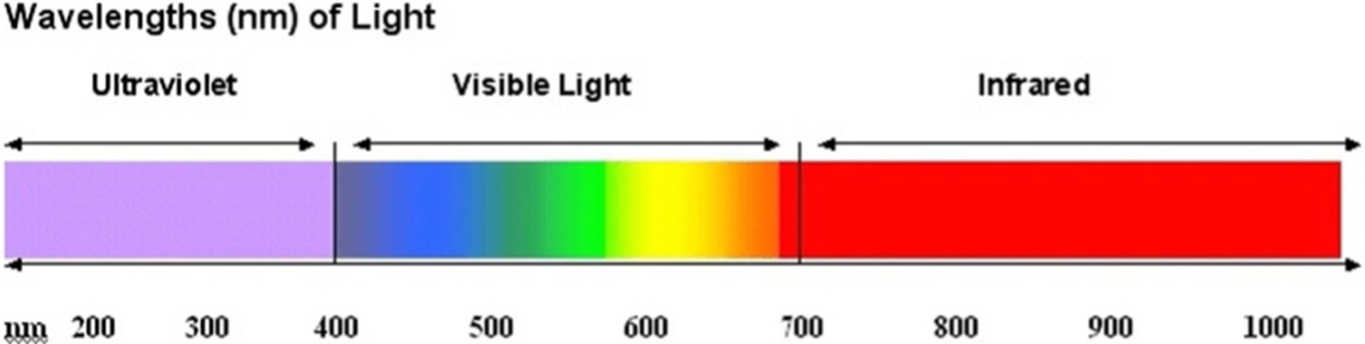
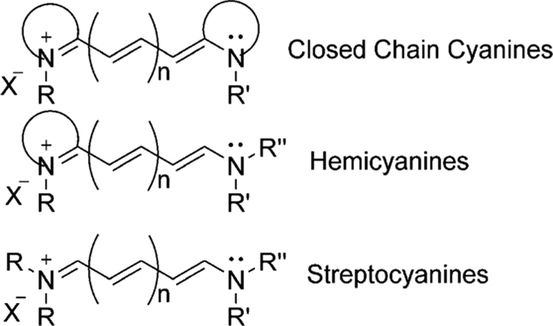
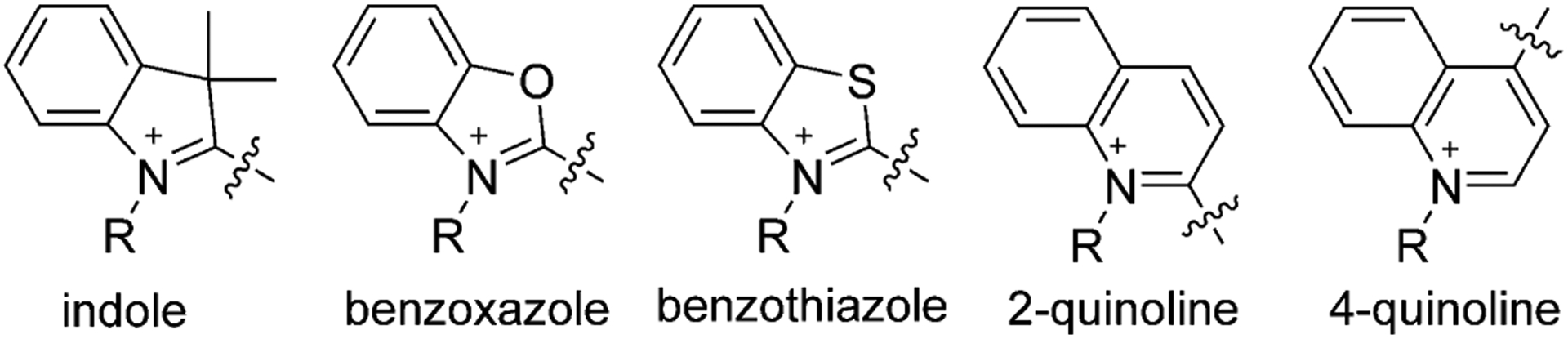
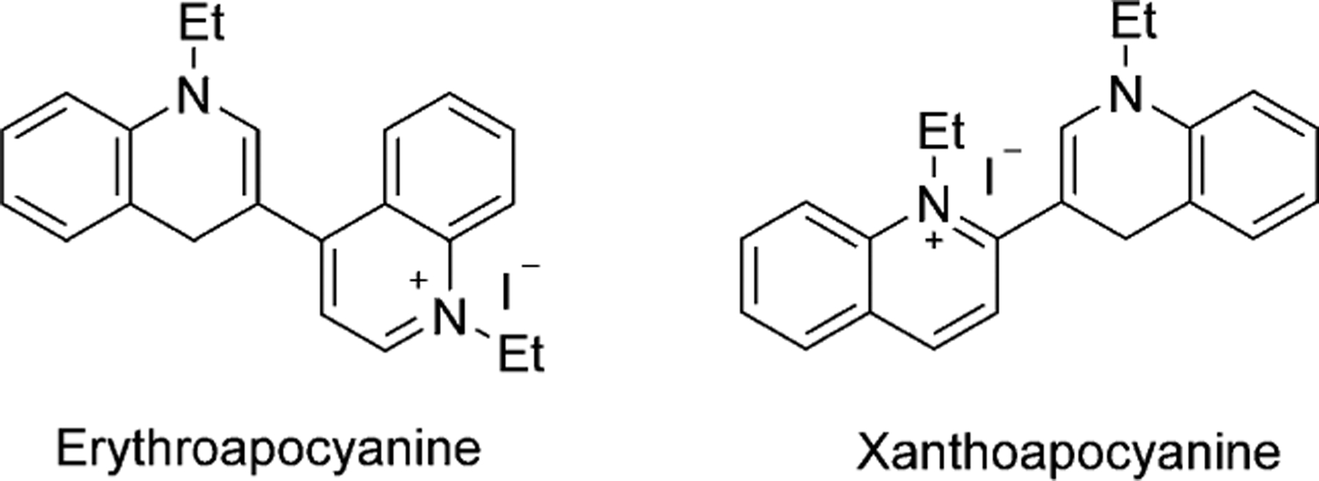
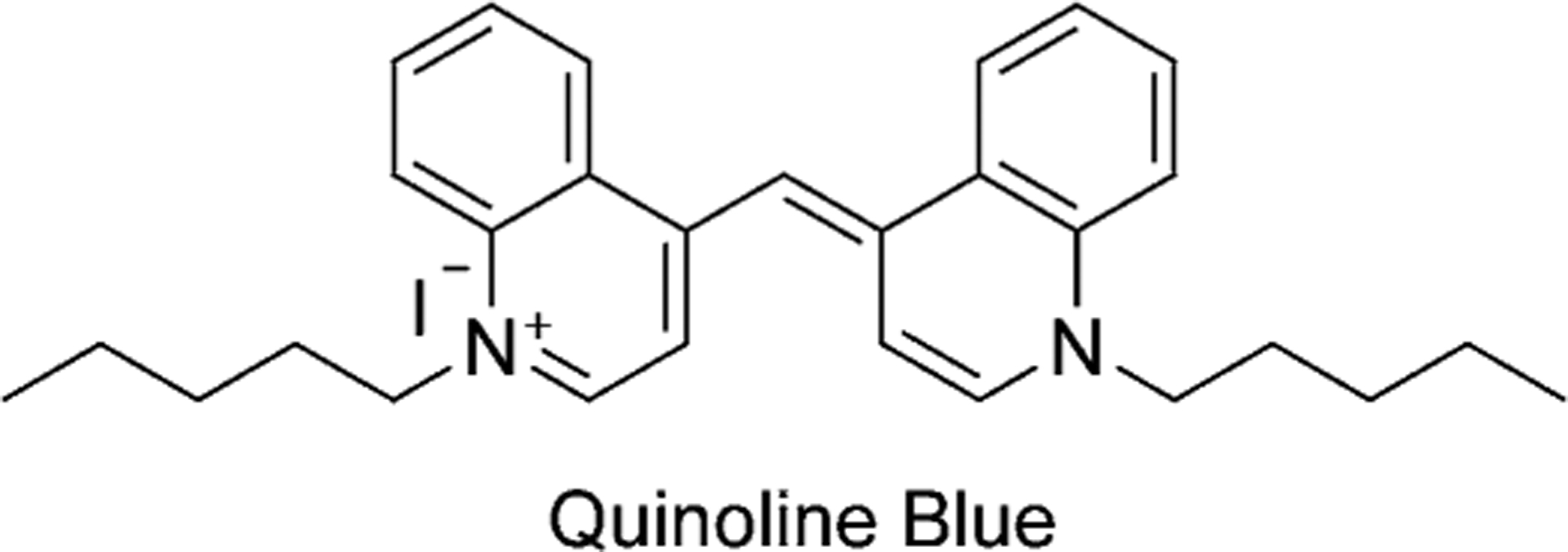
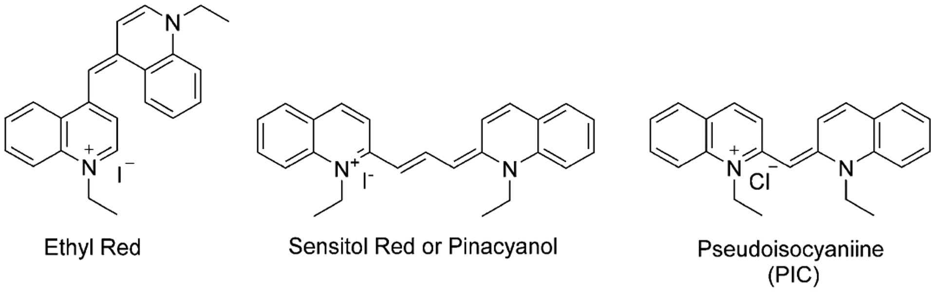
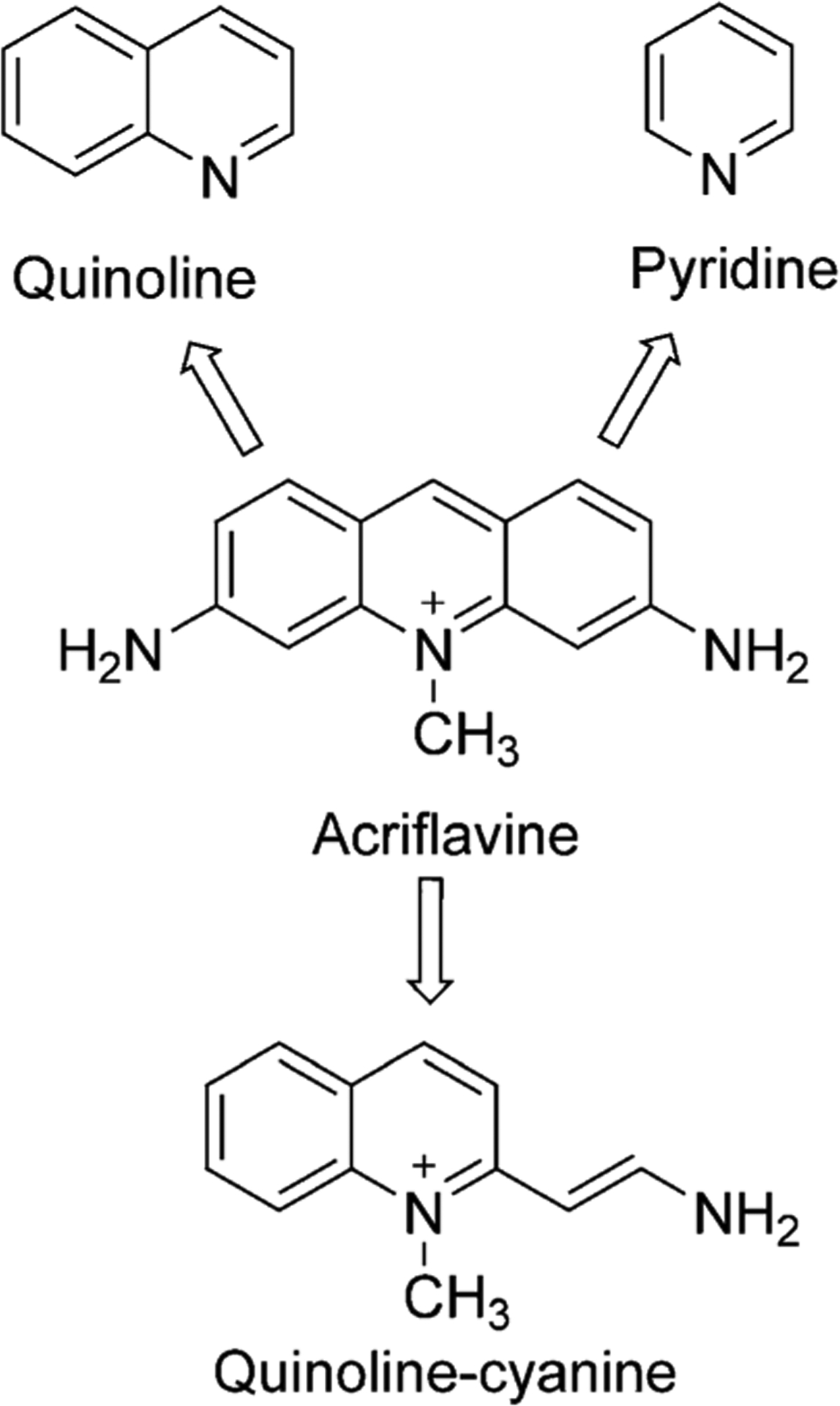
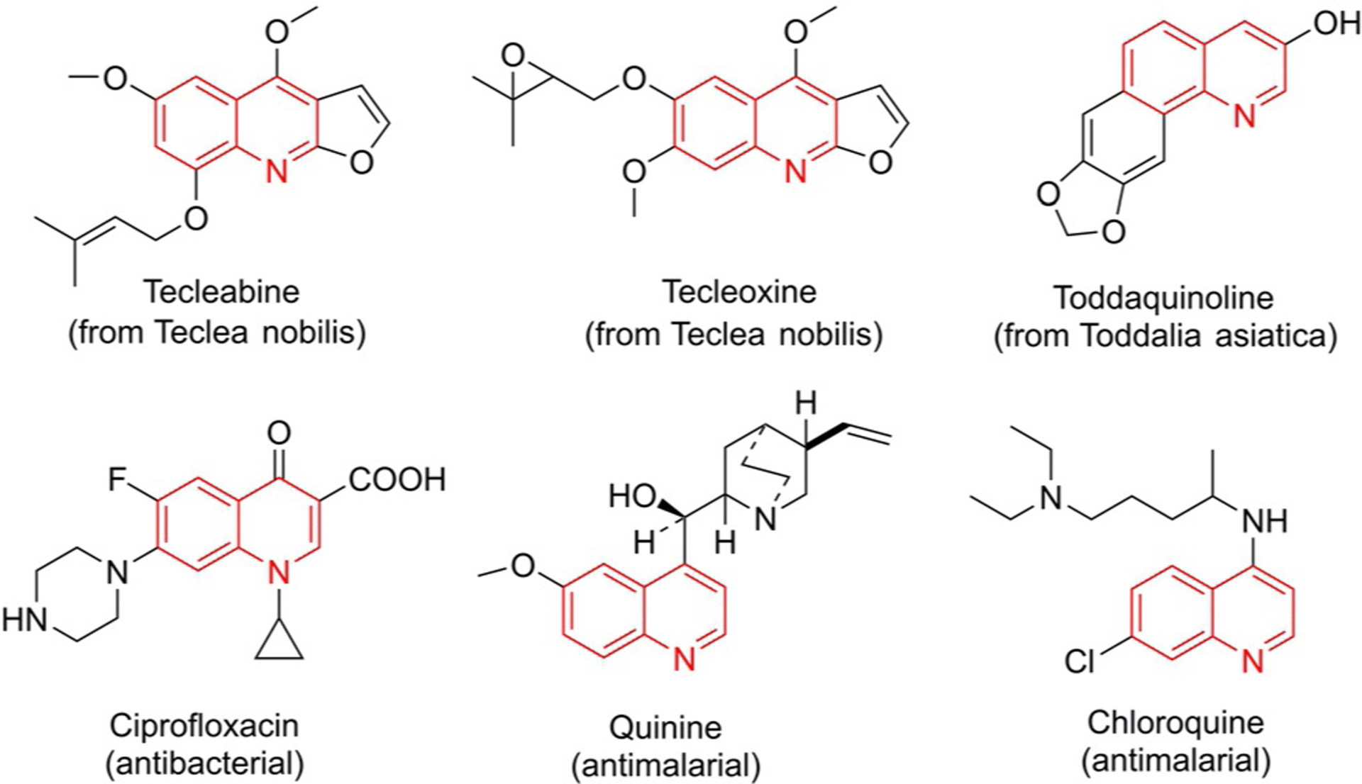
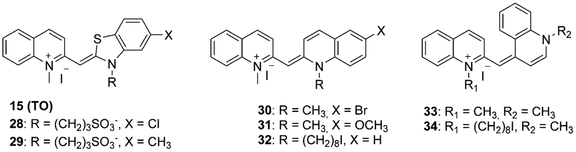
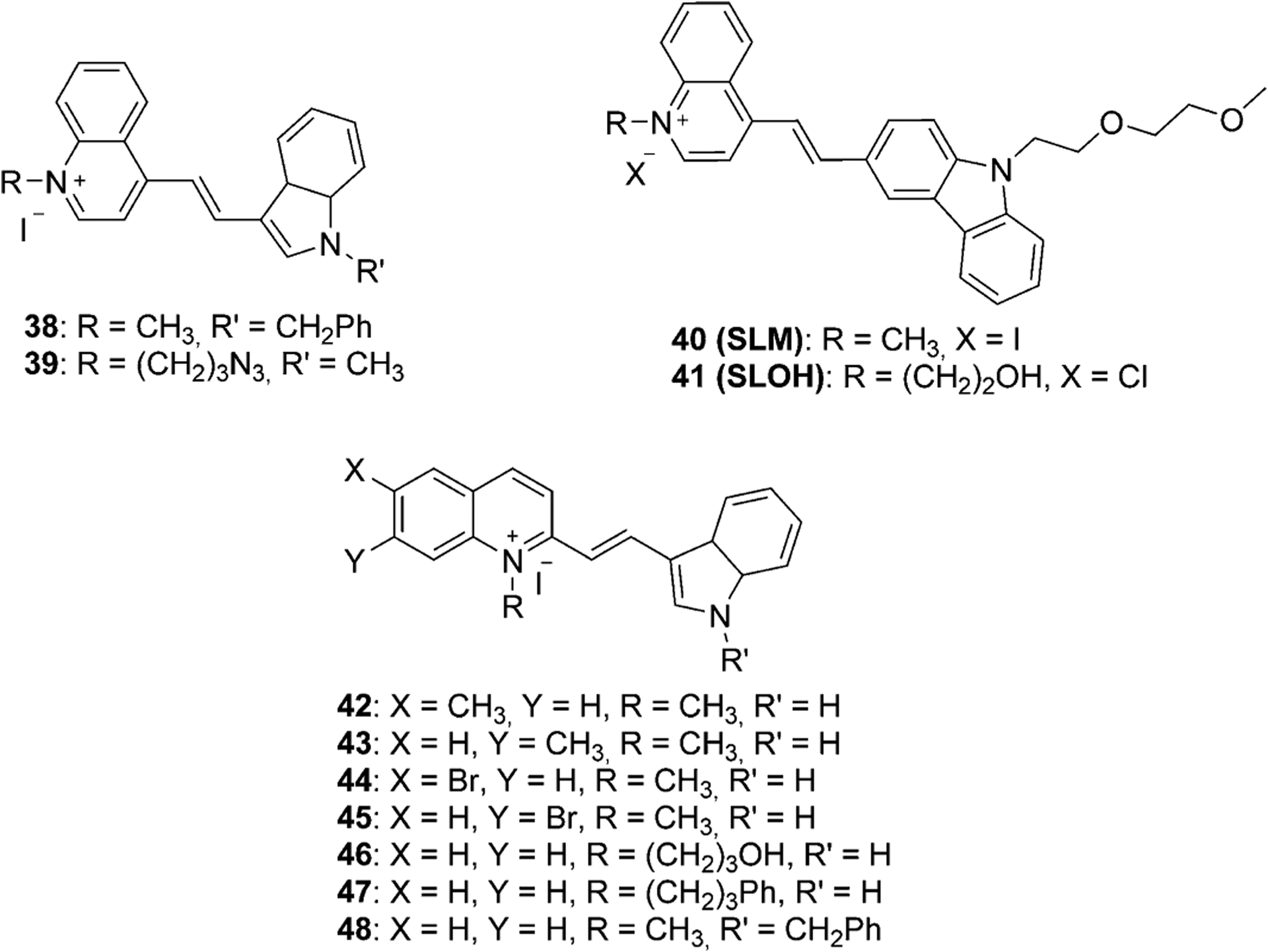
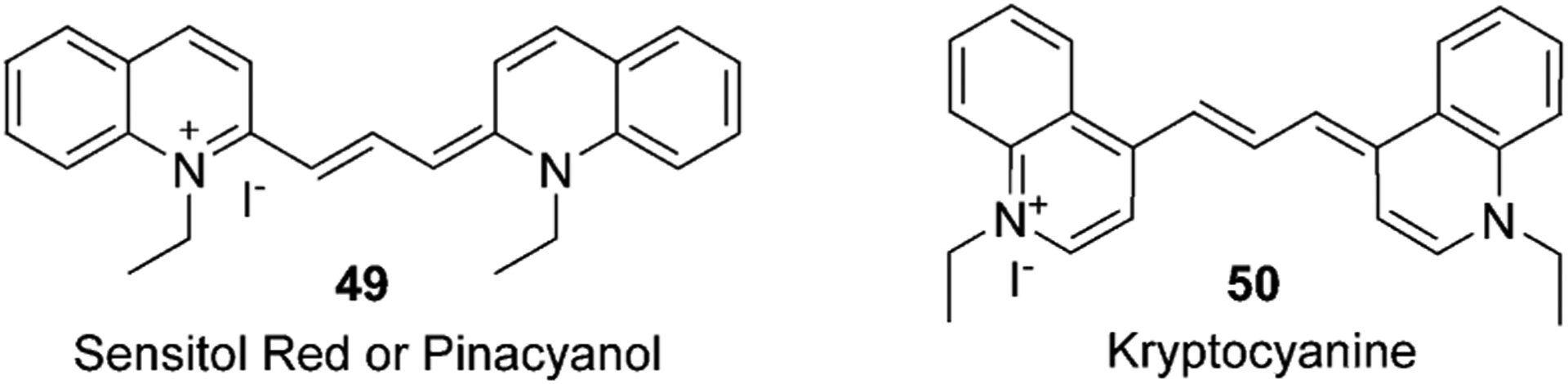
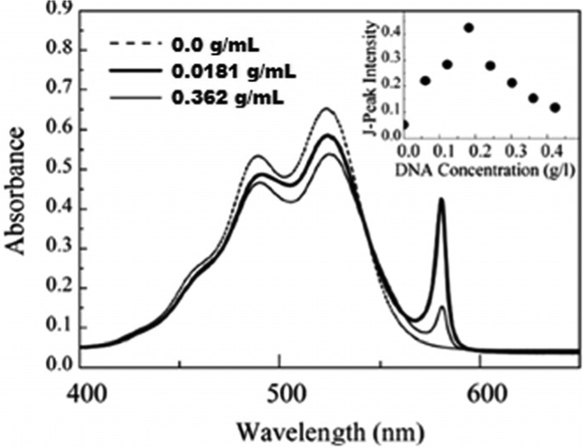
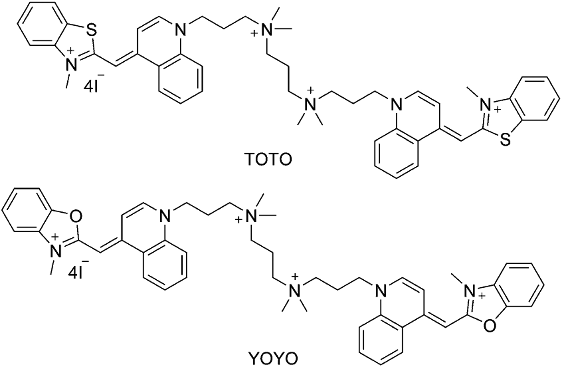
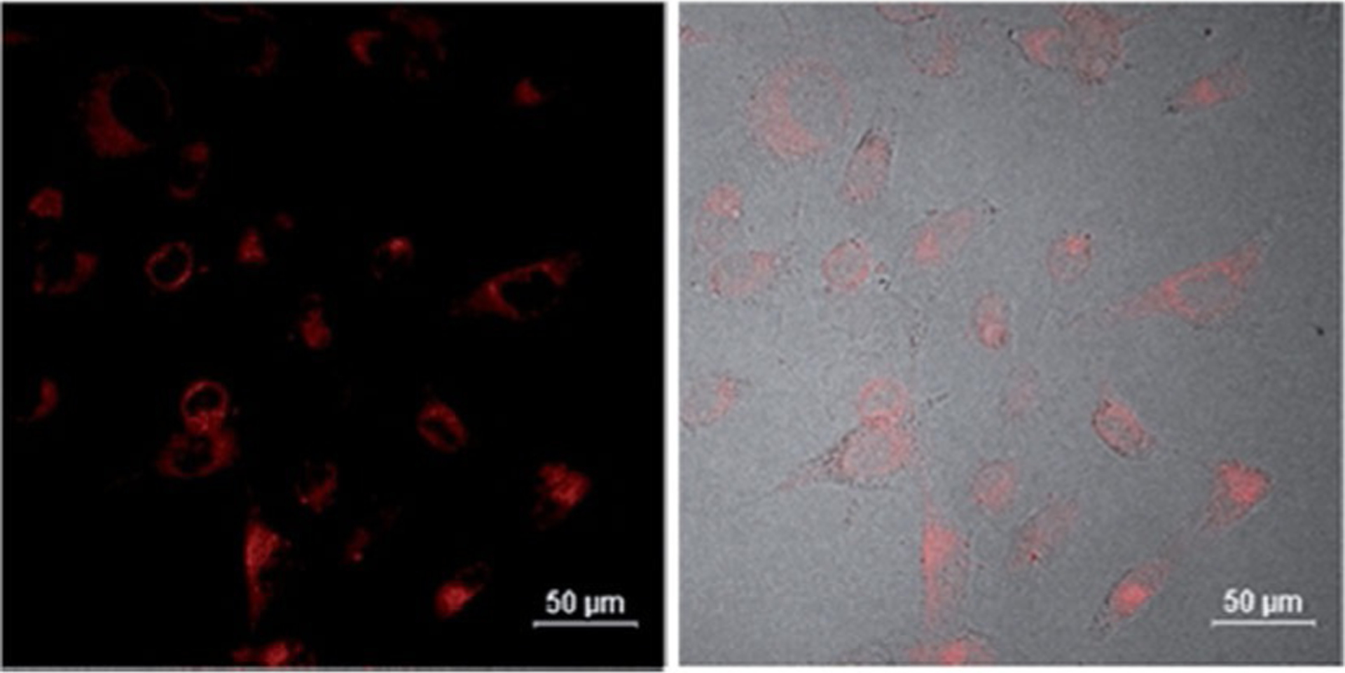
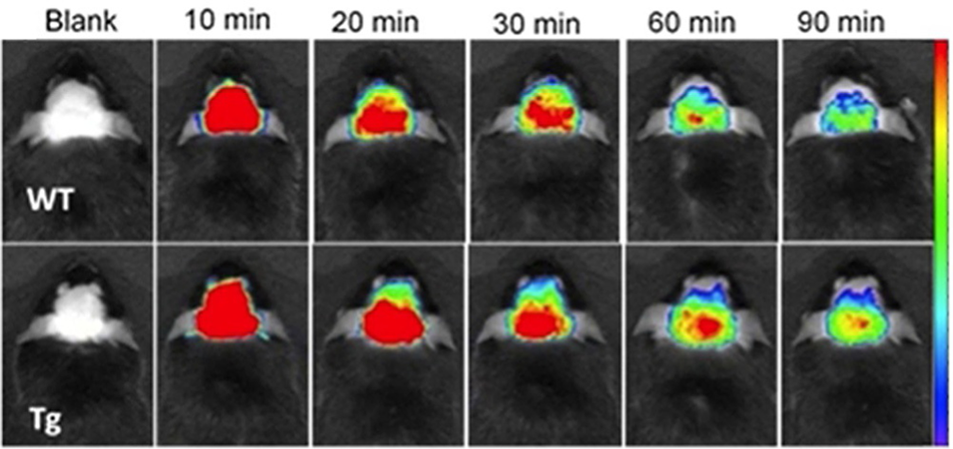
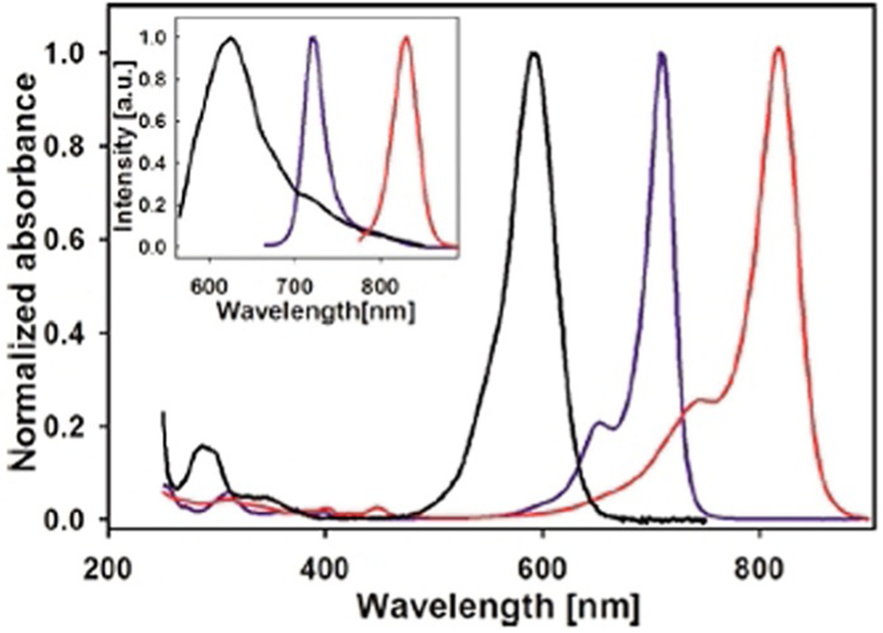
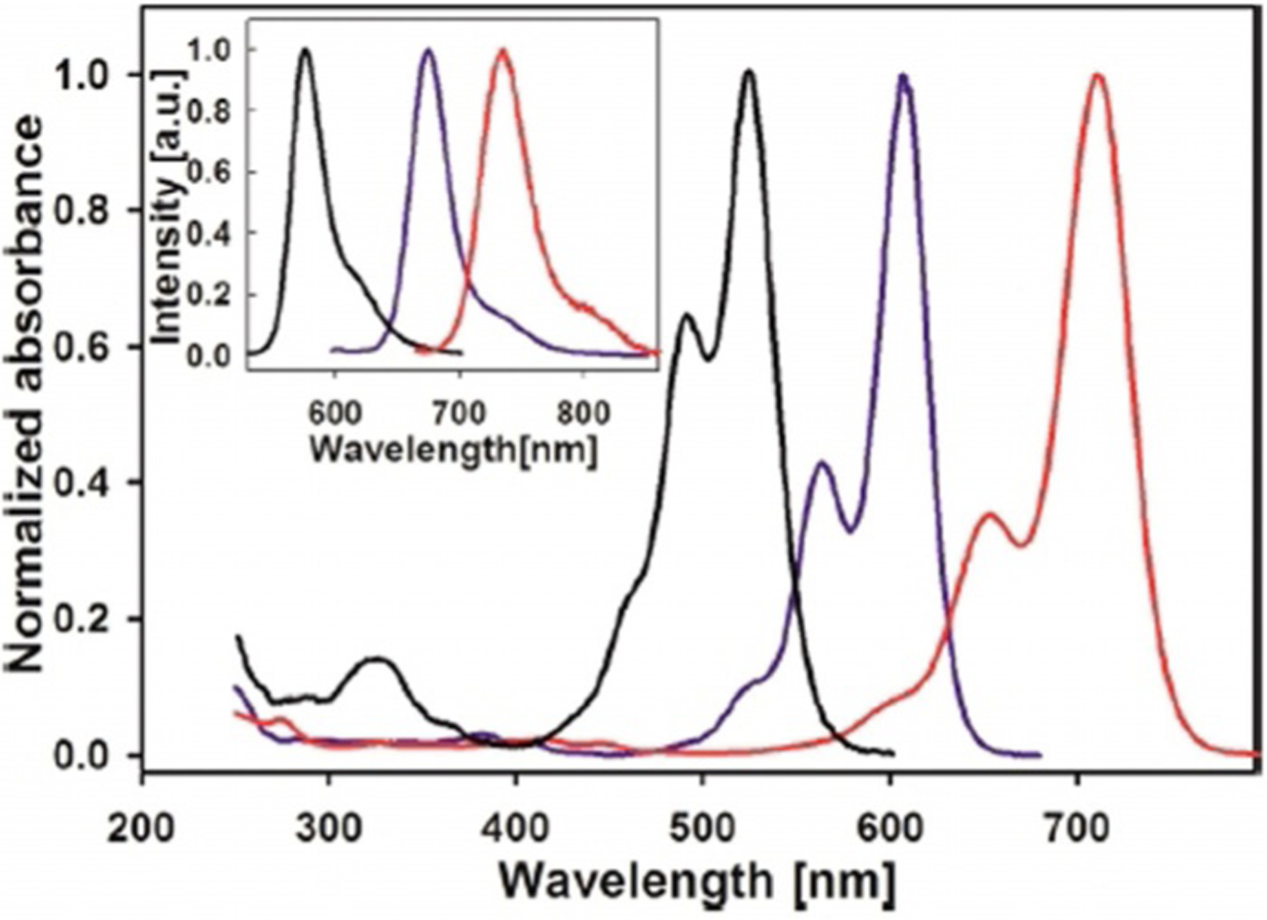
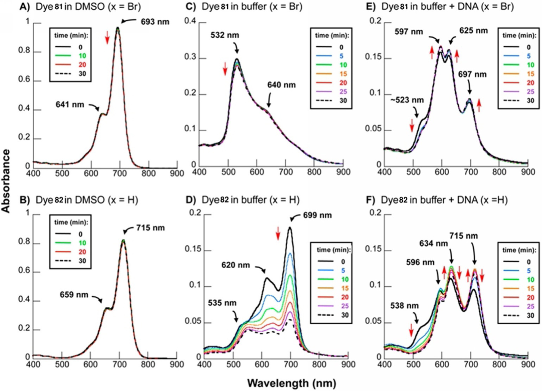
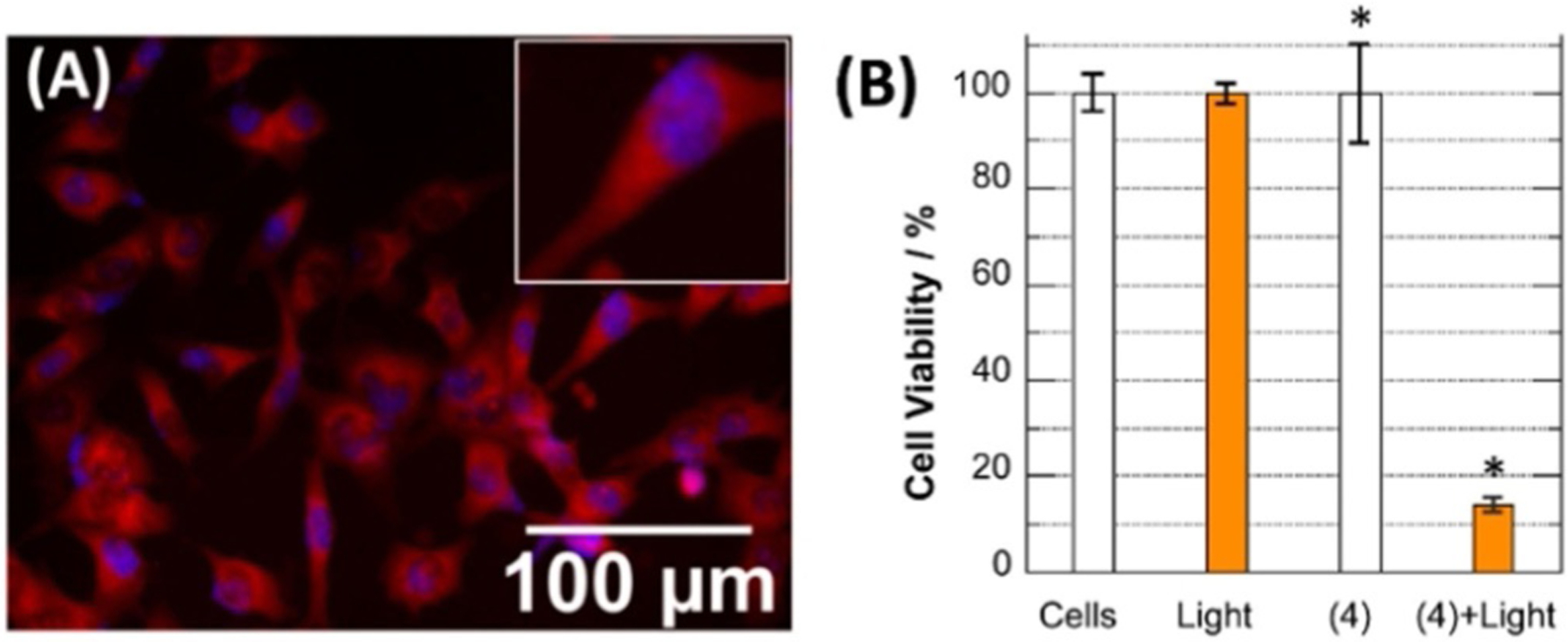
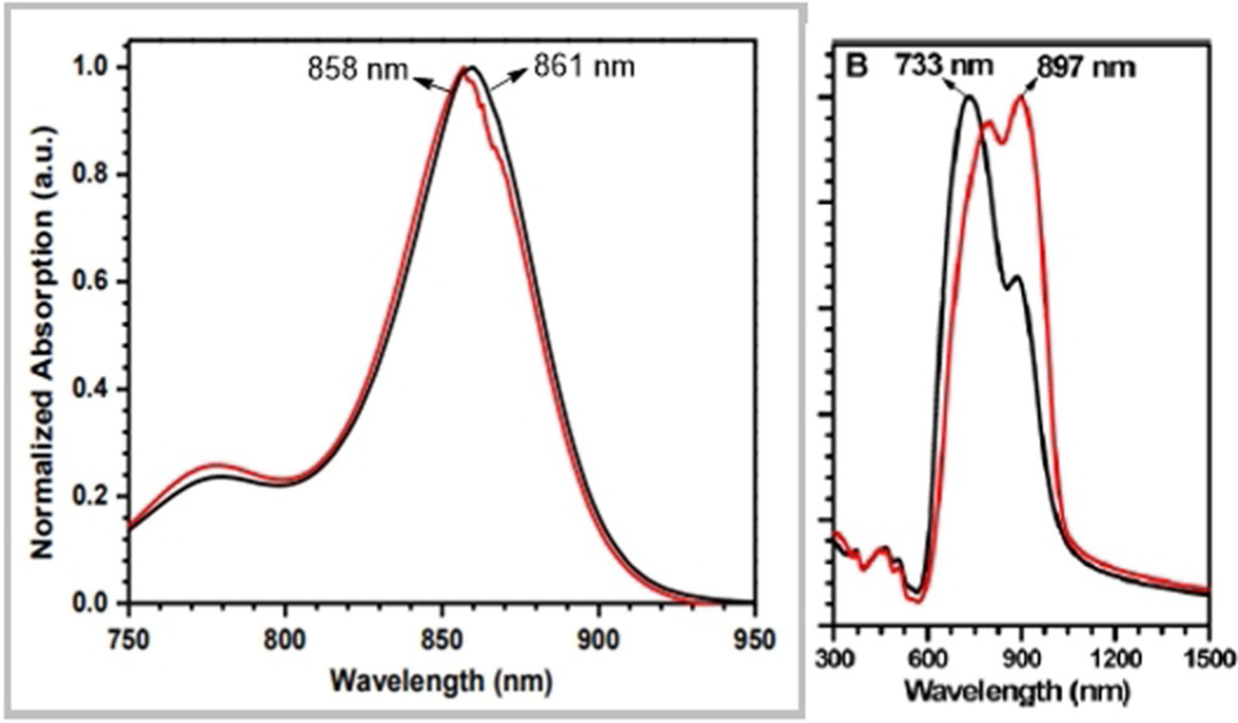
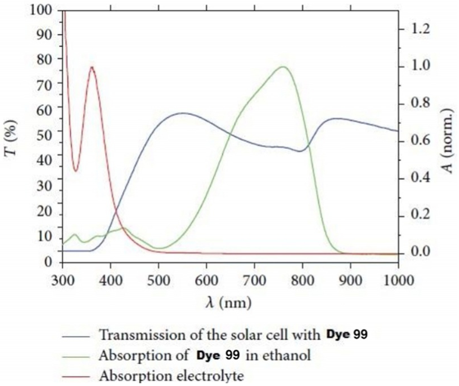
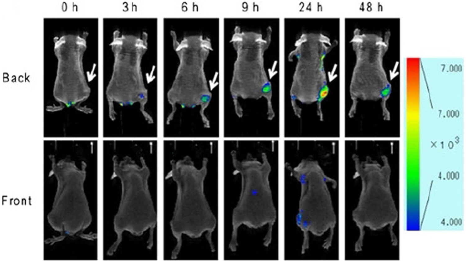
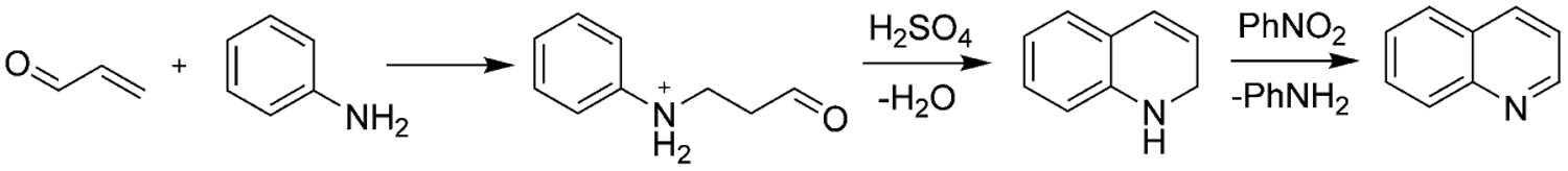
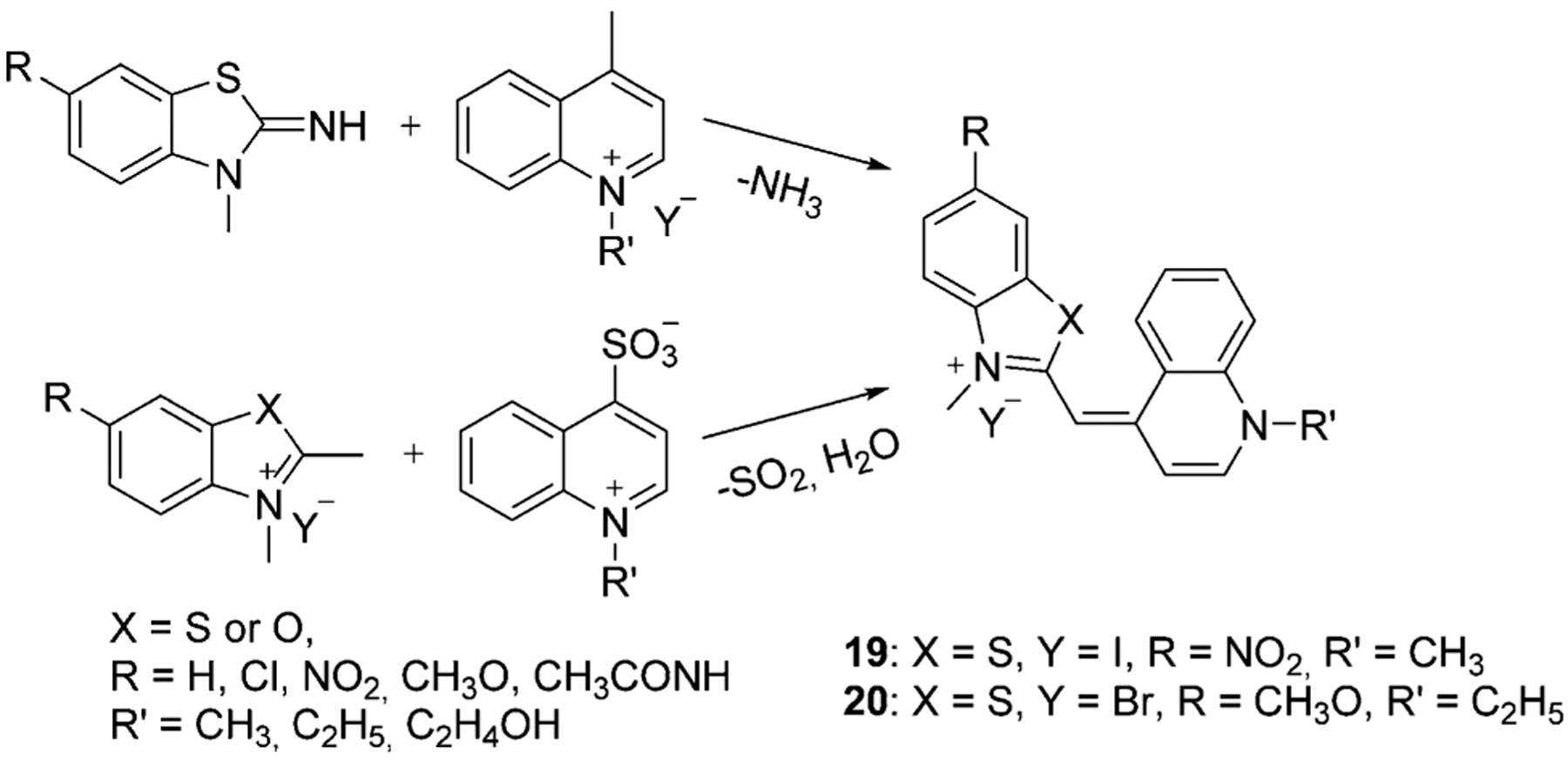
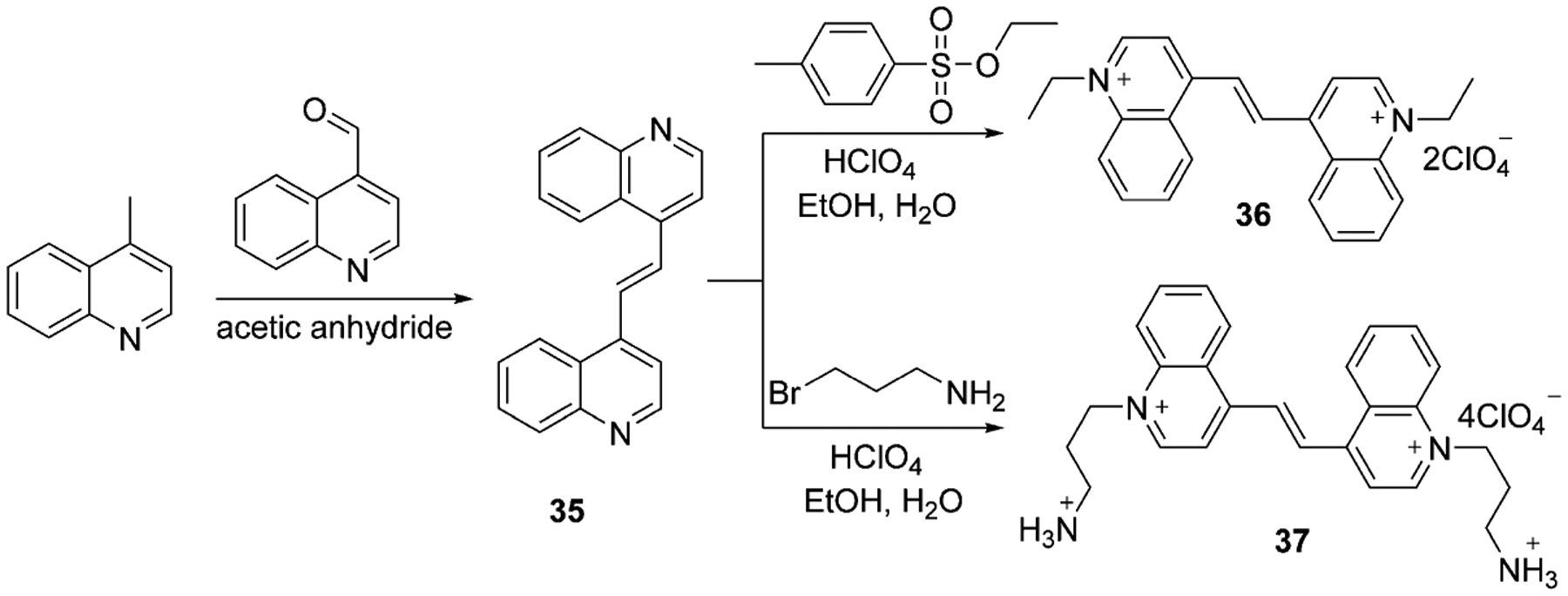
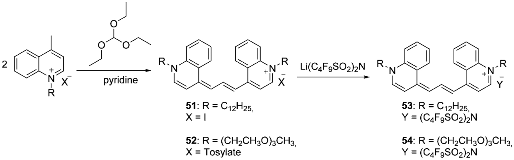
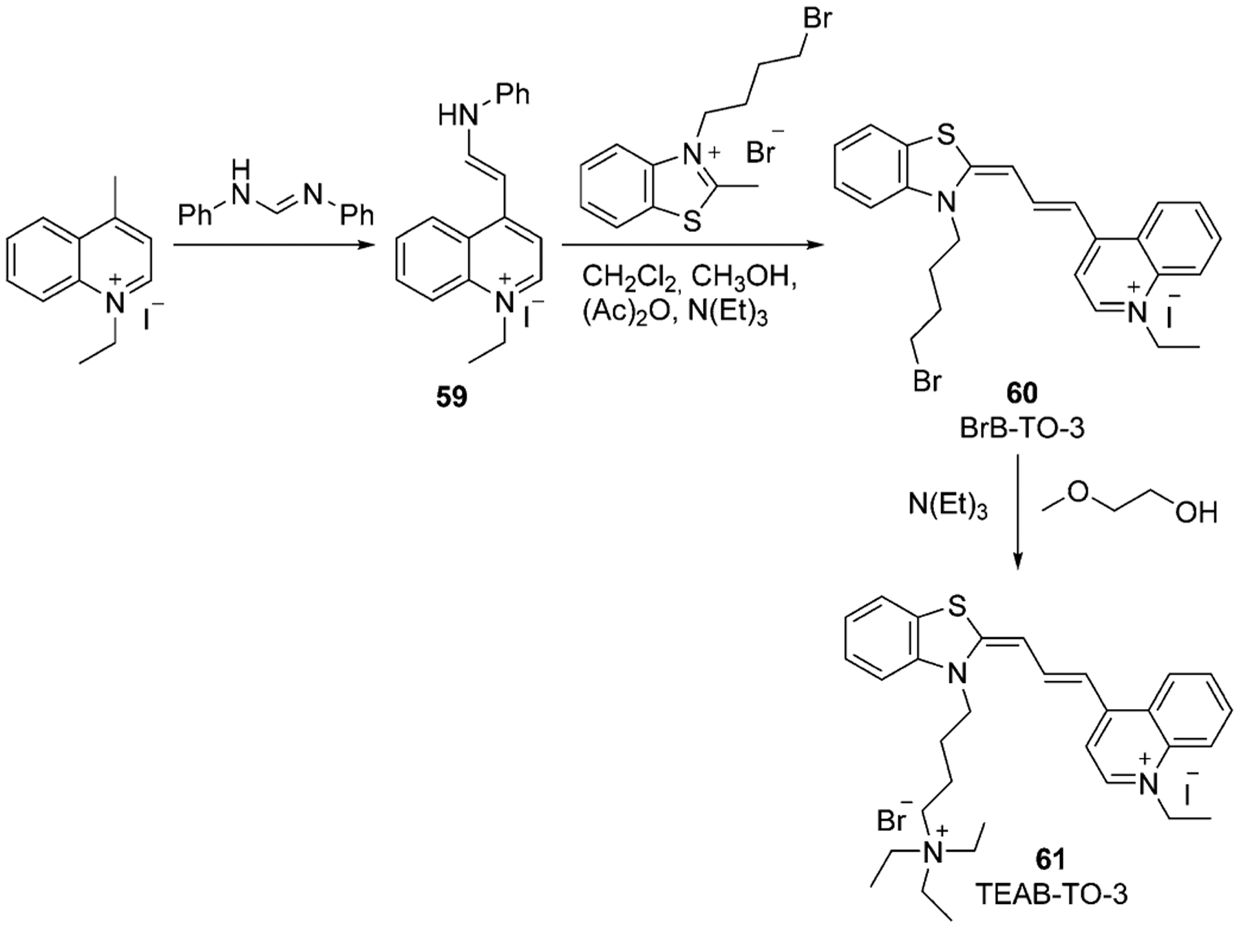
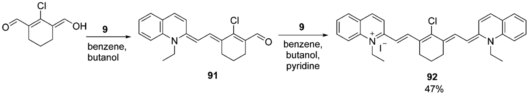
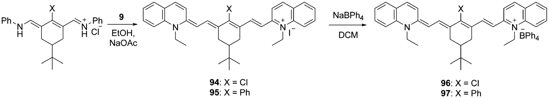
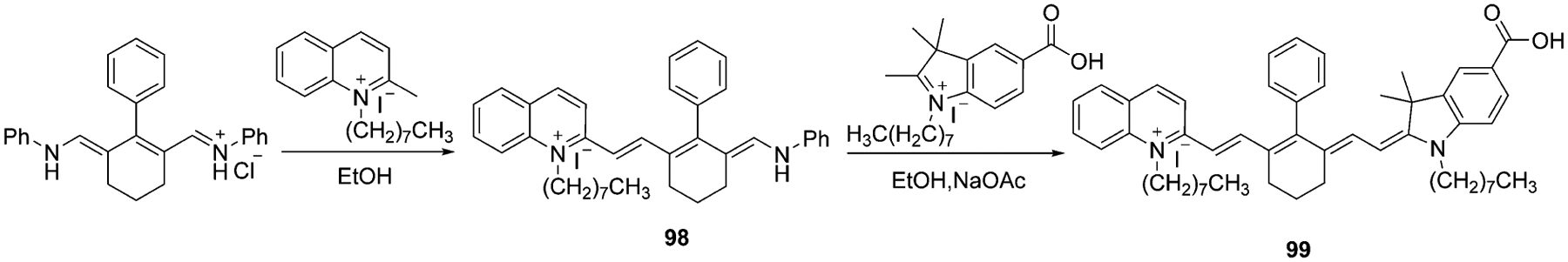
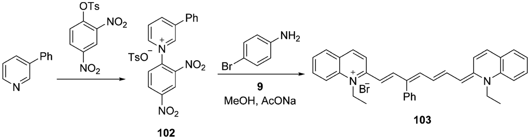
Cyanine dyes carrying quinoline moieties are an important class of organic molecules that are of great interest for applications in many fields like medicine, pharmacology, and engineering. Despite their exceptional properties, such as stability, high molar extinction coefficients, and high pH-sensitivity, this class of dyes has been less analyzed and reviewed in the last few decades. Therefore, this review article focuses on discussing the history of quinoline compounds, various synthetic routes to prepare quinolinium salts and symmetrical and asymmetrical mono-, di-, tri-, penta- and heptamethine cyanine dyes, containing quinoline moieties, together with their optical properties and applications as photosensitizers in photodynamic therapy, probes in biomolecules for labeling of nucleic acids, as well as imaging agents.

Keywords: NIR; cyanine dyes; lepidine; quinaldine; quinoline.


Conflict of interest statement

Conflict of interest

The authors declare no conflict of interest.

Figures

Figure 1.
Figure 1.
General structure of cyanine dyes.
Figure 2.
Figure 2.
Electromagnetic spectrum.[5]
Figure 3.
Figure 3.
Classification of cyanine dyes.
Figure 4.
Figure 4.
Examples of heterocyclic moieties used to synthesize cyanine dyes.
Figure 5.
Figure 5.
Structures of apocyanine dyes.
Figure 6.
Figure 6.
The structure of the first synthesized cyanine dye.
Figure 7.
Figure 7.
Dyes used as photosensitizers in photography.
Figure 8.
Figure 8.
Historic cyanine development from Acriflavine.
Figure 9.
Figure 9.
Natural products containing quinoline nucleus.
Figure 10.
Figure 10.
Monomethine quinaldine-containing cyanine dyes.[54, 55]
Figure 11.
Figure 11.
Structures of some known dimethine cyanine dyes, synthesized from lepidine and quinaldine.[, , –61]
Figure 12.
Figure 12.
Structures of first symmetrical trimethine cyanine dyes containing quinoline moieties.
Figure 13.
Figure 13.
Chemical structures of TO-3, YO-3, TO-PRO-3, and YO-PRO-3.
Figure 14.
Figure 14.
Trimethine cyanine dyes synthesized by solid-phase method.
Figure 15.
Figure 15.
Examples of symmetrical pentamethine cyanine dyes.[16, 67, 68]
Figure 16.
Figure 16.
Pentamethine cyanines synthesized by solid-phase method.
Figure 17.
Figure 17.
The absorption spectra of PIC in a mixture of water and methanol in the absence (dash) and the presence (solid) of DNA at different concentrations.[78]
Figure 18.
Figure 18.
Structure of TOTO and YOYO dimers.
Figure 19.
Figure 19.
Confocal fluorescence microscopic images of A172 cells incubated with dye 38 at concentration 20 μmolL−1 at 37 °C for 10 min.[15]
Figure 20.
Figure 20.
Fluorescence images of Tg (9 months old) and wild-type mice at different time points before (blank) and after intravenous injection of SLM 40 (dosage of 5 mgkg−1) with recordings at 10, 20, 30, 60, 90 min.[18]
Figure 21.
Figure 21.
Flow cytometry histograms of dyes 42 and 43. The x-axis presents the fluorescence intensity of stained sperm cells, and the y-axis shows the number of the stained sperm cells.[58]
Figure 22.
Figure 22.
Structure of the dimers TOTO-3 and YOYO-3.
Figure 23.
Figure 23.
Absorption and emission wavelengths of TO and YO monomers and dimers.
Figure 24.
Figure 24.
CD spectra of 4 μm G-quadruplex-forming oligonucleotides c-myc (A), HT 22 (B), and Hras (C) in the absence and presence of DIR 63 (8 μm) in 20 mm Tris-HCl buffer with 100 mm KCl, pH 7.4.[93]
Figure 25.
Figure 25.
UV/Vis (A) and fluorescence (B,C) spectra of dyes 6568 excited at 480 nm (B) and 550 nm (C).[65]
Figure 26.
Figure 26.
Absorption (main graph) and emission (inset) spectra of three symmetrical dyes: Ethyl Red (black), Kryptocyanine (blue), and 79 (red).[68]
Figure 27.
Figure 27.
Absorption (main graph) and emission (inset) spectra of three symmetrical dyes: pseudoisocyanine (black), Sensitol Red (blue), and 83 (red).[68]
Figure 28.
Figure 28.
UV/Vis spectra of 10 μm of dyes 77 (left) and 78 (right): in DMSO, 10 mm sodium phosphate buffer, pH 7.0, and in buffer with 150 μmbp CT DNA.[16]
Figure 29.
Figure 29.
(A) Superimposed fluorescence microscopy image revealing the intracellular localization of dye 78 (red) in ES2 cancer cells after 24 h incubation with the subsequent staining of nuclei Hoechst 33342 (blue). (B) ES2 cancer cell viability: with no treatment, under 808 nm laser exposure, after dye 78 incubation in the dark, after dye 78 incubation under 808 nm laser exposure.[16]
Figure 30.
Figure 30.
UV/Vis spectra of dyes 81 (top) and 82 (bottom) in DMSO (A, B), buffer pH 7.0 (C,D), and buffer pH 7.0 with 150 μm CT DNA (E,F).[94]
Figure 31.
Figure 31.
(A) Superimposed fluorescence microscopy image revealing intracellular localization of dye 81 (red) in ES2 ovarian cells after 24 h incubation with the subsequent staining of nuclei Hoechst 33342 (blue). (B) ES2 cancer cell viability: with no treatment, under 694 nm laser exposure, after dye 81 incubation in the dark, after dye 81 incubation under 694 nm laser exposure.[16]
Figure 32.
Figure 32.
The absorption spectra for dyes 96 (black) and 97 (red) in CH2Cl2 (left) and as neat thin film (right).[71]
Figure 33.
Figure 33.
Transmission of DSC with dye 99.[74]
Figure 34.
Figure 34.
Back (top) and front (bottom) sides of mouse fluorescence images of FM3A cell xenografted mouse after injection of dye 101 lactosome, recorded at 0, 3, 6, 9, 24, and 48 h.[17]
Scheme 1.
Scheme 1.
Skraup synthesis of quinolines.
Scheme 2.
Scheme 2.
Lepidine (4-methylquinoline) synthetic routes.
Scheme 3.
Scheme 3.
Quinaldine (2-methylquinoline) synthetic routes.
Scheme 4.
Scheme 4.
Synthetic routes of various lepidine salts.
Scheme 5.
Scheme 5.
Synthetic routes of various quinaldine salts.
Scheme 6.
Scheme 6.
Synthesis of TO and YO monomethine cyanine dyes.
Scheme 7.
Scheme 7.
Solid-phase synthesis of monomethine cyanines.
Scheme 8.
Scheme 8.
Synthesis of symmetrical dimethine cyanine dyes.[56, 57]
Scheme 9.
Scheme 9.
Synthesis of symmetrical trimethine cyanine dyes.
Scheme 10.
Scheme 10.
Synthesis of trimethine cyanine TEAB-TO-3.[63]
Scheme 11.
Scheme 11.
Synthesis of dimethyl indole red (DIR).[64]
Scheme 12.
Scheme 12.
Synthesis of trimethine indoline quinoline dyes.
Scheme 13.
Scheme 13.
Solid-phase synthesis of trimethine cyanine dyes.[66]
Scheme 14.
Scheme 14.
Solid-phase synthesis of pentamethine cyanine dyes.[66]
Scheme 15.
Scheme 15.
Synthesis of symmetrical heptamethine cyanine dye.[70]
Scheme 16.
Scheme 16.
Synthesis of symmetrical heptamethine cyanine dyes with substitutions at the center of the bridge.
Scheme 17.
Scheme 17.
Synthesis of asymmetrical heptamethine cyanine dye 99.[17]
Scheme 18.
Scheme 18.
Synthesis of asymmetrical heptamethine cyanine dye 101.[74]
Scheme 19.
Scheme 19.
Synthesis of heptamethine cyanine dye 103 with the substituent at the C3 position.

References

    1. Ullah S, Ahmad I, Alias Y, Yusoff I, Ashraf MA, J. Chem 2013, 136908.
    1. Ma X, Laramie M, Henary M, Bioorg. Med. Chem. Lett 2018, 28, 509–514. - PubMed
    1. Henary M, Paranjpe S, Owens EA, Heterocycl. Commun 2013, 19, 1–11.
    1. Henary M, Levitz A, Dyes Pigm. 2013, 99, 1107–1116.
    1. Visible/Infrared Spectroscopy (VIRS) as a Research Tool in Economic Geology: Background and Pilot Studies from Newfoundland and Labrador (Eds.: Kerr A, Rafuse H, Sparkes GW, Hinchey J, Sandeman H), Current Research Newfoundland and Labrador Department of Natural Resources Geological Survey, Report 11–1, 2011, pp. 145–166.

LinkOut - more resources