. 2020 Nov 2;16(11):e1009184.
doi: 10.1371/journal.pgen.1009184. eCollection 2020 Nov.

Cyclin B3 activates the Anaphase-Promoting Complex/Cyclosome in meiosis and mitosis

Affiliations

Cyclin B3 activates the Anaphase-Promoting Complex/Cyclosome in meiosis and mitosis

Damien Garrido et al. PLoS Genet. .

Abstract

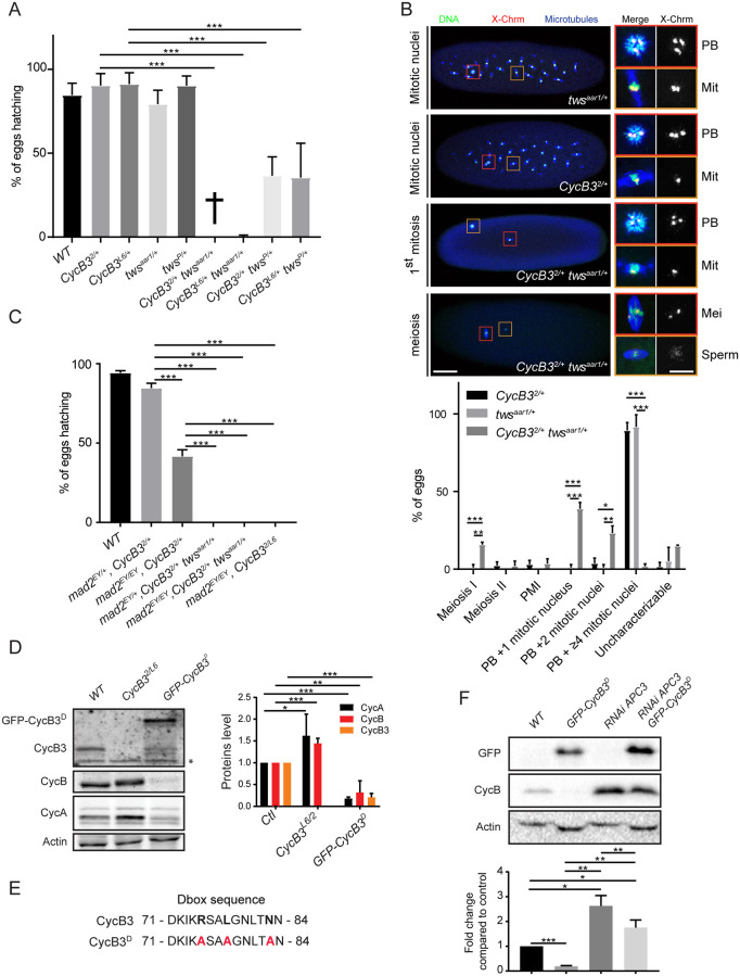
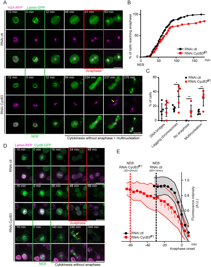
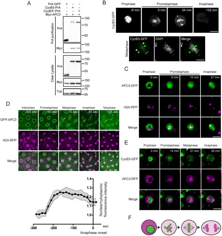
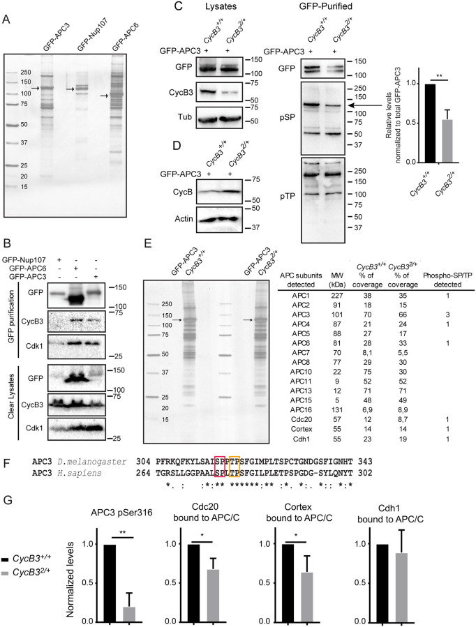
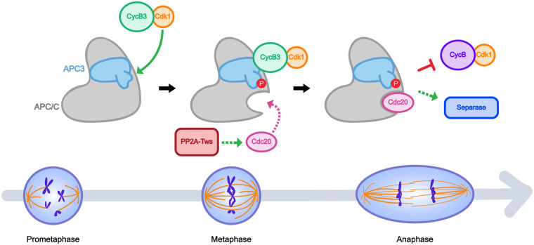
In mitosis and meiosis, chromosome segregation is triggered by the Anaphase-Promoting Complex/Cyclosome (APC/C), a multi-subunit ubiquitin ligase that targets proteins for degradation, leading to the separation of chromatids. APC/C activation requires phosphorylation of its APC3 and APC1 subunits, which allows the APC/C to bind its co-activator Cdc20. The identity of the kinase(s) responsible for APC/C activation in vivo is unclear. Cyclin B3 (CycB3) is an activator of the Cyclin-Dependent Kinase 1 (Cdk1) that is required for meiotic anaphase in flies, worms and vertebrates. It has been hypothesized that CycB3-Cdk1 may be responsible for APC/C activation in meiosis but this remains to be determined. Using Drosophila, we found that mutations in CycB3 genetically enhance mutations in tws, which encodes the B55 regulatory subunit of Protein Phosphatase 2A (PP2A) known to promote mitotic exit. Females heterozygous for CycB3 and tws loss-of-function alleles lay embryos that arrest in mitotic metaphase in a maternal effect, indicating that CycB3 promotes anaphase in mitosis in addition to meiosis. This metaphase arrest is not due to the Spindle Assembly Checkpoint (SAC) because mutation of mad2 that inactivates the SAC does not rescue the development of embryos from CycB3-/+, tws-/+ females. Moreover, we found that CycB3 promotes APC/C activity and anaphase in cells in culture. We show that CycB3 physically associates with the APC/C, is required for phosphorylation of APC3, and promotes APC/C association with its Cdc20 co-activators Fizzy and Cortex. Our results strongly suggest that CycB3-Cdk1 directly activates the APC/C to promote anaphase in both meiosis and mitosis.


Conflict of interest statement

The authors have declared that no competing interests exist.

Figures

Fig 1
Fig 1. CycB3 collaborates with PP2A-Tws and functions upstream of the APC/C to promote the metaphase-anaphase transition in meiosis and mitosis.
A. Mutations in CycB3 and tws enhance each other in a maternal effect. Eggs from mothers of the indicated genotypes were scored for their hatching rate. B. Eggs from CycB32/+ twsaar1/+ mothers arrest in metaphase of meiosis or mitosis. Green: DNA, Blue: α-Tubulin, Red: FISH for pericentric DNA of the X chromosome. Mitotic spindles (Mit) are recognized by the presence of astral microtubules, co-occurrence with a polar body (PB), and 1 or 2 foci of pericentromeric X-chromosome DNA. Meiotic I metaphase spindles (Mei) are recognized by the absence of astral microtubules, the absence of a polar body in the egg, and at least 2 foci of pericentromeric X-chromosome DNA. In a meiotic arrest, the sperm nucleus can break and nucleate a spindle (bottom images). Insets from the left are enlarged on the right with corresponding color frames. Scale bars: left panel 50 μm, right panel 10 μm. PMI: post-meiotic interphase. C. Inactivation of the SAC by mutation of mad2 does not rescue the development of embryos from CycB32/L6 or CycB32/+ twsaar1/+ mothers. Eggs from mothers of the indicated genotypes were scored for their hatching rate. D. Mutation of CycB3 results in higher CycA and CycB levels, while maternal expression of GFP-CycB3D results in lower CycA, CycB and endogenous CycB3 levels in eggs. WT (Wild type): non-fertilized eggs. *Non-specific band. E. Mutation of the destruction box motif in CycB3D. F. CycB3 negatively regulates CycB levels in an APC-dependent manner. Eggs were collected for 2 hrs from the indicated conditions and analyzed by Western blot. Expression of UASp-GFP-CycB3D and of UASp-APC3 RNAi was driven maternally by Mat α-Tubulin Gal4-VP16. The UASp-GFP-CycB3D and UASp-APC3 RNAi alone genotypes also contained a UASp-WHITE construct, to control for potential dilution of Gal4. Eggs from all conditions failed to develop (WT: unfertilized eggs). Error bars: SD. ***p < 0.001; **p < 0.01; *p < 0.05 from ANOVA for panels A and C and from paired t-tests for panels B, D and F. Numbers underlying graphs are available in supplemental file S1 Fig Numerical Data. See also S1 Fig.
Fig 2
Fig 2. CycB3 is required for normal metaphase-anaphase transition and for CycB degradation in cells in culture.
A. RNAi depletion of CycB3 delays anaphase entry and results in chromosome segregation defects in D-Mel cells. Note the cut through unsegregated chromosomes in this no-anaphase phenotype (arrow). RNAi ctl = transfection of dsRNA made from the bacterial KAN gene. B. Cumulative proportion of mitotic cells entering anaphase as a function of time after NEB. C. Frequency of cell division defects observed. Error bars: SD. For B-C, 63 and 69 cells were analyzed for RNAi CycB3 and ctl, respectively. D. RNAi depletion of CycB3 delays CycB degradation at the metaphase-anaphase transition. Examples of cell divisions are shown (Z-projection of 3 planes in focus through the nucleus). Note the abnormal persistence of CycB-GFP on the chromosomes (arrow) and centrosomes (asterisks) during cytokinesis in a cell that does not enter anaphase (bottom). E. Levels of CycB-GFP were quantified through time, relative to anaphase onset. 29 and 30 cells were analyzed for RNAi CycB3 and ctl, respectively. Error areas: SD. ***p < 0.001; **p < 0.01; *p < 0.05 from paired t-tests. Scale bars: 10 μm. Numbers underlying graphs are available in supplemental file S2 Fig Numerical Data. See also S2 Fig.
Fig 3
Fig 3. CycB3 associates with the APC/C and shows a dynamic localization relative to the APC/C in the cell cycle.
A. Myc-APC3 associates with CycB3-PrA independently from its destruction box. Cells expressing the indicated proteins were submitted to Protein A affinity purification and products were analyzed by Western blot. B. CycB3-GFP localizes to chromosomes (bracket) and spindle poles (asterisks) in early mitosis. Top: Live imaging; Bottom: a fixed cell in metaphase. C. APC3-GFP is predominantly cytoplasmic in interphase and becomes enriched on chromosomes during mitosis in D-Mel cells. D. In syncytial embryos, GFP-APC3 becomes enriched in nuclei during mitotic entry and concentrates around the metaphase plate during mitosis (arrows). Scale bar: 10 μm. Bottom: quantification of the nuclear/cytoplasmic ratio of GFP-APC3 fluorescence intensity as nuclei enter mitosis. Averages between 10 nuclei of the same embryo are shown. Error area: SD. E. CycB3-GFP and APC3-RFP are sequestered from each other in interphase and come together on chromosomes in mitosis in D-Mel cells. Scale bars: 10 μm except for the fixed cell: 5 μm. F. Schematic model of the dynamic localization of CycB3 (green) and the APC/C (magenta) from interphase to anaphase. Numbers underlying graph are available in supplemental file S3 Fig Numerical Data. See also S3 Fig.
Fig 4
Fig 4. CycB3-Cdk1 physically associates with the APC/C and promotes its phosphorylation and interaction with Cdc20 co-activators.
A. Embryos expressing GFP-APC3, GFP-APC6 and GFP-Nup107 were submitted to GFP-affinity purification and purified complexes were visualized on a silver-stained gel. Arrows: GFP-tagged proteins. B. CycB3 physically associates with the APC/C in embryos. Embryos expressing the indicated proteins were submitted to GFP-affinity purification and products were analyzed by Western blots. C. APC/C phosphorylation at SP site(s) is CycB3-dependent. GFP-affinity purifications were conducted from 0–2 hrs-old GFP-APC3 expressing embryos from CycB3+/+ and CycB32/+ mothers and products were analyzed by Western blots. Left: Western blots on lysates used in the purifications. Note that CycB3 levels are lower in CycB32/+ embryos; Center: GFP-APC3 purified products were analyzed by Western blot with antibodies against pSP or pTP sites (minimal CDK phosphorylation motifs). Arrow: presumed GFP-APC3 pSP band, less intense in CycB32/+ embryos. Right: relative intensities of the band indicated by the arrow, after normalization to the amounts of total GFP-APC3 purified. Error bar: SD for 3 experiments. **p = 0.0082 from paired t-test. D. Western blot showing that CycB levels are higher in CycB32/+ embryos (aged 0–2 hrs). E. Purification of the APC/C complex using GFP-APC3 from CycB3+/+ and CycB32/+ embryos, for mass spectrometry analysis. Table: APC/C subunits detected by mass spectrometry. Their predicted molecular weight (MW), percentage of sequence detected and number of phospho SP/TP sites detected are indicated. F. Phosphorylation sites at minimal CDK motifs identified in Drosophila APC3 are conserved in the activation loop of human APC3. Red box: Ser316; Orange box: Thr319. G. Phosphorylation at Ser316 in APC3 is reduced in CycB32/+ embryos. In addition, the amounts of Cdc20/Fizzy and Cortex co-purified with the APC/C are reduced in CycB32/+ embryos. The amount of Cdh1/Fzr co-purified is not significantly affected. Proteins and phosphopeptides abundances relative to APC3 levels were normalized to 1 in the CycB3+/+ condition (see Methods). Error bars: SD for 3 experiments. Numbers underlying graphs are available in supplemental file S4 Fig Numerical Data.
Fig 5
Fig 5. Model for APC/C activation by CycB3-Cdk1 in meiosis and mitosis.
CycB3-Cdk1 binds the APC/C and phosphorylates its APC3 subunit. This event promotes the binding of Cdc20 co-activators, likely through known mechanisms [19,20]. In addition, PP2A-Tws may help Cdc20 recruitment to the APC/C [15,17]. Activated APC/C triggers CycB degradation, separase activation and anaphase.

Similar articles

Cited by

References

    1. Morgan DO. The Cell Cycle: Principles of Control. London: New Science Press; 2007. 297 p.
    1. Holder J, Poser E, Barr FA. Getting out of mitosis: spatial and temporal control of mitotic exit and cytokinesis by PP1 and PP2A. FEBS Lett. 2019;593(20):2908–24. 10.1002/1873-3468.13595 - DOI - PubMed
    1. Schellhaus AK, De Magistris P, Antonin W. Nuclear Reformation at the End of Mitosis. J Mol Biol. 2016;428(10 Pt A):1962–85. 10.1016/j.jmb.2015.09.016 - DOI - PubMed
    1. Yamano H. APC/C: current understanding and future perspectives. F1000Res. 2019;8 10.12688/f1000research.18582.1 - DOI - PMC - PubMed
    1. Alfieri C, Zhang S, Barford D. Visualizing the complex functions and mechanisms of the anaphase promoting complex/cyclosome (APC/C). Open Biol. 2017;7(11). - PMC - PubMed

Publication types

MeSH terms

LinkOut - more resources