. 2020 Oct 29;9(11):366.
doi: 10.3390/biology9110366.

The Platelet Fraction Is a Novel Reservoir to Detect Lyme Borrelia in Blood

Affiliations

The Platelet Fraction Is a Novel Reservoir to Detect Lyme Borrelia in Blood

Victoria P Sanderson et al. Biology (Basel). .

Abstract

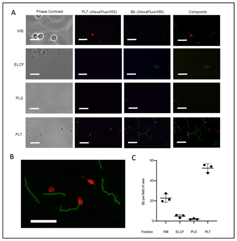
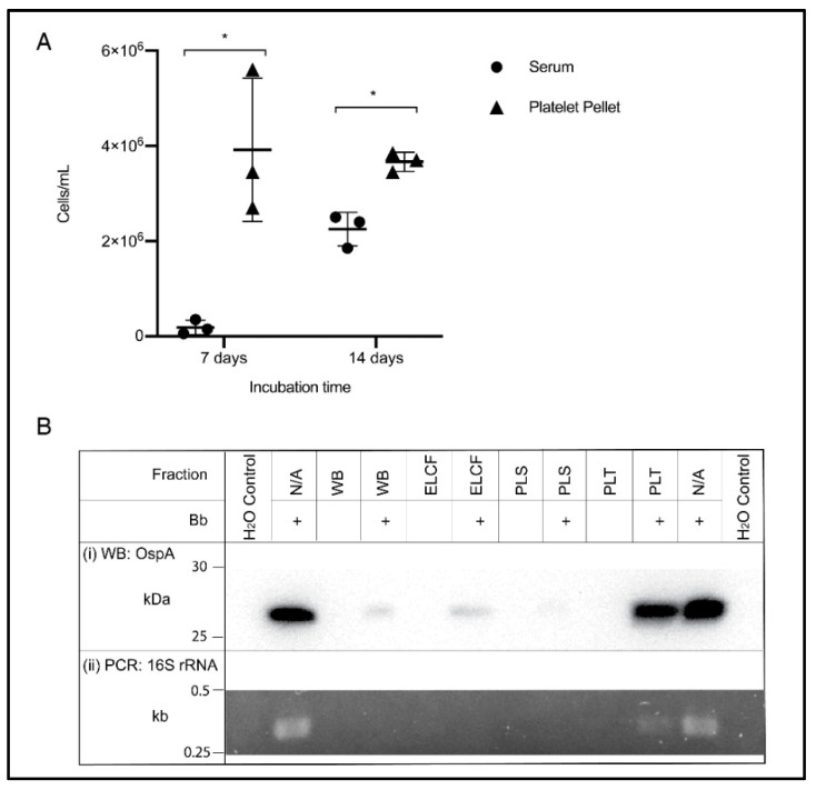
Serological diagnosis of Lyme disease suffers from considerable limitations. Yet, the technique cannot currently be replaced by direct detection methods, such as bacterial culture or molecular analysis, due to their inadequate sensitivity. The low bacterial burden in vasculature and lack of consensus around blood-based isolation of the causative pathogen, Borrelia burgdorferi, are central to this challenge. We therefore addressed methodological optimization of Borrelia recovery from blood, first by analyzing existing protocols, and then by using experimentally infected human blood to identify the processing conditions and fractions that increase Borrelia yield. In this proof-of-concept study, we now report two opportunities to improve recovery and detection of Borrelia from clinical samples. To enhance pathogen viability and cultivability during whole blood collection, citrate anticoagulant is superior to more commonly used EDTA. Despite the widespread reliance on serum and plasma as analytes, we found that the platelet fraction of blood concentrates Borrelia, providing an enriched resource for direct pathogen detection by microscopy, laboratory culture, Western blot, and PCR. The potential for platelets to serve as a reservoir for Borrelia and its diagnostic targets may transform direct clinical detection of this pathogen.

Keywords: Borrelia; EDTA; Lyme disease; OspA; blood processing; citrate; culture; diagnosis; molecular detection; platelets.


Conflict of interest statement

The authors declare no conflict of interest. The funders had no role in the design of the study; in the collection, analyses, or interpretation of data; in the writing of the manuscript, or in the decision to publish the results.

Figures

Figure 1
Figure 1
Reported use of anticoagulants to recover Borrelia from human blood. Relevant literature was curated (as described in Materials and Methods) to compare which vacutainers are commonly used when conducting Borrelia clinical culture from patient samples. Percentage calculated as number of times the vacutainer was used relative to the total experiments conducted using vacutainers in a set of applicable studies.
Figure 2
Figure 2
Influence of anticoagulants on Borrelia cultivability. To consider anticoagulant impact on Borrelia culture growth, we performed cell counts following inoculation of Borrelia into BSK broth and incubation in uncoated, EDTA, and citrate vacutainers, monitoring (A) the direct effects of anticoagulants on pathogen growth, and (B) consequences upon subcultivation. Culture was aliquoted into fresh BSK in an uncoated culture tube after 48 h in vacutainers (from A), to mimic standard shipping and laboratory culture procedure. Horizontal lines represent mean of three biological replicates and whiskers display the standard deviation. Asterisks indicate a significant difference (* = p < 0.05, ** = p < 0.0001) by one-way ANOVA and Tukey’s multiple comparisons post-hoc test (n = 3).
Figure 3
Figure 3
Stratification of Borrelia in blood following separation of cells and plasma. Quantification of Borrelia per field of view in the supernatant/liquid fraction and cellular/lower fraction following centrifugation at 400× g, 20 min. Data presented as the means with standard deviation. Asterisks (* = p < 0.05) indicate significance difference using one-tailed paired t-tests (n = 3).
Figure 4
Figure 4
Schematic of blood fractionation protocol. Multiple steps were implemented to obtain a panel of blood components for comparison by microscopy, culture and molecular analyses. The initial centrifugation step pellets red and white blood cells at 120× g for 20 min, leaving a platelet-rich plasma supernatant which is subsequently spun at 400× g for 20 min to obtain platelets (pellet) and acellular plasma (supernatant). WB = whole blood, ELCF = erythrocyte, leukocyte cellular fraction, PRP = platelet-rich plasma, PLS = plasma (platelet poor), and PLT = platelet fraction.
Figure 5
Figure 5
Microscopy of experimentally infected blood before and after fractionation. (A) Representative immunofluorescent micrographs of Borrelia (green) in fractions collected from experimentally infected whole blood (platelets labelled red). Scale bar = 13 μm. (B) Magnification of PLT fraction composite demonstrating co-localization. Scale bar = 13 μm. (C) Corresponding quantification of Borrelia per field of view from A; n = 3. All comparisons by ANOVA and Tukey’s multiple comparisons post-hoc test were significant with p < 0.005 except red blood cells and plasma. Data represented as the means with standard deviation. WB = whole blood, ELCF = erythrocyte, leukocyte cellular fraction, PRP = platelet-rich plasma, PLS = plasma (platelet poor), and PLT = platelet fraction.
Figure 6
Figure 6
Culture and molecular detection of Borrelia in blood fractions. (A) Broth culture of serum and platelet fractions collected from experimentally infected blood samples, monitored weekly for growth. Data represented as the means with standard deviation. Asterisks indicate significant difference by one-tailed paired t-test (* = p < 0.05) (n = 3). (B) Detection of OspA protein by Western blot (i) and the 16S rRNA gene by PCR (ii) in experimentally infected and matched uninfected blood, and controls. WB = whole blood, ELCF = erythrocyte, leukocyte cellular fraction, PLS = plasma, and PLT = platelet fraction.

Similar articles

Cited by

References

    1. Nelson C.A., Saha S., Kugeler K.J., Delorey M.J., Shankar M.B., Hinckley A.F., Mead P.S. Incidence of Clinician-Diagnosed Lyme Disease, United States, 2005–2010. Emerg. Infect. Dis. 2015;21:1625–1631. - PMC - PubMed
    1. Schutzer S.E., Body B.A., Boyle J., Branson B.M., Dattwyler R.J., Fikrig E., Gerald N.J., Gomes-Solecki M., Kintrup M., Ledizet M., et al. Direct Diagnostic Tests for Lyme Disease. Clin. Infect. Dis. 2019;68:1052–1057. - PMC - PubMed
    1. Feder H.M., Jr., Abeles M., Bernstein M., Whitaker-Worth D., Grant-Kels J.M. Diagnosis, treatment, and prognosis of erythema migrans and Lyme arthritis. Clin. Dermatol. 2006;24:509–520. doi: 10.1016/j.clindermatol.2006.07.012. - DOI - PubMed
    1. Godar D.A., Laniosz V., Wetter D.A. Lyme Disease Update for the General Dermatologist. Am. J. Clin. Dermatol. 2015;16:5–18. - PubMed
    1. Fix A.D., Peña C.A., Strickland G.T. Racial differences in reported lyme disease incidence. Am. J. Epidemiol. 2000;152:756–759. doi: 10.1093/aje/152.8.756. - DOI - PubMed

LinkOut - more resources