Review
. 2020 Oct 29;9(11):2372.
doi: 10.3390/cells9112372.

Autophagy and Lc3-Associated Phagocytosis in Zebrafish Models of Bacterial Infections

Affiliations
Review

Autophagy and Lc3-Associated Phagocytosis in Zebrafish Models of Bacterial Infections

Salomé Muñoz-Sánchez et al. Cells. .

Abstract

Modeling human infectious diseases using the early life stages of zebrafish provides unprecedented opportunities for visualizing and studying the interaction between pathogens and phagocytic cells of the innate immune system. Intracellular pathogens use phagocytes or other host cells, like gut epithelial cells, as a replication niche. The intracellular growth of these pathogens can be counteracted by host defense mechanisms that rely on the autophagy machinery. In recent years, zebrafish embryo infection models have provided in vivo evidence for the significance of the autophagic defenses and these models are now being used to explore autophagy as a therapeutic target. In line with studies in mammalian models, research in zebrafish has shown that selective autophagy mediated by ubiquitin receptors, such as p62, is important for host resistance against several bacterial pathogens, including Shigella flexneri, Mycobacterium marinum, and Staphylococcus aureus. Furthermore, an autophagy related process, Lc3-associated phagocytosis (LAP), proved host beneficial in the case of Salmonella Typhimurium infection but host detrimental in the case of S. aureus infection, where LAP delivers the pathogen to a replication niche. These studies provide valuable information for developing novel therapeutic strategies aimed at directing the autophagy machinery towards bacterial degradation.

Keywords: Cyba; Dram1; LAP; Optn; Rubcn; autophagy; innate immunity; p62; tuberculosis zebrafish.


Conflict of interest statement

The authors declare no conflict of interest.

Figures

Figure 1
Figure 1
Antimicrobial mechanisms dependent on the autophagy machinery. (A) Xenophagy is a subtype of selective autophagy mediated by Sequestosome1-like receptors (SLRs) that target ubiquitinated microbes in the cytosol and interact with LC3 on nascent autophagosomes. (B) SLRs also detect galectins on damaged phagosomes, which may induce the autophagic degradation of these vesicles or initiate an autophagy-mediated membrane repair process, preventing microbes from entering the cytosol. (C) The autophagolysosomal pathway processes ubiquitinated proteins into antimicrobial peptides. (D) The formation of LAPosomes, where LC3 associates with phagosomes, is an autophagy-related process linked with reactive oxygen production.
Figure 2
Figure 2
GFP-Lc3 response to M. marinum infection in the tail fin of zebrafish larvae. In the experimental setup of tail fin injections, mCrimson-labeled M. marinum bacteria (200 colony forming units) are microinjected into the tail fin area of zebrafish larvae at 3 days post fertilization (dpf), derived from a double transgenic line that contains a ubiquitously expressed CMV:GFP-Lc3 construct and an mpeg1:mCherry-F construct that labels macrophages. The injection site and region of imaging (ROI) is indicated in the schematic drawing of the larva and a representative bright field and fluorescence overlay image shows the infected area in the tail fin. Confocal laser-scanning microscopy images were acquired 1, 4, and 24 h post infection (hpi). Fluorescent excitation in the range of GFP, far-red and red were used to visualize GFP-Lc3, M. marinum, and macrophages, respectively. At 1 hpi, macrophages are already recruited at the site of infection and phagocytosis of bacteria can be observed. At 4 hpi, GFP-Lc3 signal is observed inside infected macrophages and often appears in ring-shaped patterns. At 24 hpi, the bacterial cluster sizes and numbers are increased, both intra- and extracellularly, which is associated with strong punctate GFP-Lc3 signal in the infected tissue. The boxed areas in the representative images of the different time points are shown in detail on the right. Scale bars: 10 μm.
Figure 3
Figure 3
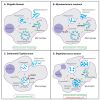
Host-protective and host-detrimental interactions of bacterial pathogens with the autophagy machinery in zebrafish infection models. (A) p62-dependent xenophagy restricts growth of S. flexneri bacteria in macrophages of the zebrafish host. (B) In the zebrafish M. marinum model, p62, Optn, and Dram1 are required for host resistance. p62 and Optn both mediate xenophagy in infected macrophages, while Dram1, an integral membrane protein of intracellular vesicles including lysosomes, promotes vesicle fusion events. (C) LAP, which requires the signaling molecule Rubicon (Rubcn) and the Cyba component of NADPH oxidase, is the predominant defense response of zebrafish macrophages to S. Typhimurium infection. (D) In zebrafish neutrophils, Cyba-mediated LAP provides a replication niche for S. aureus, while p62-mediated xenophagy counteracts the growth of bacteria that may be released from phagosomes or LAPosomes. Functions of the proteins indicated in bold have been demonstrated by knockdown, mutation, and/or overexpression analyses. See the text for references.

Similar articles

Cited by

References

    1. Levine B., Mizushima N., Virgin H.W. Autophagy in immunity and inflammation. Nature. 2011;469:323–335. doi: 10.1038/nature09782. - DOI - PMC - PubMed
    1. Deretic V., Chauhan S., Mandell M., Deretic V., Kimura T., Timmins G., Moseley P., Chauhan S., Mandell M. Immunologic manifestations of autophagy. J. Clin. Investig. 2015;125:75–84. doi: 10.1172/JCI73945. - DOI - PMC - PubMed
    1. Deretic V. Autophagy in leukocytes and other cells: Mechanisms, subsystem organization, selectivity, and links to innate immunity. J. Leukoc. Biol. 2016;100:969–978. doi: 10.1189/jlb.4MR0216-079R. - DOI - PMC - PubMed
    1. Huang J., Brumell J.H. Bacteria-autophagy interplay: A battle for survival. Nat. Rev. Microbiol. 2014;12:101–114. doi: 10.1038/nrmicro3160. - DOI - PMC - PubMed
    1. da Silva Siqueira M., de Moraes Ribeiro R., Travassos L.H. Autophagy and its interaction with intracellular bacterial pathogens. Front. Immunol. 2018;9 doi: 10.3389/fimmu.2018.00935. - DOI - PMC - PubMed

Publication types

Substances