Cornus officinalis Ethanolic Extract with Potential Anti-Allergic, Anti-Inflammatory, and Antioxidant Activities
- PMID: 33138027
- PMCID: PMC7692184
- DOI: 10.3390/nu12113317
Cornus officinalis Ethanolic Extract with Potential Anti-Allergic, Anti-Inflammatory, and Antioxidant Activities
Abstract
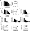
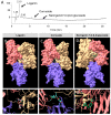
Atopic dermatitis (AD) is an allergic and chronic inflammatory skin disease. The present study investigates the anti-allergic, antioxidant, and anti-inflammatory activities of the ethanolic extract of Cornus officinalis (COFE) for possible applications in the treatment of AD. COFE inhibits the release of β-hexosaminidase from RBL-2H3 cells sensitized with the dinitrophenyl-immunoglobulin E (IgE-DNP) antibody after stimulation with dinitrophenyl-human serum albumin (DNP-HSA) in a concentration-dependent manner (IC50 = 0.178 mg/mL). Antioxidant activity determined using 2,2-diphenyl-1-picrylhydrazyl (DPPH) radical scavenging activity, ferric reducing antioxidant power assay, and 2,2'-azino-bis(3-ethylbenzothiazoline-6-sulfonic acid) (ABTS) scavenging activity, result in EC50 values of 1.82, 10.76, and 0.6 mg/mL, respectively. Moreover, the extract significantly inhibits lipopolysaccharide (LPS)-induced nitric oxide (NO) production and the mRNA expression of iNOS and pro-inflammatory cytokines (IL-1β, IL-6, and TNF-α) through attenuation of NF-κB activation in RAW 264.7 cells. COFE significantly inhibits TNF-α-induced apoptosis in HaCaT cells without cytotoxic effects (p < 0.05). Furthermore, 2-furancarboxaldehyde and loganin are identified by gas chromatography/mass spectrometry (GC-MS) and liquid chromatography with tandem mass spectrometry (LC-MS/MS) analysis, respectively, as the major compounds. Molecular docking analysis shows that loganin, cornuside, and naringenin 7-O-β-D-glucoside could potentially disrupt the binding of IgE to human high-affinity IgE receptors (FceRI). Our results suggest that COFE might possess potential inhibitory effects on allergic responses, oxidative stress, and inflammatory responses.
Keywords: Cornus officinalis; anti-inflammatory activity; antioxidant activity; atopic dermatitis; human high-affinity IgE receptors; molecular docking.
Conflict of interest statement
The authors declare no conflict of interest.
Figures


Similar articles
-
Antioxidant activities and oxidative stress inhibitory effects of ethanol extracts from Cornus officinalis on raw 264.7 cells.BMC Complement Altern Med. 2016 Jul 8;16:196. doi: 10.1186/s12906-016-1172-3. BMC Complement Altern Med. 2016. PMID: 27391600 Free PMC article.
-
Chemical composition and anti-inflammatory activity of essential oil and ethanolic extract of Campomanesia phaea (O. Berg.) Landrum leaves.J Ethnopharmacol. 2020 Apr 24;252:112562. doi: 10.1016/j.jep.2020.112562. Epub 2020 Jan 15. J Ethnopharmacol. 2020. PMID: 31954197
-
Potential anti-inflammatory, antioxidant and antimicrobial activities of Sambucus australis.Pharm Biol. 2017 Dec;55(1):991-997. doi: 10.1080/13880209.2017.1285324. Pharm Biol. 2017. PMID: 28166708 Free PMC article.
-
The Pro-Health Benefits of Morusin Administration-An Update Review.Nutrients. 2021 Aug 30;13(9):3043. doi: 10.3390/nu13093043. Nutrients. 2021. PMID: 34578920 Free PMC article. Review.
-
Active Components and Pharmacological Effects of Cornus officinalis: Literature Review.Front Pharmacol. 2021 Apr 12;12:633447. doi: 10.3389/fphar.2021.633447. eCollection 2021. Front Pharmacol. 2021. PMID: 33912050 Free PMC article. Review.
Cited by
-
Antiosteoarthritic Effect of Morroniside in Chondrocyte Inflammation and Destabilization of Medial Meniscus-Induced Mouse Model.Int J Mol Sci. 2021 Mar 15;22(6):2987. doi: 10.3390/ijms22062987. Int J Mol Sci. 2021. PMID: 33804203 Free PMC article.
-
8-Prenylnaringenin tissue distribution and pharmacokinetics in mice and its binding to human serum albumin and cellular uptake in human embryonic kidney cells.Food Sci Nutr. 2022 Jan 22;10(4):1070-1080. doi: 10.1002/fsn3.2733. eCollection 2022 Apr. Food Sci Nutr. 2022. PMID: 35432956 Free PMC article.
-
Optimizing Processing Technology of Cornus officinalis: Based on Anti-Fibrotic Activity.Front Nutr. 2022 May 3;9:807071. doi: 10.3389/fnut.2022.807071. eCollection 2022. Front Nutr. 2022. PMID: 35592634 Free PMC article.
-
Ethanol-Induced Hepatotoxicity and Alcohol Metabolism Regulation by GABA-Enriched Fermented Smilax china Root Extract in Rats.Foods. 2021 Oct 8;10(10):2381. doi: 10.3390/foods10102381. Foods. 2021. PMID: 34681429 Free PMC article.
-
Mechanism Prediction of Astragalus membranaceus against Cisplatin-Induced Kidney Damage by Network Pharmacology and Molecular Docking.Evid Based Complement Alternat Med. 2021 Aug 19;2021:9516726. doi: 10.1155/2021/9516726. eCollection 2021. Evid Based Complement Alternat Med. 2021. PMID: 34457031 Free PMC article.
References
MeSH terms
Substances
Grants and funding
LinkOut - more resources
Full Text Sources
Medical
Miscellaneous

