. 2020 Oct 29;21(21):8079.
doi: 10.3390/ijms21218079.

Developmental Roles of FUSE Binding Protein 1 (Fubp1) in Tooth Morphogenesis

Affiliations

Developmental Roles of FUSE Binding Protein 1 (Fubp1) in Tooth Morphogenesis

Yam Prasad Aryal et al. Int J Mol Sci. .

Abstract

FUSE binding protein 1 (Fubp1), a regulator of the c-Myc transcription factor and a DNA/RNA-binding protein, plays important roles in the regulation of gene transcription and cellular physiology. In this study, to reveal the precise developmental function of Fubp1, we examined the detailed expression pattern and developmental function of Fubp1 during tooth morphogenesis by RT-qPCR, in situ hybridization, and knock-down study using in vitro organ cultivation methods. In embryogenesis, Fubp1 is obviously expressed in the enamel organ and condensed mesenchyme, known to be important for proper tooth formation. Knocking down Fubp1 at E14 for two days, showed the altered expression patterns of tooth development related signalling molecules, including Bmps and Fgf4. In addition, transient knock-down of Fubp1 at E14 revealed changes in the localization patterns of c-Myc and cell proliferation in epithelium and mesenchyme, related with altered tooth morphogenesis. These results also showed the decreased amelogenin and dentin sialophosphoprotein expressions and disrupted enamel rod and interrod formation in one- and three-week renal transplanted teeth respectively. Thus, our results suggested that Fubp1 plays a modulating role during dentinogenesis and amelogenesis by regulating the expression pattern of signalling molecules to achieve the proper structural formation of hard tissue matrices and crown morphogenesis in mice molar development.

Keywords: Fubp1; amelogenesis; dentinogenesis; tooth development; transcriptional regulator.


Conflict of interest statement

The authors declare no potential conflicts of interest with respect to the authorship and/or publication of this article.

Figures

Figure 1
Figure 1
Expression of Fubp1 in developing tooth. H&E staining of developing tooth (ac) and section in situ hybridization of Fubp1 at E14-E16 (df). At E14 and E15, Fubp1 is expressed broadly at epithelium and mesenchyme. At E16, Fubp1 is expressed in IEE and DP, however, more intense expression is observed in IEE. The dotted lines demarcate the boundary of tooth epithelium (af). EK, enamel knot; IEE, inner enamel epithelium; OEE, outer enamel epithelium; SR, stellate reticulum; DP, dental papilla. Scale bars: 50 μm (af).
Figure 2
Figure 2
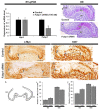
Altered c-Myc localization and cellular proliferation after Fubp1 knock-down. Fubp1 siRNA (100 nM) downregulates the expression of Fubp1 during in vitro organ cultivation at E14 for 1 day as determined by quantitative PCR (a). Hematoxylin and eosin staining of E14 + 2 control and Fubp1 siRNA-transfected tooth germ showing IEE and DP (b,c). Immunohistochemistry of c-Myc and Ki67 showing increased number of c-Myc and ki67 positive cells at IEE and decreased number of c-Myc and Ki67 cells at OEE in the Fubp1 knock-down specimen compared to control (dg). Schematic diagram showing region of interest for cell count (h). Statistical analysis showing increased and decreased c-Myc and Ki67 positive cell numbers along the IEE and OEE respectively in the Fubp1 knock-down specimen (i,j). Arrows and arrowheads indicate c-Myc positive cells (d,f) and Ki67 positive cells (e,g). EK; enamel knot, IEE; inner enamel epithelium, OEE; outer enamel epithelium, DP; dental Papilla. Scale bars: 50 μm (bg).
Figure 3
Figure 3
Altered epithelial cell rearrangements in Fubp1 knock-down molar. Knocking down Fubp1 at E14 for 2 days shows decreased localization of E-cadherin and ROCK1 along the inner enamel epithelium (ae). Similarly, weak phalloidin staining is observed in the basement membrane of inner enamel epithelium along the secondary enamel-knot in the Fubp1 knock-down tooth compared with control (c,f). The dotted lines demarcate the boundary of tooth epithelium (af).* in control indicates comparatively increased localization of respective proteins along inner enamel epithelium when compared with the same region of Fubp1 knock-down tooth (af). Scale bars: 20 μm (af).
Figure 4
Figure 4
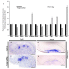
Expression patterns of tooth related signalling molecules after Fubp1 knock-down. Knock-down of Fubp1 results altered expression of tooth development related signalling molecules including Bmp, Wnt, Shh and Fgf4 as detected by qPCR (a) and section in situ hybridization (be). Especially, Bmp2, Bmp4 and Rock1 are downregulated, whereas β-catenin, Fgf4, Lef1 and Shh are upregulated. Enamel- and dentin-specific signalling molecules: Ambn, Amelx and Dspp are downregulated in Fubp1 knock-down tooth. Similarly, section in situ hybridization shows increased expression of Fgf4 and decreased expression of Bmp4 along enamel and mesenchyme of the Fubp1 siRNA transfected tooth germ respectively compared with the control (be). * indicates p < 0.05. Scale bars: 50 μm (be).
Figure 5
Figure 5
Renal capsule transplanted teeth for one-week and three-week. One-week (af) and three-week (gl) renal calcified tooth. H&E staining showing ameloblast and odontoblast layer with enamel and dentin matrix in one-week calcified tooth. (a,d). Amelx and Dspp expression is relatively weaker in the Fubp1 knock-down tooth compared with the control (b,c,e,f). Dspp and Amelx mRNA expression is comparatively weaker in knock-down tooth especially along cusp region (arrows, (ef)). Three-week renal calcified control and Fubp1 knock-down tooth (g,j). SEM showing patterned enamel rod and interrod in the control specimen (h) which is abrogated in the knock-down tooth (k). Resin section shows well organized dentinal tubules in the control specimen (i), while dentinal tubules are not well arranged (indicated by double astericks) especially along the cusp of thee Fubp1 knock-down tooth (l). Square box indicates the magnified view (il). SEM, scanning electron micrograph; HE, hematoxylin and eosin staining; R, rod; Ir, interrod; Amelx, amelogenin; Dspp, dentin sialophosphoprotein; De, dentin; Am, Ameloblast; Od, odontoblast. Arrow in ‘d’ indicates comparatively thinner dentin layer in the Fubp1 knock-down tooth. Scale bars: 50 μm (al).

References

    1. Gilbert S.F., Barresi M.J.F. Developmental Biology, 11th Edition 2016. Am. J. Med. Genet. Part A. 2017;173:1430. doi: 10.1002/ajmg.a.38166. - DOI
    1. Kuure S., Vuolteenaho R., Vainio S. Kidney morphogenesis: Cellular and molecular regulation. Mech. Dev. 2000;92:31–45. doi: 10.1016/S0925-4773(99)00323-8. - DOI - PubMed
    1. Pispa J., Thesleff I. Mechanisms of ectodermal organogenesis. Dev. Biol. 2003;262:195–205. doi: 10.1016/S0012-1606(03)00325-7. - DOI - PubMed
    1. Hogan B.L.M., Yingling J.M. Epithelial/mesenchymal interactions and branching morphogenesis of the lung. Curr. Opin. Genet. Dev. 1998;8:481–486. doi: 10.1016/S0959-437X(98)80121-4. - DOI - PubMed
    1. Mousavi S.J., Hamdy Doweidar M. Role of mechanical cues in cell differentiation and proliferation: A 3D numerical model. PLoS ONE. 2015;10:e0124529. doi: 10.1371/journal.pone.0124529. - DOI - PMC - PubMed