. 2020 Oct 29;9(11):3498.
doi: 10.3390/jcm9113498.

Spinal Flexibility Is an Important Factor for Improvement in Spinal and Knee Alignment after Total Knee Arthroplasty: Evaluation Using a Whole Body EOS System

Affiliations

Spinal Flexibility Is an Important Factor for Improvement in Spinal and Knee Alignment after Total Knee Arthroplasty: Evaluation Using a Whole Body EOS System

Seong Chan Kim et al. J Clin Med. .

Abstract

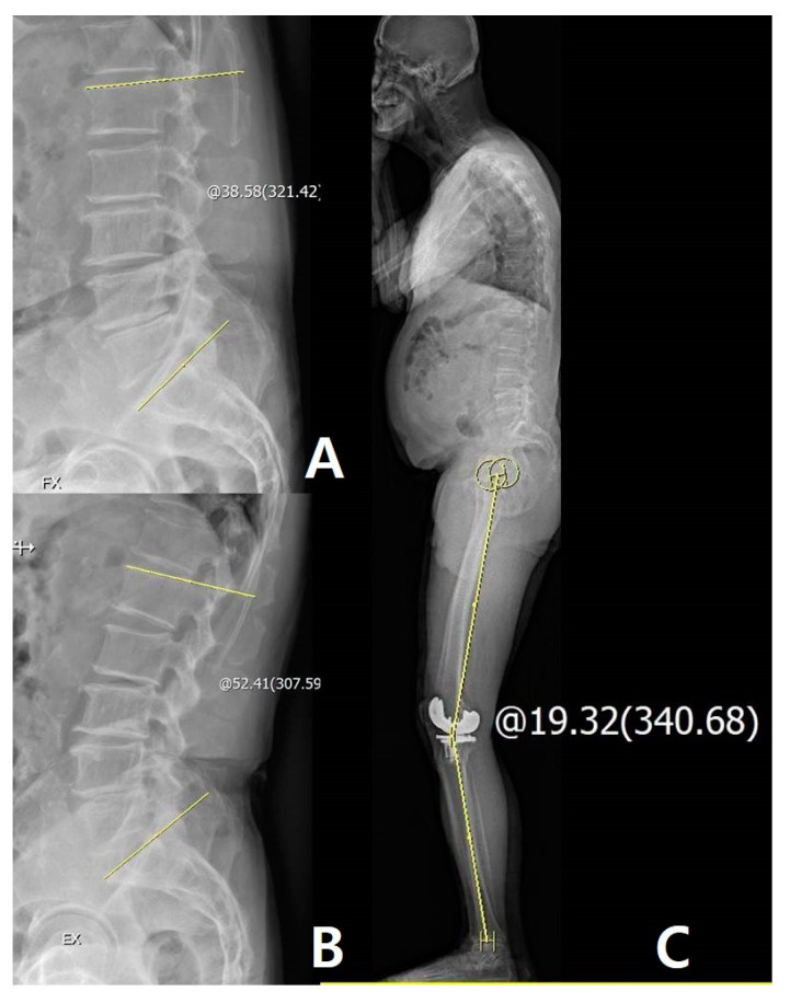
The purposes of this study were (1) to evaluate the relationship between lumbosacral flexibility and the effects of total knee arthroplasty (TKA) on whole-body alignment; and (2) to determine the prerequisites of the adjacent joints for successful TKA. A total of 116 patients (156 cases) who had whole-body X-ray and flexion-extension lumbar radiograph available were enrolled. For the sagittal alignment evaluation, hip-knee-ankle (HKA) angle, pelvic tilt (PT), sacral slope (SS), lumbar lordosis (LL), thoracic kyphosis (TK), and C7 plumb line-sacrum distance (SVA) were evaluated on the whole-body radiographs. Lumbar flexibility (LF) was evaluated using the flexion-extension lumbar radiographs, and pelvic flexibility (PF) was evaluated using the pelvic incidence (PI). The disparities in the knee joint between postoperative passive motion and weight-bearing posture were assessed. LF was significantly correlated with ΔLL and ΔSVA (LL: p = 0.039, SVA: p = 0.040; Pearson correlation coefficient (PCC): -0.206 and 0.205, respectively). There were correlations between PF and ΔSS (p < 0.001, PCC: -0.362), and between the disparity and LF (p = 0.005, PCC = -0.275). Linear regression analysis demonstrated that LF was significantly associated with the presence of disparity (p = 0.005, β = -0.205). LF is an important factor for improved spinal and lower limb alignment after TKA. Additionally, reduced LF may result in knee joint disparity between passive extension and standing extension status. Therefore, surgeons should consider spinopelvic alignment, including lower limb alignment preoperatively, to be able to predict possible changes in whole-body alignment following TKA.

Keywords: EOS; knee; osteoarthritis; sagittal alignment; spinal flexibility; total knee arthroplasty.


Conflict of interest statement

The authors declare no conflict of interest.

Figures

Figure 1
Figure 1
(A) Measurement of hip–knee–ankle (HKA) angle: +22.12°. (B) Measurement of PT: +27.02°. (C) Measurement of SS: +41.33°. (D) Measurement of TK and LL: +33.37° and +42.11°, respectively. (E) Measurement of C7 plumb line-sacrum distance: +13.64 mm. The sagittal HKA angle was defined as the angle between two lines: One joining the center of the femoral head and the center of the knee and the other joining the center of the knee and the center of the superiaor articular surface of the talus.
Figure 2
Figure 2
(A) Measurement of lumbar lordosis (LL) on extension view: 65.09°. (B) Measurement of LL on flexion view: 35.32°. (C) Measurement of pelvic incidence: 47.17°. The yellow line and circle are the measurement about lumbar lordosis and pelvic incidence
Figure 3
Figure 3
Patient with larger lumbar flexibility (LF) improved sagittal spinal alignment after TKA: Measurement of LF on flexion-extension view: 58.81°. (A) Measurement of LL on flexion view: 14.88°. (B) Measurement of LL on extension view: 73.69°, improved spinal alignment increased LL and decreased line-sacrum distance (SVA). (C) Measurement of the preoperative LL: 47.67° and SVA: 45.87 mm. (D) Measurement of postoperative LL: 66.48° and SVA: 15.73 mm. The yellow line and circle are the measurement about lumbar lordosis, SVA and sagittal HKA.
Figure 4
Figure 4
Patient with larger pelvic flexibility (PF) improved sagittal pelvic alignment, such as increased SS, after TKA. (A) Measurement of PF: 53.33°. (B) Measurement of preoperative SS: 28.57°. (C) Measurement of postoperative SS: 38.07°. The yellow line and circle are the measurement about pelvic incidence and sacral slope.
Figure 5
Figure 5
Patients with lesser LF have disparities in the knee joint between passive extension and real standing extension after TKA. (A) Measurement of LL on flexion view: 38.58°. (B) Measurement of LL on extension view: 52.41°. (C) Measurement of disparity is 19.32° with passive full extension. The yellow line and circle are the measurement about lumbar flexibility and sagittal HKA.
Figure 6
Figure 6
Cervical deformity showed little effect on the lower limb and spinal alignment itself. (A) Preoperative cervical lordosis: 3.35° and HKA angle: 8.63°. (B) Postoperative cervical lordosis: 3.55° and HKA angle: 2.37°. Thoracic deformity showed little effect on the lower limb and spinal alignment itself. (C) Preoperative thoracic kyphosis: 54.14° and HKA angle: 0.73°. (D) Postoperative thoracic kyphosis: 54.57° and HKA angle: 0.76°. The yellow line and circle are the measurement about sagittal HKA and cervical lordosis and thoracic kyphosis.

References

    1. Roussouly P., Pinheiro-Franco J.L. Biomechanical analysis of the spino-pelvic organization and adaptation in pathology. Eur. Spine J. 2011;5:609. doi: 10.1007/s00586-011-1928-x. - DOI - PMC - PubMed
    1. Chaleat-Valayer E., Mac-Thiong J.M., Paquet J., Berthonnaud E., Siani F., Roussouly P. Sagittal spino-pelvic alignment in chronic low back pain. Eur. Spine J. 2011;5:634. doi: 10.1007/s00586-011-1931-2. - DOI - PMC - PubMed
    1. Murata Y., Takahashi K., Yamagata M., Hanaoka E., Moriya H. The knee-spine syndrome. Association between lumbar lordosis and extension of the knee. J. Bone Jt. Surg. Br. 2003;85:95. doi: 10.1302/0301-620X.85B1.13389. - DOI - PubMed
    1. Cho M.R., Lee Y.S., Choi W.K. Relationship between Lateral Femoral Bowing and Varus Knee Deformity Based on Two-Dimensional Assessment of Side-to-Side Differences. Knee Surg. Relat. Res. 2018;30:58. doi: 10.5792/ksrr.17.007. - DOI - PMC - PubMed
    1. Shetty G.M., Mullaji A., Khalifa A.A., Ray A., Nikumbha V. The Effect of Sagittal Knee Deformity on Preoperative Measurement of Coronal Mechanical Alignment during Total Knee Arthroplasty. Knee Surg. Relat. Res. 2017;29:110. doi: 10.5792/ksrr.17.003. - DOI - PMC - PubMed