

miRNA-126-3p carried by human umbilical cord mesenchymal stem cell enhances endothelial function through exosome-mediated mechanisms in vitro and attenuates vein graft neointimal formation in vivo
- PMID: 33138861
- PMCID: PMC7607661
- DOI: 10.1186/s13287-020-01978-z
miRNA-126-3p carried by human umbilical cord mesenchymal stem cell enhances endothelial function through exosome-mediated mechanisms in vitro and attenuates vein graft neointimal formation in vivo
Abstract
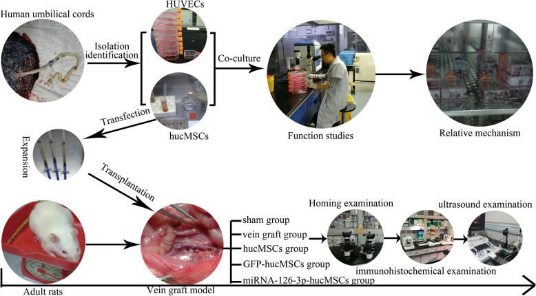
Background: The aim of this study was to determine whether the combination of MSC implantation with miRNA-126-3p overexpression would further improve the surgical results after vein grafting.
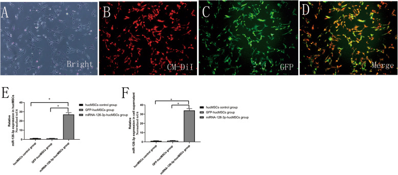
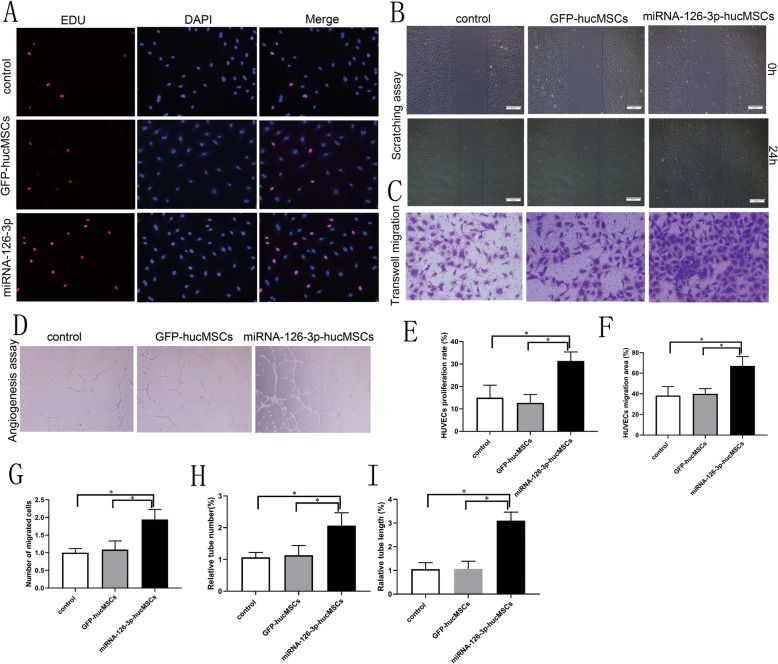
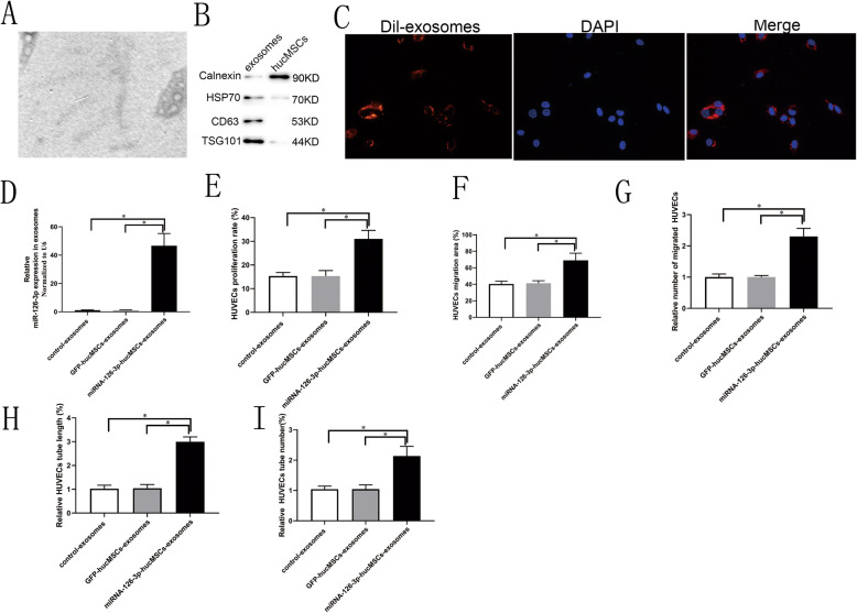
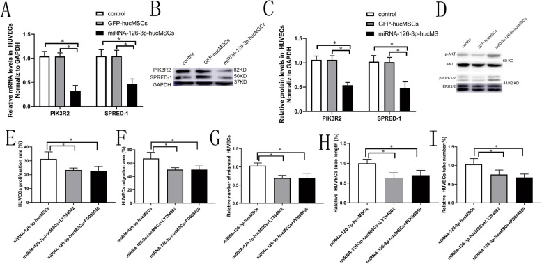
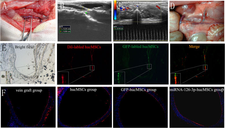
Methods: human umbilical cord MSCs (hucMSCs) and human umbilical vein endothelial cells (HUVECs) were isolated from human umbilical cords and characterized by a series of experiments. Lentivirus vector encoding miRNA-126-3p was transfected into hucMSCs and verified by PCR. We analyzed the miRNA-126-3p-hucMSC function in vascular endothelial cells by using a series of co-culture experiments. miRNA-126-3p-hucMSCs-exosomes were separated from cell culture supernatants and identified by WB and TEM. We validated the role of miRNA-126-3p-hucMSCs-exosomes on HUVECs proliferative and migratory and angiogenic activities by using a series of function experiments. We further performed co-culture experiments to detect downstream target genes and signaling pathways of miRNA-126-3p-hucMSCs in HUVECs. We established a rat vein grafting model, CM-Dil-labeled hucMSCs were injected intravenously into rats, and the transplanted cells homing to the vein grafts were detected by fluorescent microscopy. We performed historical and immunohistochemical experiments to exam miRNA-126-3p-hucMSC transplantation on vein graft neointimal formation and reendothelialization in vitro.
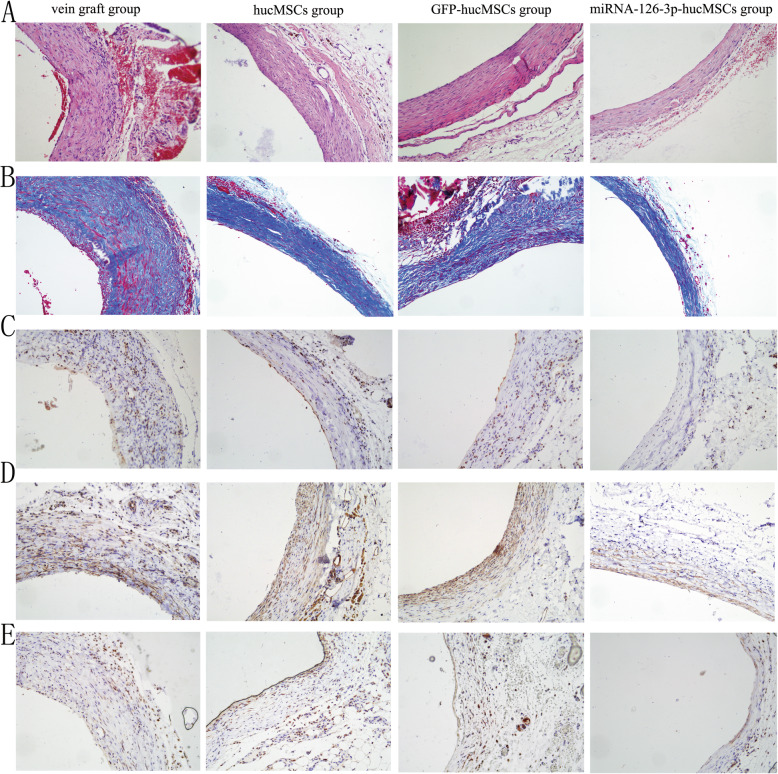
Results: We successfully isolated and identified primary hucMSCs and HUVECs. Primary hucMSCs were transfected with lentiviral vectors carrying miRNA-126-3p at a MOI 75. Co-culture studies indicated that overexpression of miRNA-126-3p in hucMSCs enhanced HUVECs proliferation, migration, and tube formation in vivo. We successfully separated hucMSCs-exosomes and found that miRNA-126-3p-hucMSCs-exosomes can strengthen the proliferative, migratory, and tube formation capacities of HUVECs. Further PCR and WB analysis indicated that, SPRED-1/PIK3R2/AKT/ERK1/2 pathways are involved in this process. In the rat vein arterialization model, reendothelialization analysis showed that transplantation with hucMSCs modified with miRNA-126-3p had a higher reendothelialization of the vein grafts. The subsequent historical and immunohistochemical examination revealed that delivery with miRNA-126-3p overexpressed hucMSCs significantly reduced vein graft intimal hyperplasia in rats.
Conclusion: These results suggest hucMSC-based miRNA-126-3p gene therapy may be a novel option for the treatment of vein graft disease after CABG.
Keywords: Exosomes; Mesenchymal stem cell; Neointimal hyperplasia; Reendothelialization; Vein graft; miRNA-126-3p.
Conflict of interest statement
On behalf of all authors, the corresponding author states that there is no conflict of interest.
Figures

Similar articles
-
Enhancement of endothelial function and attenuation of portal vein injury using mesenchymal stem cells carrying miRNA-25-3p.Sci Rep. 2024 Jul 2;14(1):15113. doi: 10.1038/s41598-024-64263-6. Sci Rep. 2024. PMID: 38956421 Free PMC article.
-
Exosomes derived from human umbilical cord mesenchymal stem cells inhibit vein graft intimal hyperplasia and accelerate reendothelialization by enhancing endothelial function.Stem Cell Res Ther. 2020 Mar 23;11(1):133. doi: 10.1186/s13287-020-01639-1. Stem Cell Res Ther. 2020. PMID: 32293542 Free PMC article.
-
miR-126-3p containing exosomes derived from human umbilical cord mesenchymal stem cells promote angiogenesis and attenuate ovarian granulosa cell apoptosis in a preclinical rat model of premature ovarian failure.Stem Cell Res Ther. 2022 Jul 26;13(1):352. doi: 10.1186/s13287-022-03056-y. Stem Cell Res Ther. 2022. PMID: 35883161 Free PMC article.
-
The Therapeutic Potential of Human Umbilical Cord Mesenchymal Stromal Cells Derived Exosomes for Wound Healing: Harnessing Exosomes as a Cell-free Therapy.J Stem Cells Regen Med. 2024 May 31;20(1):14-23. doi: 10.46582/jsrm.2003003. eCollection 2024. J Stem Cells Regen Med. 2024. PMID: 39044811 Free PMC article. Review.
-
Research Progress on the Osteogenesis-Related Regulatory Mechanisms of Human Umbilical Cord Mesenchymal Stem Cells.Stem Cell Rev Rep. 2023 Jul;19(5):1252-1267. doi: 10.1007/s12015-023-10521-5. Epub 2023 Mar 14. Stem Cell Rev Rep. 2023. PMID: 36917312 Review.
Cited by
-
microRNA-301a-3p is a potential biomarker in venous ulcers vein and gets involved in endothelial cell dysfunction.Bioengineered. 2022 Jun;13(6):14138-14158. doi: 10.1080/21655979.2022.2083821. Bioengineered. 2022. PMID: 35734851 Free PMC article.
-
miR-1307-5p regulates proliferation and apoptosis of chondrocytes in osteoarthritis by specifically inhibiting transforming growth factor beta-induced gene.Am J Transl Res. 2021 Jul 15;13(7):7756-7766. eCollection 2021. Am J Transl Res. 2021. PMID: 34377252 Free PMC article.
-
Enhancement of endothelial function and attenuation of portal vein injury using mesenchymal stem cells carrying miRNA-25-3p.Sci Rep. 2024 Jul 2;14(1):15113. doi: 10.1038/s41598-024-64263-6. Sci Rep. 2024. PMID: 38956421 Free PMC article.
-
Exosomal microRNAs from Mesenchymal Stem Cells: Novel Therapeutic Effect in Wound Healing.Tissue Eng Regen Med. 2023 Aug;20(5):647-660. doi: 10.1007/s13770-023-00542-z. Epub 2023 May 2. Tissue Eng Regen Med. 2023. PMID: 37131016 Free PMC article. Review.
-
Mesenchymal Stromal Cell-Derived Extracellular Vesicles for Vasculopathies and Angiogenesis: Therapeutic Applications and Optimization.Biomolecules. 2023 Jul 12;13(7):1109. doi: 10.3390/biom13071109. Biomolecules. 2023. PMID: 37509145 Free PMC article. Review.
References
-
- Hillis L, Smith P, Anderson J, et al. ACCF/AHA guideline for coronary artery bypass graft surgery. A report of the American College of Cardiology Foundation/American Heart Association Task Force on Practice Guidelines. Developed in collaboration with the American Association for Thoracic Surgery, Society of Cardiovascular Anesthesiologists, and Society of Thoracic Surgeons. J Am College Cardiol. 2011;58:e123–e210. doi: 10.1016/j.jacc.2011.08.009. - DOI - PubMed
-
- Gaudino M, Benedetto U, Fremes S, et al. Association of radial artery graft vs saphenous vein graft with long-term cardiovascular outcomes among patients undergoing coronary artery bypass grafting: a systematic review and meta-analysis. JAMA. 2020;324:179–187. doi: 10.1001/jama.2020.8228. - DOI - PMC - PubMed
MeSH terms
Substances
LinkOut - more resources
Full Text Sources
Miscellaneous
